Effects and Mechanisms of Metformin on the Proliferation of Esophageal Cancer Cells In Vitro and In Vivo
- Affiliations
-
- 1Department of Biochemistry, North of Sichuan Medical University, Nanchong, China. tangjiancai1980@163.com
- 2School of Basic Medical Sciences, North of Sichuan Medical University, Nanchong, China.
- 3Pathogenic Biology and Immunology Experiment Teaching Center, North of Sichuan Medical University, Nanchong, China.
- KMID: 2388323
- DOI: http://doi.org/10.4143/crt.2015.485
Abstract
- PURPOSE
The purpose of this study was to observe the effects of metformin on human esophageal cancer cell and to investigate its possible mechanisms.
MATERIALS AND METHODS
Cell viability was detected by using a Cell Counting Kit-8, while cell cycle and apoptosis were assessed by flow cytometry and western blot was used to measure the expression of the related proteins. RNAi was used to knockout pyruvate kinase muscle isozyme 2 (PKM2). An Eca109 tumor model was established to evaluate the antitumor effect in vivo. Immunohistochemistry was determined based on the expression of PKM2 and Bim in tumor tissues. Tunnel was used to assess tumor cell apoptosis.
RESULTS
Esophageal cancer cells viability was reduced after metformin treatment. The cell cycle was arrested in the G0/G1 phase, apoptosis was induced, caspase 3 was activated, caspase 9 was downregulated, and the pro-apoptotic protein Bim increased. Further study revealed that metformin could suppress the expression of insulin-like growth factor 1 receptor and its downstream proteins, phosphoinositide 3-kinase (PI3K), protein kinase B (AKT/PKB), phosphorylation of AKT (pAKT), mammalian target of rapamycin (mTOR), p70S6K, and PKM2. Insulin-like growth factor 1 partly reversed metfromin-induced apoptosis and attenuated the repression effect of metfomin to PI3K, pAKT, and PKM2. Knockout PKM2 resulted in the activation of caspase 3, down-regulation of caspase 9, and increased expression of Bim. In the Eca109 xenograft model, metformin significantly reduced tumor growth. Furthermore, we found that metformin treatment increased the rate of apoptosis, down-regulation of PKM2, and up-regulation of Bim in tumor tissues.
CONCLUSION
Metformin restrained esophageal cancer cell proliferation partly by suppressing the PI3K/AKT/mTOR pathway.
Keyword
MeSH Terms
-
Apoptosis
Blotting, Western
Caspase 3
Caspase 9
Cell Count
Cell Cycle
Cell Proliferation
Cell Survival
Down-Regulation
Esophageal Neoplasms*
Flow Cytometry
Heterografts
Humans
Immunohistochemistry
In Vitro Techniques*
Metformin*
Phosphorylation
Proto-Oncogene Proteins c-akt
Pyruvate Kinase
Repression, Psychology
Ribosomal Protein S6 Kinases, 70-kDa
RNA Interference
Sirolimus
Up-Regulation
Caspase 3
Caspase 9
Metformin
Proto-Oncogene Proteins c-akt
Pyruvate Kinase
Ribosomal Protein S6 Kinases, 70-kDa
Sirolimus
Figure
-
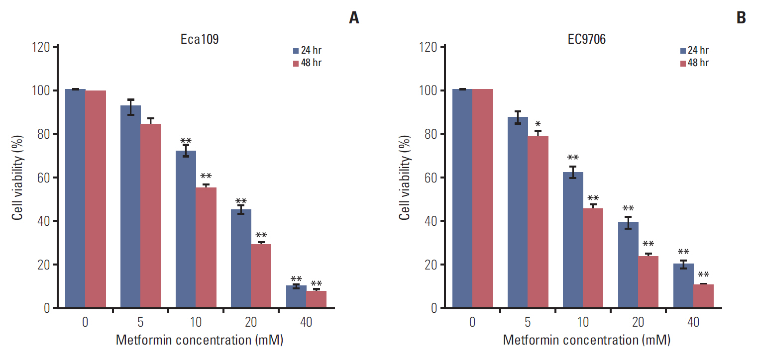
Fig. 1. Metformin inhibited the proliferation of esophageal cancer cells. Eca109 and EC9706 cells were treated with metformin (0, 5, 10, 20, and 40 mM) for 24 hours and 48 hours. Cell viability was determined by Cell Counting Kit-8. (A) Metformin reduced Eca109 cell viability. (B) Metformin decreased EC9706 cell viability. Data represent the mean±standard deviation of three independent experiments (*p < 0.05, **p < 0.01).
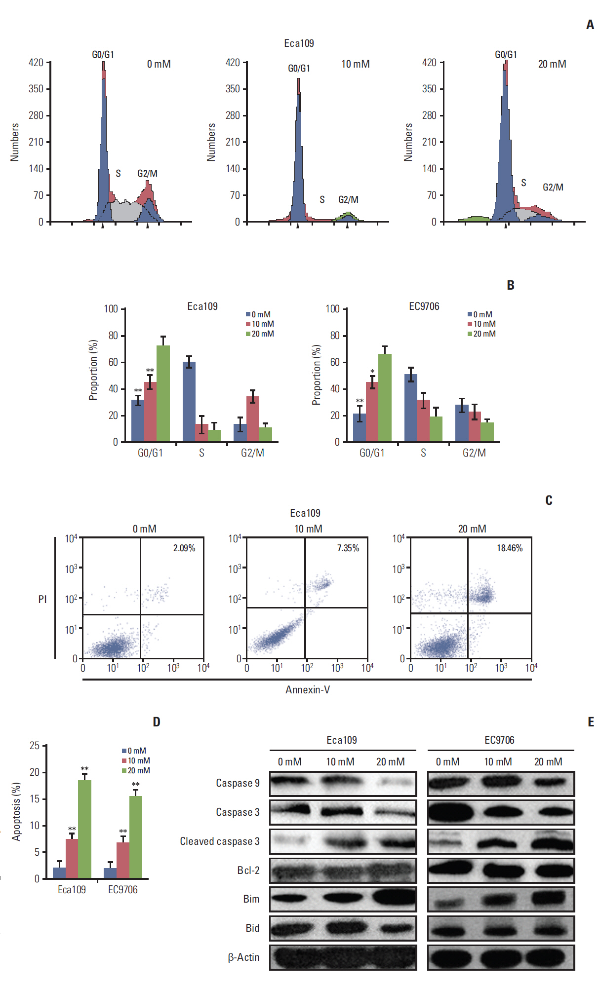
Fig. 2. Metformin induced cell cycle arrest and apoptosis in esophageal cancer cell cells. Eca109 and EC9706 Cells were treated with metformin (10 and 20 mM) for 24 hours. To investigate the cell cycle, cells were stained with propidium iodide (PI) according to the manufacturer’s instructions. For apoptosis determination, cells were analyzed by flow cytometry after staining with Annexin-V/PI according to the manufacturer’s instructions. (A) Typical flow cytometric graph of cell cycle. (B) The percentage of cells in each cell cycle phase. The results showed that metformin blocked cell arrest in the G0/G1 phrase in Eca109 and EC9706. (C) Typical flow cytometric graph of apoptosis. (D) The percentage of apoptotic cells. The results showed that metformin increased the rate of apoptosis in Eca109 and EC9706. (E) The expression of apoptosis-related proteins. Eca109 and EC9706 were treated with metformin. At 24 hours after treatment, the cells were lysed and subjected to immunoblot analysis. The levels of caspase 3, caspase 9, cleaved caspase 3, Bcl-2, Bim, and Bid were detected. Data represent the mean±standard deviation from three independent experiments (*p < 0.05 and **p < 0.01 compared to control, n=3).
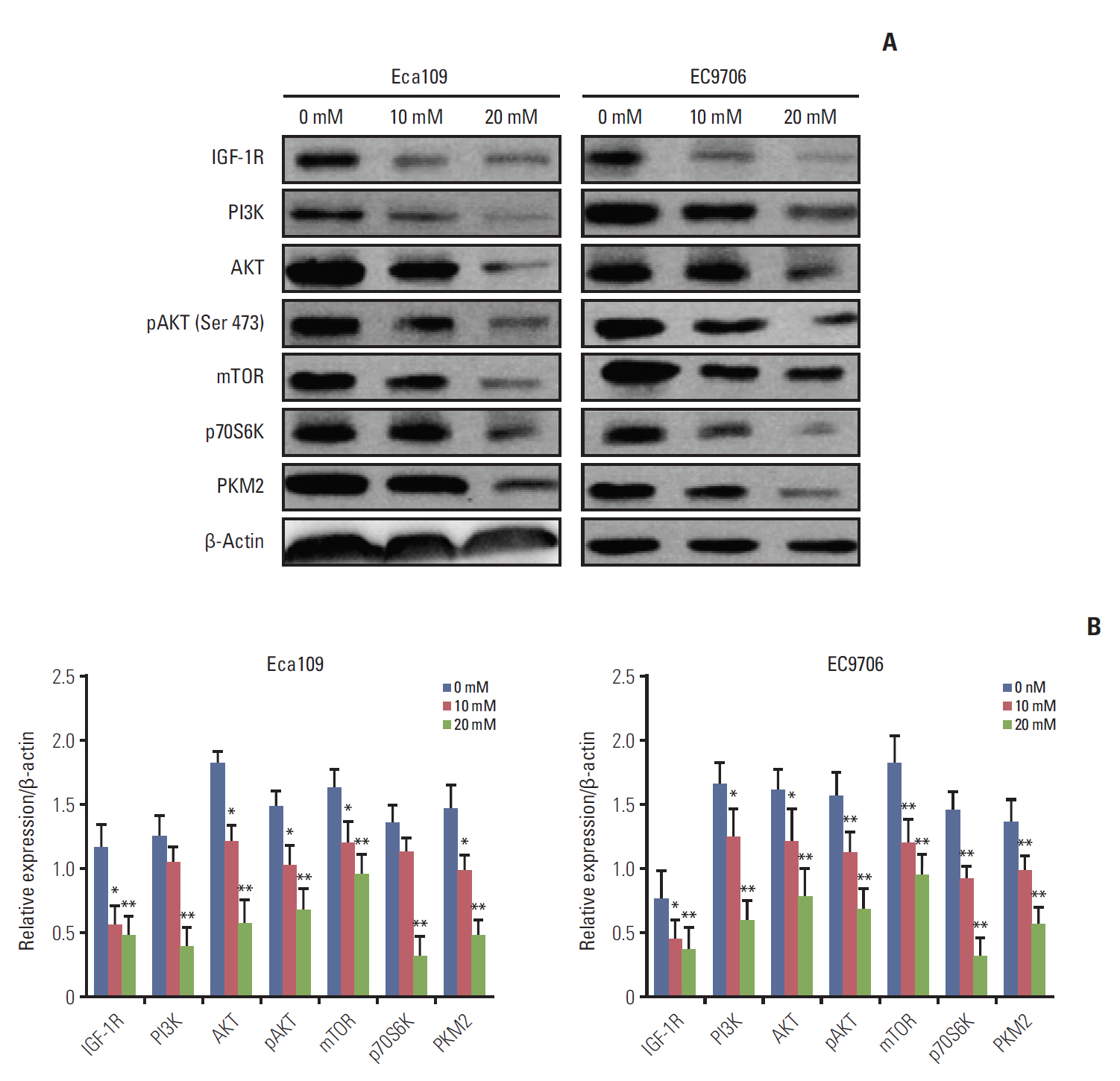
Fig. 3. Metformin suppressed the PI3K/AKT/mTOR signaling pathway. Eca109 and EC9706 cells were treated with metformin (0, 10, and 20 mM) for 24 hours. Total proteins were extracted as previously described. (A) Western blot analyses of IGF-1R, PI3K, AKT, pAKT, mTOR, p70S6K, and PKM2. Metformin reduced the IGF-1R expression and suppressed the PI3K/AKT/mTOR/PKM2 pathway. (B) Graph depicts densitometric analyses of IGF-1R, PI3K, AKT, pAKT, mTOR, p70S6K, and PKM2 normalized by β-actin (*p < 0.05 and **p < 0.01 compared to control, n=3). PI3K, phosphoinositide 3-kinase; pAKT, phosphorylation of AKT; mTOR, mammalian target of rapamycin; PKM2, pyruvate kinase muscle isozyme 2; IGF-1R, insulin-like growth factor 1 receptor.
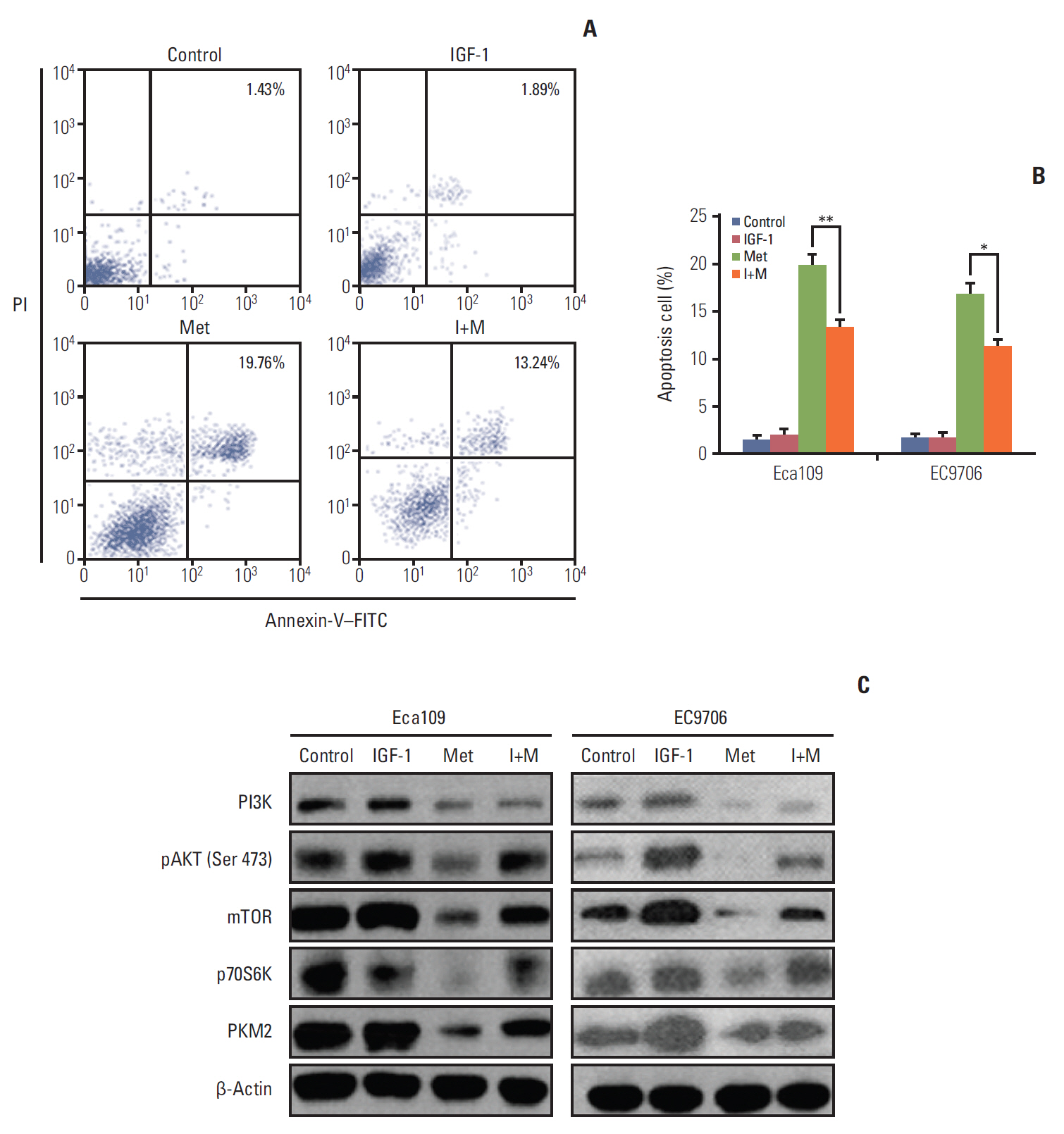
Fig. 4. IGF partly reversed the effect of metformin-induced apoptosis and attenuated the repression action of metfomin toward PI3K, pAKT, mTOR, p70S6K, and PKM2 in esophageal cancer cells. Eca109 and EC9706 cells were pre-treated with IGF-1 (100 ng/mL) for 2 hours, then with metformin for 24 hours. Apoptosis was determined by flow cytometry. (A) Typical flow cytometric graph of apoptosis. (B) The average percentage of apoptosis in Eca109 and EC9706 cells after treatment with metformin. The results showed that metformin partly prevented the metformin-induced apoptosis. (C) The expression of PI3K, pAKT, mTOR, p70S6K, and PKM2 was detected by western blot. The outcome indicates that IGF-1 attenuates the repression action of metformin toward PI3K, pAKT, mTOR, p70S6K, and PKM2 in Eca109 and EC9706 cells (*p < 0.05 and **p < 0.01 compared to control, n=3). PI3K, phosphoinositide 3-kinase; pAKT, phosphorylation of AKT; PKM2, pyruvate kinase muscle isozyme 2; IGF-1, insulin-like growth factor 1; mTOR, mammalian target of rapamycin; PI, propidium iodide; Met, metformin; I+M, insulin-like growth factors+metformin; FITC, fluorescein isothiocyanate.

Fig. 5. Knockdown of PKM2 altered the expression of apoptosis-related proteins. Eca109 or EC9706 cells (3×10⁵ cells/well) were plated in a 12-well plate. PKM2 was knocked down in Eca109 and EC9706 cells by siRNA (50 nM final concentration for both PKM2-specific and NC siRNA). At 48 hours after transfection, the whole cell extracts were collected for western blot. Total RNA was extracted from cultured cells (Eca109 and EC9706) using Trizol reagent according to the manufacturer’s instructions. β-Actin and Bim mRNA were quantified in duplicate by SYBR Green 2-step, real-time reverse transcription polymerase chain reaction. (A) Caspase 9, caspase 3, cleaved caspase 3, Bcl-2, and Bim were detected by western blot. The results showed that knockout PKM2 activated caspase 3 and downregulated caspase 9. Bim protein expression was evidently increased. (B) Bim mRNA level was increased in depletion PKM2 esophageal cancer cells. PKM2, pyruvate kinase muscle isozyme 2; NC, normal control.
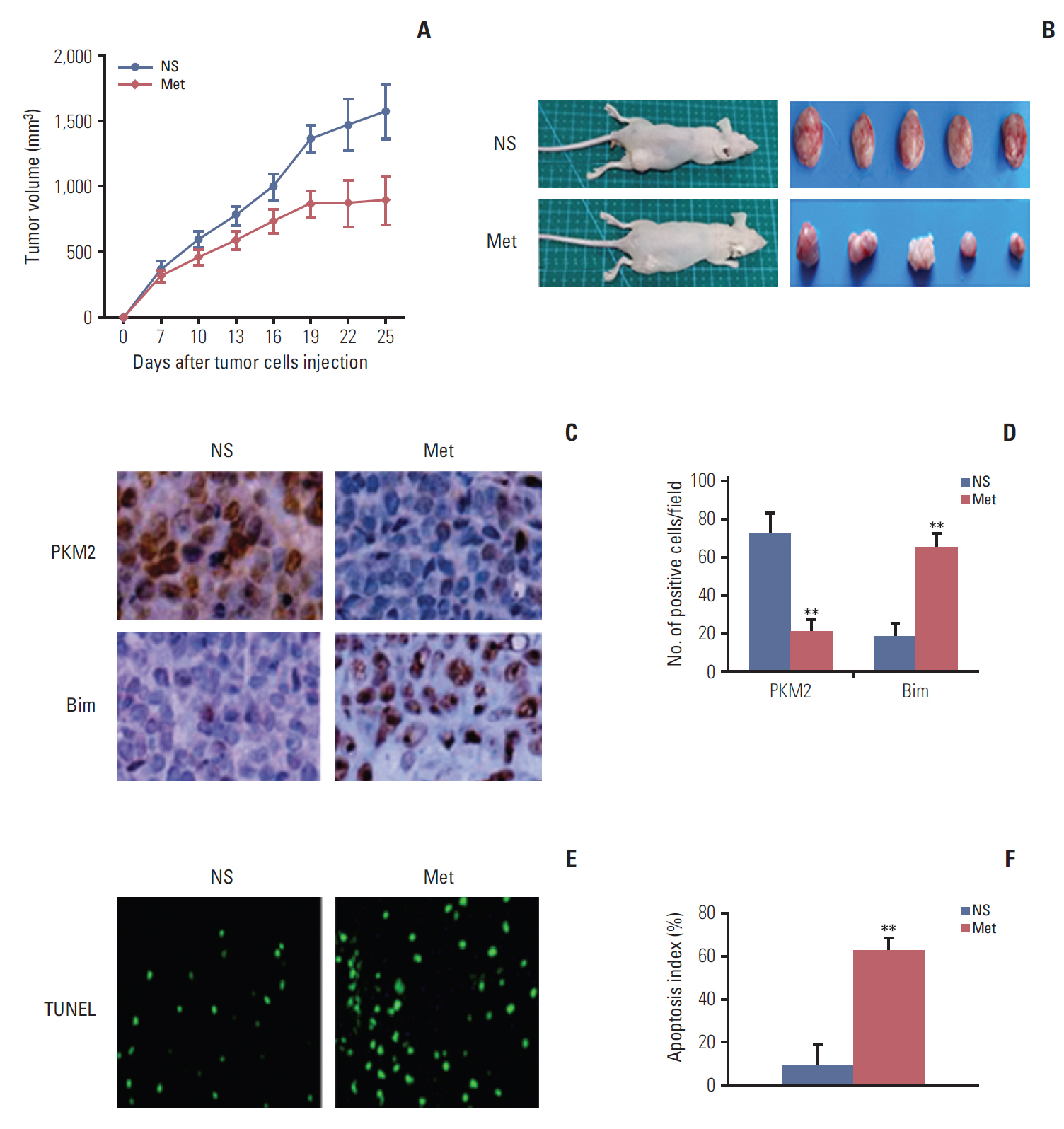
Fig. 6. Metformin inhibited tumor growth in vivo. Mice were treated with NS or metformin (300 mg/kg/day) for 21 days at the same time (n=5 per group). (A) Suppressed growth of tumor in mice. The results showed that metformin inhibited tumor growth (n=5, p < 0.01). Data are shown as the mean±standard deviation. (B) Representation of photos of tumors from mice bearing Eca109 receiving different treatments. Mice were sacrificed when they were moribund. (C) Typical graph of immunohistochemical staining of PKM2 and Bim in tumor tissues (×400). (D) Quantification of the number of PKM2 and Bim positive cells per field. Metformin treatment significantly decreased the number of PKM2-positive cells and increased the number of Bim-positive cells. (E) Typical sections from tumor tissue. (F) Apoptotic index within tissues. Metformin showed a significant increase of apoptotic cells in tumor tissues compared to the control (**p < 0.001). Data are expressed as the mean apoptotic index±standard deviation of cancer cells. NS, normal saline; PKM2, pyruvate kinase muscle isozyme 2; Met, metformin; TUNEL, terminal deoxynucleotidyl transferase dUTP nick end labeling.
Reference
-
References
1. Zhuang Y, Miskimins WK. Cell cycle arrest in Metformin treated breast cancer cells involves activation of AMPK, down-regulation of cyclin D1, and requires p27Kip1 or p21Cip1. J Mol Signal. 2008; 3:18.2. Huang Y, Guo W, Shi S, He J. Evaluation of the 7(th) edition of the UICC-AJCC tumor, node, metastasis classification for esophageal cancer in a Chinese cohort. J Thorac Dis. 2016; 8:1672–80.
Article3. Xie YE, Tang EJ, Zhang DR, Ren BX. Down-regulation of BclXL by RNA interference suppresses cell growth and induces apoptosis in human esophageal cancer cells. World J Gastroenterol. 2006; 12:7472–7.4. Tan R, Yao SZ, Huang ZQ, Li J, Li X, Tan HH, et al. Combination of FDG PET/CT and contrast-enhanced MSCT in detecting lymph node metastasis of esophageal cancer. Asian Pac J Cancer Prev. 2014; 15:7719–24.
Article5. Kato K, Gong J, Iwama H, Kitanaka A, Tani J, Miyoshi H, et al. The antidiabetic drug metformin inhibits gastric cancer cell proliferation in vitro and in vivo. Mol Cancer Ther. 2012; 11:549–60.
Article6. Shi WY, Xiao D, Wang L, Dong LH, Yan ZX, Shen ZX, et al. Therapeutic metformin/AMPK activation blocked lymphoma cell growth via inhibition of mTOR pathway and induction of autophagy. Cell Death Dis. 2012; 3:e275.
Article7. Zakikhani M, Dowling R, Fantus IG, Sonenberg N, Pollak M. Metformin is an AMP kinase-dependent growth inhibitor for breast cancer cells. Cancer Res. 2006; 66:10269–73.
Article8. Klil-Drori AJ, Azoulay L, Pollak MN. Cancer, obesity, diabetes, and antidiabetic drugs: is the fog clearing? Nat Rev Clin Oncol. 2017; 14:85–99.
Article9. Mu L, Zhu N, Zhang J, Xing F, Li D, Wang X. Type 2 diabetes, insulin treatment and prognosis of breast cancer. Diabetes Metab Res Rev. 2017; 33:e2823.
Article10. Vigneri R, Frasca F, Sciacca L, Vigneri P, Frittitta L. Re: Insulin, insulin-like growth factor-I, and risk of breast cancer in postmenopausal women. J Natl Cancer Inst. 2009; 101:1030–1.
Article11. Skinner HD, McCurdy MR, Echeverria AE, Lin SH, Welsh JW, O'Reilly MS, et al. Metformin use and improved response to therapy in esophageal adenocarcinoma. Acta Oncol. 2013; 52:1002–9.
Article12. Wild SH. Diabetes, treatments for diabetes and their effect on cancer incidence and mortality: attempts to disentangle the web of associations. Diabetologia. 2011; 54:1589–92.
Article13. Walker JJ, Johnson JA, Wild SH. Diabetes treatments and cancer risk: the importance of considering aspects of drug exposure. Lancet Diabetes Endocrinol. 2013; 1:132–9.
Article14. Memmott RM, Mercado JR, Maier CR, Kawabata S, Fox SD, Dennis PA. Metformin prevents tobacco carcinogen-induced lung tumorigenesis. Cancer Prev Res (Phila). 2010; 3:1066–76.
Article15. Iliopoulos D, Hirsch HA, Struhl K. Metformin decreases the dose of chemotherapy for prolonging tumor remission in mouse xenografts involving multiple cancer cell types. Cancer Res. 2011; 71:3196–201.
Article16. Rocha GZ, Dias MM, Ropelle ER, Osorio-Costa F, Rossato FA, Vercesi AE, et al. Metformin amplifies chemotherapy-induced AMPK activation and antitumoral growth. Clin Cancer Res. 2011; 17:3993–4005.
Article17. Zhuo Z, Wang A, Yu H. Metformin targeting autophagy overcomes progesterone resistance in endometrial carcinoma. Arch Gynecol Obstet. 2016; 294:1055–61.
Article18. Poli G, Cantini G, Armignacco R, Fucci R, Santi R, Canu L, et al. Metformin as a new anti-cancer drug in adrenocortical carcinoma. Oncotarget. 2016; 7:49636–48.
Article19. Towler MC, Hardie DG. AMP-activated protein kinase in metabolic control and insulin signaling. Circ Res. 2007; 100:328–41.
Article20. Ma W, Zhang T, Pan J, Shi N, Fan Q, Wang L, et al. Assessment of insulin-like growth factor 1 receptor as an oncogene in esophageal squamous cell carcinoma and its potential implication in chemotherapy. Oncol Rep. 2014; 32:1601–9.
Article21. Imsumran A, Adachi Y, Yamamoto H, Li R, Wang Y, Min Y, et al. Insulin-like growth factor-I receptor as a marker for prognosis and a therapeutic target in human esophageal squamous cell carcinoma. Carcinogenesis. 2007; 28:947–56.
Article22. Christofk HR, Vander Heiden MG, Harris MH, Ramanathan A, Gerszten RE, Wei R, et al. The M2 splice isoform of pyruvate kinase is important for cancer metabolism and tumour growth. Nature. 2008; 452:230–3.
Article23. Harris I, McCracken S, Mak TW. PKM2: a gatekeeper between growth and survival. Cell Res. 2012; 22:447–9.
Article24. Jain M, Nilsson R, Sharma S, Madhusudhan N, Kitami T, Souza AL, et al. Metabolite profiling identifies a key role for glycine in rapid cancer cell proliferation. Science. 2012; 336:1040–4.
Article25. Yang W, Xia Y, Hawke D, Li X, Liang J, Xing D, et al. PKM2 phosphorylates histone H3 and promotes gene transcription and tumorigenesis. Cell. 2012; 150:685–96.
Article26. Cai X, Hu X, Tan X, Cheng W, Wang Q, Chen X, et al. Metformin induced AMPK activation, G0/G1 phase cell cycle arrest and the inhibition of growth of esophageal squamous cell carcinomas in vitro and in vivo. PLoS One. 2015; 10:e0133349.
Article27. Banerjee J, Bruckbauer A, Zemel MB. Activation of the AMPK/Sirt1 pathway by a leucine-metformin combination increases insulin sensitivity in skeletal muscle, and stimulates glucose and lipid metabolism and increases life span in Caenorhabditis elegans. Metabolism. 2016; 65:1679–91.
Article28. Pollak MN. Investigating metformin for cancer prevention and treatment: the end of the beginning. Cancer Discov. 2012; 2:778–90.
Article29. Munoz-Pinedo C, El Mjiyad N, Ricci JE. Cancer metabolism: current perspectives and future directions. Cell Death Dis. 2012; 3:e248.
Article30. Feng Y, Ke C, Tang Q, Dong H, Zheng X, Lin W, et al. Metformin promotes autophagy and apoptosis in esophageal squamous cell carcinoma by downregulating Stat3 signaling. Cell Death Dis. 2014; 5:e1088.
Article31. Ye P, Qu CF, Hu XL. Impact of IGF-1, IGF-1R, and IGFBP-3 promoter methylation on the risk and prognosis of esophageal carcinoma. Tumour Biol. 2016; 37:6893–904.
Article32. Iqbal MA, Gupta V, Gopinath P, Mazurek S, Bamezai RN. Pyruvate kinase M2 and cancer: an updated assessment. FEBS Lett. 2014; 588:2685–92.
Article33. Cheong JH, Park ES, Liang J, Dennison JB, Tsavachidou D, Nguyen-Charles C, et al. Dual inhibition of tumor energy pathway by 2-deoxyglucose and metformin is effective against a broad spectrum of preclinical cancer models. Mol Cancer Ther. 2011; 10:2350–62.
Article34. Giovannucci E, Harlan DM, Archer MC, Bergenstal RM, Gapstur SM, Habel LA, et al. Diabetes and cancer: a consensus report. Diabetes Care. 2010; 33:1674–85.
Article35. Silvestri A, Palumbo F, Rasi I, Posca D, Pavlidou T, Paoluzi S, et al. Metformin induces apoptosis and downregulates pyruvate kinase M2 in breast cancer cells only when grown in nutrient-poor conditions. PLoS One. 2015; 10:e0136250.
Article
- Full Text Links
-
- Actions
-
Cited
- CITED
-
- Close
- Share
-
- Similar articles
-
- Metformin displays in vitro and in vivo antitumor effect against osteosarcoma
- Sanghuangporus sanghuang extract inhibits the proliferation and invasion of lung cancer cells in vitro and in vivo
- Metformin Suppresses Both PD-L1 Expression in Cancer Cells and Cancer-Induced PD-1 Expression in Immune Cells to Promote Antitumor Immunity
- Rethinking about Metformin: Promising Potentials
- The Metformin Use and Gastric Cancer Risk
