Immunomodulatory Effects of Dioscoreae Rhizome Against Inflammation through Suppressed Production of Cytokines Via Inhibition of the NF-kappaB Pathway
- Affiliations
-
- 1College of Pharmacy, Sahmyook University, Seoul 139-742, Korea. kimkj@syu.ac.kr
- 2College of Pharmacy, Chungbuk University, Cheongju 361-763, Korea.
- KMID: 2150747
- DOI: http://doi.org/10.4110/in.2012.12.5.181
Abstract
- Dioscoreae Rhizome (DR) has been used in traditional medicine to treat numerous diseases and is reported to have anti-diabetes and anti-tumor activities. To identify a bioactive traditional medicine with anti-inflammatory activity of a water extract of DR (EDR), we determined the mRNA and protein levels of proinflammatory cytokines in macrophages through RT-PCR and western blot analysis and performed a FACS analysis for measuring surface molecules. EDR dose-dependently decreased the production of NO and pro-inflammatory cytokines such as IL-1beta, IL-6, TNF-alpha, and PGE2, as well as mRNA levels of iNOS, COX-2, and pro-inflammatory cytokines, as determined by western blot and RT-PCR analysis, respectively. The expression of co-stimulatory molecules such as B7-1 and B7-2 was also reduced by EDR. Furthermore, activation of the nuclear transcription factor, NF-kappaB, but not that of IL-4 and IL-10, in macrophages was inhibited by EDR. These results show that EDR decreased pro-inflammatory cytokines via inhibition of NF-kappaB-dependent inflammatory protein level, suggesting that EDR could be a useful immunomodulatory agent for treating immunological diseases.
MeSH Terms
-
Blotting, Western
Cytokines
Dinoprostone
Dioscorea
Immune System Diseases
Inflammation
Interleukin-10
Interleukin-4
Interleukin-6
Macrophages
Medicine, Traditional
NF-kappa B
Rhizome
RNA, Messenger
Transcription Factors
Tumor Necrosis Factor-alpha
Water
Cytokines
Dinoprostone
Interleukin-10
Interleukin-4
Interleukin-6
NF-kappa B
RNA, Messenger
Transcription Factors
Tumor Necrosis Factor-alpha
Water
Figure
-
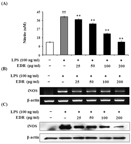
Figure 1 EDR inhibits the production of NO (A), expression of iNOS mRNA (B), and protein (C) in LPS-stimulated RAW 264.7 cells. RAW 264.7 cells were treated with various concentrations (25, 50, 100, 200 µg/ml) of EDR in the absence or presence of LPS (100 ng/ml) overnight. Culture supernatants were then collected and NO concentrations were measured using Griess reagent (A). Cell lysates were extracted, and protein levels of iNOS were then analyzed by Western blotting (B). RAW 264.7 cells were incubated with EDR in the absence or presence of LPS for 24 h. Total RNA was isolated, and levels of iNOS mRNA were then measured by RT-PCR (C). Each value represents the mean±S.D. of three independent experiments. ††p<0.01 vs. cells only based on Student's t-test. **p<0.01 vs. LPS only based on Student's t-test.
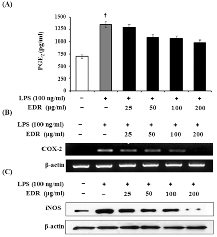
Figure 2 EDR inhibits the production of PGE2 (A), COX-2 mRNA (B) and protein (C) in LPS-stimulated RAW 264.7 cells. RAW 264.7 cells were treated with various concentrations (25, 50, 100, 200 µg/ml) of EDR in the absence or presence of LPS (100 ng/ml) for 48 h. Culture supernatants were then collected and PGE2 concentrations were measured using ELISA kits (A). Cell lysates were extracted, and protein levels of COX-2 were then analyzed by Western blotting (B). RAW 264.7 cells were incubated with EDR in the absence or presence of LPS for 24 h. Total RNA was isolated, and levels of COX-2 mRNA were then measured by RT-PCR (C). Each value represents the mean±S.D. of three independent experiments. †p<0.01 vs. cells only based on Student's t-test.
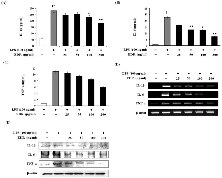
Figure 3 EDR inhibits pro-inflammatory cytokine production in LPS-stimulated RAW 264.7 cells. RAW 264.7 cells were treated with various concentrations (25, 50, 100, 200 µg/ml) of EDR in the absence or presence of LPS (100 ng/ml) for 48 h. Culture supernatants were then collected and cytokine concentrations were measured using ELISA kits (A~C). RAW 264.7 cells were incubated with EDR in the absence or presence of LPS for 24 h. Cell lysates were extracted, and protein levels of each cytokine were then analyzed by Western blotting (D). Total RNA was isolated, and mRNA levels of each cytokine were then measured by RT-PCR (E). Each value represents the mean±S.D. of three independent experiments. ††p<0.01 vs. cells only based on Student's t-test. *p<0.05, **p<0.01 vs. LPS only based on Student's t-test.
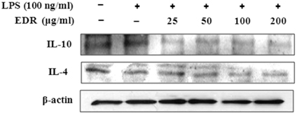
Figure 4 Effects of EDR on the anti-inflammatory cytokine production in LPS-stimulated RAW 264.7 cells. RAW 264.7 cells were treated with various concentrations (25, 50, 100, 200 µg/ml) of EDR in the absence or presence of LPS (100 ng/ml) for 24 h. Cell lysates were extracted, and protein levels of each cytokine were then analyzed by Western blotting.
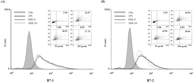
Figure 5 EDR inhibits the expression of co-stimulatory molecules in LPS-stimulated RAW 264.7 cells. RAW 264.7 cells were cultured and activated with LPS (100 ng/ml) in the absence or presence of various EDR concentrations for 24 hrs. The surface B7-1 (A) and B7-2 (B) were labeled with anti-B7-1/-2 antibodies and the cells were then stained using anti-Vβ8.1+8.2-FITC, anti-Vβ2-PE, or anti-Vβ2-FITC, which served as an isotype control for nonspecific binding.
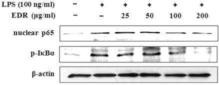
Figure 6 EDR inhibits the IκBα phosphorylation in the cytoplasm and the nuclear translocation of NF-κB p65 in LPS-stimulated RAW 264.7 cells. RAW 264.7 cells were treated with various concentrations (25, 50, 100, 200 µg/ml) of EDR in the absence or presence of LPS (100 ng/ml) for 24 hrs. Cell lysates were extracted, and protein levels of each cytokine were then analyzed by Western blotting.
Reference
-
1. Duncan BB, Schmidt MI, Pankow JS, Ballantyne CM, Couper D, Vigo A, Hoogeveen R, Folsom AR, Heiss G. Atherosclerosis Risk in Communities Study. Low-grade systemic inflammation and the development of type 2 diabetes: the atherosclerosis risk in communities study. Diabetes. 2003. 52:1799–1805.2. Jacobs M, van Greevenbroek MM, van der Kallen CJ, Ferreira I, Blaak EE, Feskens EJ, Jansen EH, Schalkwijk CG, Stehouwer CD. Low-grade inflammation can partly explain the association between the metabolic syndrome and either coronary artery disease or severity of peripheral arterial disease: the CODAM study. Eur J Clin Invest. 2009. 39:437–444.
Article3. Vane JR, Mitchell JA, Appleton I, Tomlinson A, Bishop-Bailey D, Croxtall J, Willoughby DA. Inducible isoforms of cyclooxygenase and nitric-oxide synthase in inflammation. Proc Natl Acad Sci USA. 1994. 91:2046–2050.
Article4. Lee TH, Kwak HB, Kim HH, Lee ZH, Chung DK, Baek NI, Kim J. Methanol extracts of Stewartia koreana inhibit cyclooxygenase-2 (COX-2) and inducible nitric oxide synthase (iNOS) gene expression by blocking NF-kappaB transactivation in LPS-activated RAW 264.7 cells. Mol Cells. 2007. 23:398–404.5. Nathan C. what difference does it make? J Clin Invest. 1997. 100:2417–2423.6. Bogdan C. Nitric oxide and the immune response. Nat Immunol. 2001. 2:907–916.
Article7. Vila-del Sol V, Fresno M. Involvement of TNF and NF-kappa B in the transcriptional control of cyclooxygenase-2 expression by IFN-gamma in macrophages. J Immunol. 2005. 174:2825. 2833.
Article8. Lind L. Circulating markers of inflammation and atherosclerosis. Atherosclerosis. 2003. 169:203. 214.
Article9. Tilg H, Wilmer A, Vogel W, Herold M, Nöolchen B, Judmaier G, Huber C. Serum levels of cytokines in chronic liver diseases. Gastroenterology. 1992. 103:264–274.
Article10. Coker RK, Laurent GJ. Pulmonary fibrosis: cytokines in the balance. Eur Respir J. 1998. 11:1218–1221.
Article11. Siebenlist U, Franzoso G, Brown K. Structure, regulation and function of NF-kappa B. Annu Rev Cell Biol. 1994. 10:405. 455.12. Renard P, Raes M. The proinflammatory transcription factor NFkappaB: a potential target for novel therapeutical strategies. Cell Biol Toxicol. 1999. 15:341–344.13. Tian B, Brasier AR. Identification of a nuclear factor kappa B-dependent gene network. Recent Prog Horm Res. 2003. 58:95–130.
Article14. Zhong X, Nishino E, Okagami N. Temperature dependence of seedling establishment of a perennial, Dioscorea tokoro. J Plant Res. 2002. 115:55–57.
Article15. Lee SC, Tsai CC, Chen JC, Lin CC, Hu ML, Lu S. The evaluation of reno- and hepatoprotective effects of huai-shan-yao (Rhizome Dioscoreae). Am J Chin Med. 2002. 30:609–616.
Article16. Tewtrakul S, Itharat A. Nitric oxide inhibitory substances from the rhizomes of Dioscorea membranacea. J Ethnopharmacol. 2007. 109:412. 416.
Article17. Stuehr DJ, Nathan CF. Nitric oxide. A macrophage product responsible for cytostasis and respiratory inhibition in tumor target cells. J Exp Med. 1989. 169:1543. 1555.
Article18. Lee GI, Ha JY, Min KR, Nakagawa H, Tsurufuji S, Chang IM, Kim Y. Inhibitory effects of oriental herbal medicines on IL-8 induction in lipopolysaccha-rideactivated rat macrophages. Planta Med. 1995. 61:26–30.
Article19. Laskin DL, Pendino KJ. Macrophages and inflammatory mediators in tissue injury. Annu Rev Pharmacol Toxicol. 1995. 35:655–677.
Article20. Furchgott R, Cherry P, Zawadzki J, Jothianandan D. Endothelial cells as mediators of vasodilation of arteries. J Cardiovasc Pharmacol. 1984. 6:S336–S343.
Article21. Wei X, Charles I, Smith A, Ure J, Feng G, Huang H. Altered immune responses in mice lacking inducible nitric oxide synthase. Nature. 1995. 375:408–411.
Article22. Ahmad N, Chen LC, Gordon MA, Laskin JD, Laskin DL. Regulation of cyclooxygenase-2 by nitric oxide in activated hepatic macrophages during acute endotoxemia. J Leukoc Biol. 2002. 71:1005–1011.23. Li Q, Verma IM. NFkappa B regulation in the immune system. Nat Rev Immunol. 2002. 2:725–734.24. Richmond A. NF-kappa B, chemokine gene transcription and tumour growth. Nat Rev Immunol. 2002. 2:664. 674.25. Lee S, Shin S, Kim H, Han S, Kim K, Kwon J, Kwak JH, Lee CK, NJ Ha, Yim D, Kim K. Antipin-flammatory function of arctiin by inhibiting COX-2 expression via NF-kappaB pathways. Inflammation. 2011. 8:16.26. Karin M, Ben-Neriah Y. The control of NF-kappa B activity. Annu Rev Immunol. 2000. 18:621–663.
- Full Text Links
-
- Actions
-
Cited
- CITED
-
- Close
- Share
-
- Similar articles
-
- Silymarin Inhibits Morphological Changes in LPS-Stimulated Macrophages by Blocking NF-kappaB Pathway
- Acetylshikonin Inhibits Human Pancreatic PANC-1 Cancer Cell Proliferation by Suppressing the NF-kappaB Activity
- Differential Regulation of NF-kappaB Signaling during Human Cytomegalovirus Infection
- Cordycepin Suppresses Expression of Diabetes Regulating Genes by Inhibition of Lipopolysaccharide-induced Inflammation in Macrophages
- Anticolitic Effect of the Rhizome Mixture of Anemarrhena asphodeloides and Coptidis chinensis (AC-mix) in Mice
