Induction of Melanoma Cell-Selective Apoptosis Using Anti-HER2 Antibody-Conjugated Gold Nanoparticles
- Affiliations
-
- 1Department of Oral & Maxillofacial Surgery, School of Dentistry, Pusan National University, Yangsan, Korea. kuksjs@pusan.ac.kr
- 2Department of Oral Anatomy, School of Dentistry, Pusan National University, Yangsan, Korea. ki91000m@pusan.ac.kr
- 3Feagle Co., Ltd., Yangsan, Korea.
- KMID: 2446955
- DOI: http://doi.org/10.3349/ymj.2019.60.6.509
Abstract
- PURPOSE
This study was conducted to verify the induction and mechanism of selective apoptosis in G361 melanoma cells using anti-HER2 antibody-conjugated gold nanoparticles (GNP-HER2).
MATERIALS AND METHODS
Following GNP-HER2 treatment of G361 cells, cell cycle arrest and apoptosis were measured by WST-1 assay, Hemacolor staining, Hoechst staining, immunofluorescence staining, fluorescence-activated cell sorting analysis, and Western blotting.
RESULTS
G361 cells treated with GNP-HER2 showed condensation of nuclei, which is an apoptotic phenomenon, and translocation of apoptosis-inducing factor and cytochrome c from mitochondria into the nucleus and cytoplasm, respectively. Increases in BAX in cells undergoing apoptosis, activation of caspase-3 and -9, and fragmentation of poly (ADP-ribose) polymerase and DNA fragmentation factor 45 (inhibitor of caspase-activated DNase) were observed upon GNP-HER2 treatment. Following GNP-HER2 treatment, an increase of cells in sub-G1 phase, which is a signal of cell apoptosis, was observed. This resulted in the down-regulation of cyclin A, cyclin D1, cyclin E, cdk2, cdk4, and cdc2 and the up-regulation of p21. Thus, GNP-HER2 treatment was confirmed to induce the cessation of cell cycle progression. Also, decreases in phospho-focal adhesion kinase and phospho-human epidermal growth factor receptor, which activate cellular focal adhesion, and decreases in phospho-paxillin, which stimulates the disassembly of filamentous actin, were observed. Reduced cell adhesion and disassembly of the intracellular structure indicated cell deactivation.
CONCLUSION
GNP-HER2 can selectively kill G361 melanoma cells without affecting normal cells. The mechanism of G361 cell death upon treatment with GNP-HER2 was apoptosis accompanied by activation of caspases.
Keyword
MeSH Terms
-
Actins
Apoptosis Inducing Factor
Apoptosis*
Blotting, Western
Caspase 3
Caspases
Cell Adhesion
Cell Cycle
Cell Cycle Checkpoints
Cell Death
Cyclin A
Cyclin D1
Cyclin E
Cyclins
Cytochromes c
Cytoplasm
DNA Fragmentation
Down-Regulation
Flow Cytometry
Fluorescent Antibody Technique
Focal Adhesions
Melanoma*
Mitochondria
Nanoparticles*
Phosphotransferases
Receptor, Epidermal Growth Factor
Up-Regulation
Actins
Apoptosis Inducing Factor
Caspase 3
Caspases
Cyclin A
Cyclin D1
Cyclin E
Cyclins
Cytochromes c
Phosphotransferases
Receptor, Epidermal Growth Factor
Figure
-

Fig. 1 (A) Schematic diagram of GNP-HER2 with tumor marker attached to gold nanoparticles (NPs). (B) Comparison of expression levels of HER2 protein by Western blot analysis. The expression level of melanoma cells (G361) was significantly higher than in keratinocytes (HaCaT). GNP-HER2, gold nanoparticles combined with the antibody against HER2.

Fig. 2 The effects of GNP-HER2 on the proliferation of G361 cells (A), HaCaT cells (B), and HER2 antibody-only cell lines (C). Cells were incubated with GNP-HER2 for 24, 48, and 72 hours and then analyzed by the WST-1 method. GNP-HER2, gold nanoparticles combined with the antibody against HER2.

Fig. 3 Apoptosis induced by GNP-HER2. G361 was treated with GNP-HER2 and Hemacolor staining after 24-hour incubation. (A) G361 cells treated with GNP-HER2 (lower) and untreated cells (upper). (B) Cells treated with GNP-HER2 (right) and apoptotic bodies and untreated cells (left) after Hoechst staining (×1000). GNP-HER2, gold nanoparticles combined with the antibody against HER2.
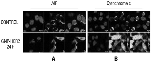
Fig. 4 Photomicrograph of GNP-HER2 for 24 h (lower end) and non-use (upper end). Immunofluorescent staining followed by confocal microscopy (×800). (A) Localization changes of intracellular AIF were observed through immunocytochemistry using anti-AIF-antibodies. (B) Changes in the localization of cytochrome c due to GNP-HER2 treatment for 24 hours were observed by immunocytochemistry using anti-cytochrome c antibody. GNP-HER2, gold nanoparticles combined with the antibody against HER2; AIF, apoptosis inducing factor.
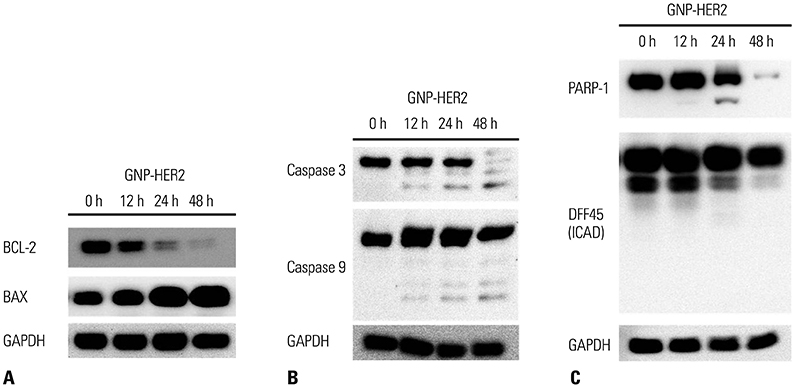
Fig. 5 The activity of GNP-HER2-induced apoptosis-related proteins in G361 cells was observed by Western blotting (A–C). G361 cells were incubated with GNP-HER2 at the scheduled time. Total protein lysates were Bax/Bcl-2 (A), caspase-3/caspase-9N (B), and PARP-1/DFF45 (C) antibodies. Glyceraldehyde-3-phosphate dehydrogenase was used for protein quantification. GNP-HER2, gold nanoparticles combined with the antibody against HER2.

Fig. 6 G361 cells treated with GNP-HER2 induce apoptosis. G361 cells were treated with GNP-HER2 for 12 to 48 hours. At each time point, fluorescence-activated cell sorting analysis was performed using Pl. GNP-HER2, gold nanoparticles combined with the antibody against HER2.
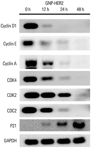
Fig. 7 Western blot analysis showing that GNP-HER2 effectively reduced protein expression for cell cycle arrest. When G361 cells and GNP-HER2 were co-cultured, expression of p21; cyclin D, E, and A; and the cyclin partners cdk2, cdk4, and cdc2 was observed. GNP-HER2, gold nanoparticles combined with the antibody against HER2.
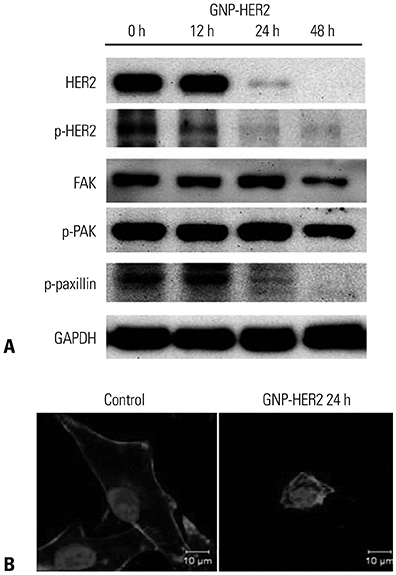
Fig. 8 GNP-HER2 treatment significantly decreased focal adhesion proteins in the cells over time. G361 cells were incubated for the indicated time with GNP-HER2, and then, Western blot tests were performed using p-paxillin, p-FAK, p-HER2, FAK, and HER2 antibodies. (A) GNP-HER2 severely induces the disintegration of filamentous actin. (B) Control group was not treated with GNP-HER2 (left). F-actin staining of G361 cells was treated with GNP-HER2 for 24 h (right). GNP-HER2, gold nanoparticles combined with the antibody against HER2; FAK, focal adhesion kinase.
Reference
-
1. Kim SH, Hashimoto Y, Cho SN, Roszik J, Milton DR, Dal F, et al. Microsomal PGE2 synthase-1 regulates melanoma cell survival and associates with melanoma disease progression. Pigment Cell Melanoma Res. 2016; 29:297–308.
Article2. Soengas MS, Lowe SW. Apoptosis and melanoma chemoresistance. Oncogene. 2003; 22:3138–3151.
Article3. Service RF. Materials and biology. Nanotechnology takes aim at cancer. Science. 2005; 310:1132–1134.
Article4. Lam CW, James JT, McCluskey R, Hunter RL. Pulmonary toxicity of single-wall carbon nanotubes in mice 7 and 90 days after intratracheal instillation. Toxicol Sci. 2004; 77:126–134.
Article5. Shi Kam NW, Jessop TC, Wender PA, Dai H. Nanotube molecular transporters: internalization of carbon nanotube-protein conjugates into Mammalian cells. J Am Chem Soc. 2004; 126:6850–6851.
Article6. Magrez A, Kasas S, Salicio V, Pasquier N, Seo JW, Celio M, et al. Cellular toxicity of carbon-based nanomaterials. Nano Lett. 2006; 6:1121–1125.
Article7. Derfus AM, Chan WCW, Bhatia SN. Probing the cytotoxicity of semiconductor quantum dots. Nano Lett. 2004; 4:11–18.
Article8. Wu X, Liu H, Liu J, Haley KN, Treadway JA, Larson JP, et al. Immunofluorescent labeling of cancer marker Her2 and other cellular targets with semiconductor quantum dots. Nat Biotechnol. 2003; 21:41–46.
Article9. Ahamed M, Akhtar MJ, Raja M, Ahmad I, Siddiqui MK, AlSalhi MS, et al. ZnO nanorod-induced apoptosis in human alveolar adenocarcinoma cells via p53, survivin and bax/bcl-2 pathways: role of oxidative stress. Nanomedicine. 2011; 7:904–913.
Article10. Arya G, Vandana M, Acharya S, Sahoo SK. Enhanced antiproliferative activity of Herceptin (HER2)-conjugated gemcitabine-loaded chitosan nanoparticle in pancreatic cancer therapy. Nanomedicine. 2011; 7:859–870.
Article11. Connor EE, Mwamuka J, Gole A, Murphy CJ, Wyatt MD. Gold nanoparticles are taken up by human cells but do not cause acute cytotoxicity. Small. 2005; 1:325–327.
Article12. Thomas M, Klibanov AM. Conjugation to gold nanoparticles enhances polyethylenimine's transfer of plasmid DNA into mammalian cells. Proc Natl Acad Sci U S A. 2003; 100:9138–9143.
Article13. Rosenberg SJ, Loening SA, Hawtrey CE, Narayana AS, Culp DA. Radical prostatectomy with adjuvant radioactive gold for prostatic cancer: a preliminary report. J Urol. 1985; 133:225–227.
Article14. Patra HK, Banerjee S, Chaudhuri U, Lahiri P, Dasgupta AK. Cell selective response to gold nanoparticles. Nanomedicine. 2007; 3:111–119.
Article15. El-Sayed IH, Huang X, El-Sayed MA. Surface plasmon resonance scattering and absorption of anti-EGFR antibody conjugated gold nanoparticles in cancer diagnostics: applications in oral cancer. Nano Lett. 2005; 5:829–834.
Article16. El-Sayed IH, Huang X, El-Sayed MA. Selective laser photo-thermal therapy of epithelial carcinoma using anti-EGFR antibody conjugated gold nanoparticles. Cancer Lett. 2006; 239:129–135.
Article17. Bargmann CI, Hung MC, Weinberg RA. The neu oncogene encodes an epidermal growth factor receptor-related protein. Nature. 1986; 319:226–230.
Article18. Rubin I, Yarden Y. The basic biology of HER2. Ann Oncol. 2001; 12:Suppl 1. S3–S8.
Article19. Neve RM, Lane HA, Hynes NE. The role of overexpressed HER2 in transformation. Ann Oncol. 2001; 12:Suppl 1. S9–S13.
Article20. Ullrich A, Schlessinger J. Signal transduction by receptors with tyrosine kinase activity. Cell. 1990; 61:203–212.
Article21. Kämmerer U, Thanner F, Kapp M, Dietl J, Sütterlin M. Expression of tumor markers on breast and ovarian cancer cell lines. Anticancer Res. 2003; 23:1051–1055.22. Nakamura H, Saji H, Ogata A, Hosaka M, Hagiwara M, Kawasaki N, et al. Correlation between encoded protein overexpression and copy number of the HER2 gene with survival in non-small cell lung cancer. Int J Cancer. 2003; 103:61–66.
Article23. Ross JS, McKenna BJ. The HER-2/neu oncogene in tumors of the gastrointestinal tract. Cancer Invest. 2001; 19:554–568.24. Oehler MK, Brand A, Wain GV. Molecular genetics and endometrial cancer. J Br Menopause Soc. 2003; 9:27–31.
Article25. Jacobson MD, Weil M, Raff MC. Programmed cell death in animal development. Cell. 1997; 88:347–354.
Article26. Nagata S. Apoptosis by death factor. Cell. 1997; 88:355–365.
Article27. Alberts B, Johnson A, Lewis J, Raff M, Roberts K, Walter P. Apoptosis: programmed cell death eliminates unwanted cells. Molecular biology of the cell. 5th ed. New York: Garland Science;2008.28. Sabour Alaoui S, Dessirier V, de Araujo E, Alexaki VI, Pelekanou V, Lkhider M, et al. TWEAK affects keratinocyte G2/M growth arrest and induces apoptosis through the translocation of the AIF protein to the nucleus. PLoS One. 2012; 7:e33609.
Article29. Arias-González I, García-Carrancà AM, Cornejo-Garrido J, Ordaz-Pichardo C. Cytotoxic effect of Kalanchoe flammea and induction of intrinsic mitochondrial apoptotic signaling in prostate cancer cells. J Ethnopharmacol. 2018; 222:133–147.
Article30. Fan Y, Chiu JF, Liu J, Deng Y, Xu C, Zhang J, et al. Resveratrol induces autophagy-dependent apoptosis in HL-60 cells. BMC Cancer. 2018; 18:581.
Article31. Luo T, Yuan Y, Yu Q, Liu G, Long M, Zhang K, et al. PARP-1 overexpression contributes to Cadmium-induced death in rat proximal tubular cells via parthanatos and the MAPK signalling pathway. Sci Rep. 2017; 7:4331.
Article32. Errami Y, Brim H, Oumouna-Benachour K, Oumouna M, Naura AS, Kim H, et al. ICAD deficiency in human colon cancer and predisposition to colon tumorigenesis: linkage to apoptosis resistance and genomic instability. PLoS One. 2013; 8:e57871.
Article
- Full Text Links
-
- Actions
-
Cited
- CITED
-
- Close
- Share
-
- Similar articles
-
- The Study of EGFR Expression on Malignant Skin Tumor and Molecular Labelling Using Gold Nanoparticles
- Induction of Selective Cell Death of Oral Squamous Carcinoma Cells by Integrin alpha2 Antibody and EGFR Antibody
- Clinical Application of Gold Nanoparticles for Diagnosis and Treatment
- Trastuzumab-Conjugated Liposome-Coated Fluorescent Magnetic Nanoparticles to Target Breast Cancer
- Immunohistochemical Study of Malignant Melanoma with HMB - 45 Monoclonal Antibody and Anti S - 100 Protein Antibody
