Ferulic acid prevents the injury-induced decrease of gamma-enolase expression in brain tissue and HT22 cells
- Affiliations
-
- 1Department of Anatomy, College of Veterinary Medicine, Research Institute of Life Science, Gyeongsang National University, Jinju, Korea. pokoh@gnu.ac.kr
- KMID: 2312114
- DOI: http://doi.org/10.5625/lar.2014.30.1.8
Abstract
- Ferulic acid is known to act as a protective agent in cerebral ischemia through its anti-oxidant activity. gamma-Enolase is a neuron-specific enolase that also exerts a neuroprotective effect. Here, we investigated whether ferulic acid regulates the expression level of gamma-enolase in middle cerebral artery occlusion (MCAO)-induced brain injury and glutamate exposure-induced neuronal cell death. Adult male rats were treated with either vehicle or ferulic acid (100 mg/kg, i.v.) after MCAO and cerebral cortex tissues were collected 24 h after MCAO. Using a proteomics approach, we found that gamma-enolase expression was decreased in MCAO-injured animals treated with vehicle alone, whereas ferulic acid treatment attenuated this decrease. Reverse-transcription PCR and Western blot analyses confirmed that ferulic acid treatment prevented MCAO injury-induced decrease in gamma-enolase. Furthermore, in hippocampal-derived cell lines, glutamate exposure also decreased gamma-enolase expression and ferulic acid treatment attenuated this glutamate-induced decrease in gamma-enolase. These findings suggest that ferulic acid mediates a neuroprotective effect by attenuating injury-induced decreases of gamma-enolase expression in neuronal cells.
Keyword
MeSH Terms
-
Adult
Animals
Blotting, Western
Brain Injuries
Brain Ischemia
Brain*
Cell Death
Cell Line
Cerebral Cortex
Glutamic Acid
Humans
Infarction, Middle Cerebral Artery
Male
Neurons
Neuroprotective Agents
Phosphopyruvate Hydratase*
Polymerase Chain Reaction
Proteomics
Rats
Glutamic Acid
Neuroprotective Agents
Phosphopyruvate Hydratase
Figure
-
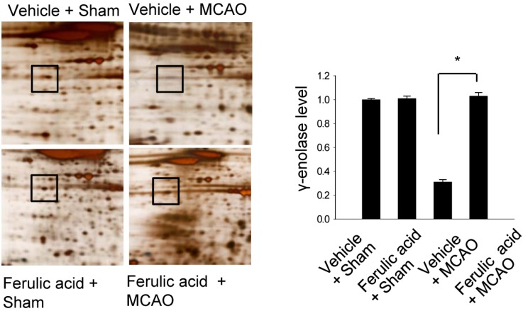
Figure 1 γ-Enolase protein spots identified by MALDI-TOF in the vehicle + sham, ferulic acid + sham, vehicle + middle cerebral artery occlusion (MCAO), ferulic acid + MCAO groups. Squares indicate the γ-enolase protein spots. Spot intensities were measured using PDQuest software. Intensity ratios represent the average spot intensities of treated animals relative to the average spot intensity of control (vehicle + sham) animals. Data (n=3) are shown as mean±SEM. *P<0.05.
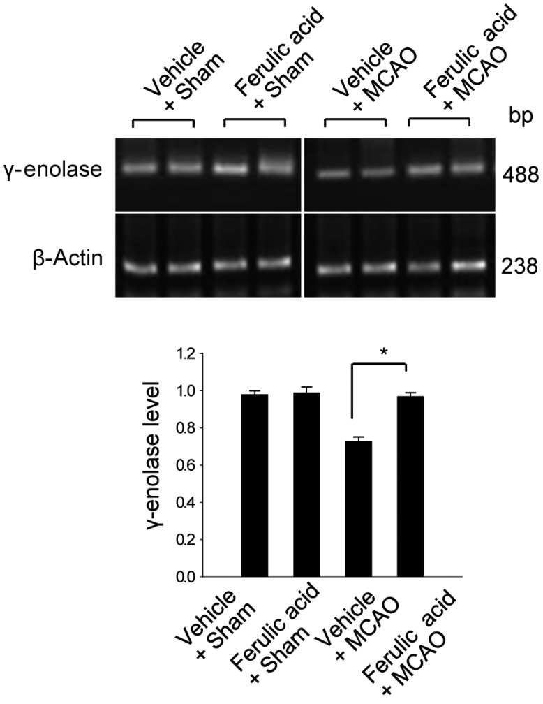
Figure 2 Reverse-transcription PCR analysis of γ-enolase expression levels in the vehicle + sham, ferulic acid + sham, vehicle + middle cerebral artery occlusion (MCAO), ferulic acid + MCAO groups. Each lane represents an individual animal. Densitometric analysis was performed to express the intensity of each γ-enolase band relative to the intensity of the corresponding actin control band. Data (n=5) are shown as mean±SEM. *P<0.05.
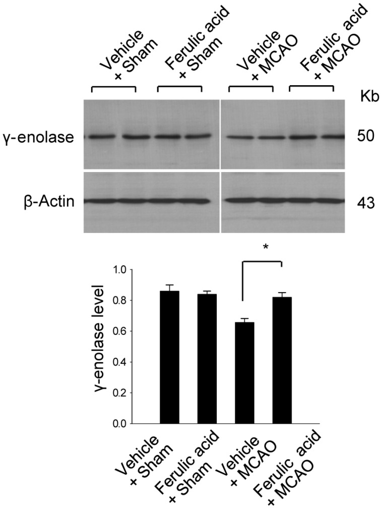
Figure 3 Western blot analysis of γ-enolase protein levels in the vehicle + sham, ferulic acid + sham, vehicle + middle cerebral artery occlusion (MCAO), ferulic acid + MCAO groups. Each lane represents an individual animal. Densitometric analysis was performed to express the intensity of each γ-enolase band relative to the intensity of the corresponding actin control band. Data (n=5) are shown as mean±SEM. *P<0.05.
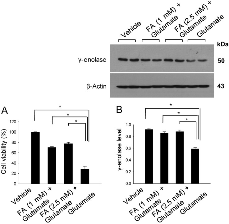
Figure 4 Cell viability (A) and Western blot analyses of γ-enolase protein levels (B) in hippocampal neuronal HT22 cells. Glutamate (5 mM) was added to HT22 cells for 24 h, with ferulic acid (FA, 1 or 2.5 mM) added to cells 30 min before glutamate exposure. Cell viability was assessed using the MTT assay (A). Cell survival is expressed as the percentage of viable treated cells out of the percentage of viable vehicle-treated cells (set to 100%). Densitometric analysis was performed to express the intensity of each γ-enolase band relative to the intensity of the corresponding actin control band (B). Data (n=5) are represented as mean±SEM. *P<0.05.
Cited by 1 articles
-
Curcumin attenuates the middle cerebral artery occlusion-induced reduction in γ-enolase expression in an animal model
Sang-Ah Gim, So-Ra Lee, Fawad-Ali Shah, Phil-Ok Koh
Lab Anim Res. 2015;31(4):198-203. doi: 10.5625/lar.2015.31.4.198.
Reference
-
1. Rider CC, Taylor CB. Enolase isoenzymes in rat tissues. Electrophoretic, chromatographic, immunological and kinetic properties. Biochim Biophys Acta. 1974; 365(1):285–300. PMID: 4413246.
Article2. Schmechel DE, Brightman MW, Marangos PJ. Neurons switch from non-neuronal enolase to neuron-specific enolase during differentiation. Brain Res. 1980; 190(1):195–214. PMID: 6769533.
Article3. Hattori T, Takei N, Mizuno Y, Kato K, Kohsaka S. Neurotrophic and neuroprotective effects of neuron-specific enolase on cultured neurons from embryonic rat brain. Neurosci Res. 1995; 21(3):191–198. PMID: 7753500.
Article4. Hafner A, Obermajer N, Kos J. γ-Enolase C-terminal peptide promotes cell survival and neurite outgrowth by activation of the PI3K/Akt and MAPK/ERK signalling pathways. Biochem J. 2012; 443(2):439–450. PMID: 22257123.
Article5. Cheng CY, Ho TY, Lee EJ, Su SY, Tang NY, Hsieh CL. Ferulic acid reduces cerebral infarct through its antioxidative and anti-inflammatory effects following transient focal cerebral ischemia in rats. Am J Chin Med. 2008; 36(6):1105–1119. PMID: 19051339.
Article6. Gim SA, Sung JH, Shah FA, Kim MO, Koh PO. Ferulic acid regulates the AKT/GSK-3β/CRMP-2 signaling pathway in a middle cerebral artery occlusion animal model. Lab Anim Res. 2013; 29(2):63–69. PMID: 23825478.
Article7. Koh PO. Ferulic acid modulates nitric oxide synthase expression in focal cerebral ischemia. Lab Anim Res. 2012; 28(4):273–278. PMID: 23326288.
Article8. Jin Y, Yan EZ, Fan Y, Guo XL, Zhao YJ, Zong ZH, Liu Z. Neuroprotection by sodium ferulate against glutamate-induced apoptosis is mediated by ERK and PI3 kinase pathways. Acta Pharmacol Sin. 2007; 28(12):1881–1890. PMID: 18031600.
Article9. Cheng CY, Su SY, Tang NY, Ho TY, Chiang SY, Hsieh CL. Ferulic acid provides neuroprotection against oxidative stress-related apoptosis after cerebral ischemia/reperfusion injury by inhibiting ICAM-1 mRNA expression in rats. Brain Res. 2008; 1209:136–150. PMID: 18400211.
Article10. Ou L, Kong LY, Zhang XM, Niwa M. Oxidation of ferulic acid by Momordica charantia peroxidase and related anti-inflammation activity changes. Biol Pharm Bull. 2003; 26(11):1511–1516. PMID: 14600392.
Article11. Joshi G, Perluigi M, Sultana R, Agrippino R, Calabrese V, Butterfield DA. In vivo protection of synaptosomes by ferulic acid ethyl ester (FAEE) from oxidative stress mediated by 2,2-azobis(2-amidino-propane)dihydrochloride (AAPH) or Fe(2+)/H(2)O(2): insight into mechanisms of neuroprotection and relevance to oxidative stress-related neurodegenerative disorders. Neurochem Int. 2006; 48(4):318–327. PMID: 16386335.
Article12. Srinivasan M, Sudheer AR, Menon VP. Ferulic Acid: therapeutic potential through its antioxidant property. J Clin Biochem Nutr. 2007; 40(2):92–100. PMID: 18188410.
Article13. Longa EZ, Weinstein PR, Carlson S, Cummins R. Reversible middle cerebral artery occlusion without craniectomy in rats. Stroke. 1989; 20(1):84–91. PMID: 2643202.
Article14. Sung JH, Kim MO, Koh PO. Proteomic identification of proteins differentially expressed by nicotinamide in focal cerebral ischemic injury. Neuroscience. 2011; 174:171–177. PMID: 21111033.
Article
- Full Text Links
-
- Actions
-
Cited
- CITED
-
- Close
- Share
-
- Similar articles
-
- Ferulic acid modulates nitric oxide synthase expression in focal cerebral ischemia
- Ferulic acid regulates the AKT/GSK-3beta/CRMP-2 signaling pathway in a middle cerebral artery occlusion animal model
- Curcumin attenuates the middle cerebral artery occlusion-induced reduction in gamma-enolase expression in an animal model
- Focal Cerebral Ischemia Induces Decrease of Astrocytic Phosphoprotein PEA-15 in Brain Tissue and HT22 Cells
- Combined Method of Neuronal Cell-Inducible Vector and Valproic Acid for Enhanced Gene Expression under Hypoxic Conditions
