miR-195/miR-497 Regulate CD274 Expression of Immune Regulatory Ligands in Triple-Negative Breast Cancer
- Affiliations
-
- 1The 1st Ward of the Medical Department, Affiliated Cancer Hospital and Institute of Guangzhou Medical University, Guangzhou, China. chhpan@163.com
- 2Radiotherapy Department, Central Hospital of Guangdong Nongken, Zhanjiang, China.
- 3State Key Laboratory of Oncology in South China, Collaborative Innovation Center for Cancer Medicine, Sun Yat-sen University Cancer Center, Guangzhou, China.
- KMID: 2429815
- DOI: http://doi.org/10.4048/jbc.2018.21.e60
Abstract
- PURPOSE
Immune suppression is common in patients with advanced breast cancer but the mechanisms underlying this phenomenon have not been sufficiently studied. In this study, we aimed to identify B7 family members that were able to predict the immune status of patients, and which may serve as potential targets for the treatment of breast cancer. We also aimed to identify microRNAs that may regulate the expression of B7 family members.
METHODS
The Cancer Genome Atlas data from 1,092 patients with breast cancer, including gene expression, microRNA expression and survival data, were used for statistical and survival analyses. Polymerase chain reaction and Western blot were used to measure messenger RNA and protein expression, respectively. Luciferase assay was used to investigate direct microRNA target.
RESULTS
Bioinformatic analysis predicted that microRNA (miR)-93, miR-195, miR-497, and miR-340 are potential regulators of the immune evasion of breast cancer cells, and that they exert this function by targeting CD274, PDCD1LG2, and NCR3LG1. We chose CD274 for further investigations. We found that miR-195, miR-497, and CD274 expression levels were inversely correlated in MDA-MB-231 cells, and miR-195 and miR-497 expressions mimic inhibited CD274 expression in vitro. Mechanistic investigations demonstrated that miR-195 and miR-497 directly target CD274 3"² untranslated region.
CONCLUSION
Our data indicated that the level of B7 family members can predict the prognosis of breast cancer patients, and miR-195/miR-497 regulate CD274 expression in triple negative breast cancer. This regulation may further influence tumor progression and the immune tolerance mechanism in breast cancer and may be able to predict the effect of immunotherapy on patients.
Keyword
MeSH Terms
-
Antigens, CD274
B7 Antigens
Blotting, Western
Breast Neoplasms
Computational Biology
Gene Expression
Genome
Humans
Immune Evasion
Immune Tolerance
Immunotherapy
In Vitro Techniques
Ligands*
Luciferases
MicroRNAs
Polymerase Chain Reaction
Prognosis
RNA, Messenger
Triple Negative Breast Neoplasms*
Untranslated Regions
Antigens, CD274
B7 Antigens
Ligands
Luciferases
MicroRNAs
RNA, Messenger
Untranslated Regions
Figure
-
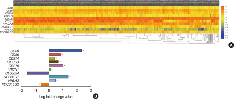
Figure 1 Identification of differentially expressed messenger RNAs (mRNAs) in the The Cancer Genome Atlas (TCGA) BRCA database. (A) Heat map of the log2-fold expression changes in 10 B7 family members mRNAs in the TCGA BRCA database (n=1,092). The horizontal row represents different mRNAs, and the vertical column represents different patients. Green squares indicate increases, and red squares indicate decreases. (B) Statistical analysis of altered expression of B7 family members in the TCGA BRCA data. The false discovery rate for all data is less than 0.01.CD80=cluster of differentiation 80; CD86=cluster of differentiation 86; CD274=cluster of differentiation 274; ICOSLG=inducible T-cell costimulator ligand; CD276=cluster of differentiation 276; VTCN1=V-set domain containing T cell activation inhibitor 1; C10orf54=chromosome 10 open reading frame 54; NCR3LG1=natural killer cell cytotoxicity receptor 3 ligand 1; HHLA2=HERV-H LTR-associating 2; PDCD1LG2=programmed cell death 1 ligand 2. *p<0.05.
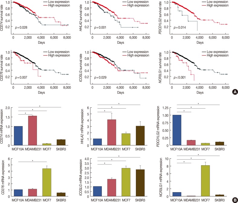
Figure 2 Relationship between six B7 family members and overall survival in breast cancer patients. (A) Using The Cancer Genome Atlas survival data and Cutoff Finder (http://molpath.charite.de/cutoff/), we correlated the messenger RNA (mRNA) expression level of B7 family members with overall survival. Six B7 family members, CD274, CD276, HHLA2, ICOSLG, NCR3LG1, and PDCD1LG2, had prognostic value in patients with breast cancer (log-rank p<0.05). Notably, high CD274, HHLA2, and PDCD1LG2 expression correlated with longer survival, whereas high CD276, ICOSLG, and NCR3LG1 expression correlated with shorter survival. (B) Real-time polymerase chain reaction was performed to determine CD274, CD276, HHLA2, ICOSLG, NCR3LG1, and PDCD1LG2 expression in MCF7, MDA-MB-231, SK-BR-3 and human fibrocystic disease epithelium cell lines MCF10A.CD274=cluster of differentiation 274; HHLA2=HERV-H LTR-associating 2; PDCD1LG2=programmed cell death 1 ligand 2; CD276=cluster of differentiation 276; ICOSLG=inducible T-cell costimulator ligand; NCR3LG1=natural killer cell cytotoxicity receptor 3 ligand 1. *p<0.05.
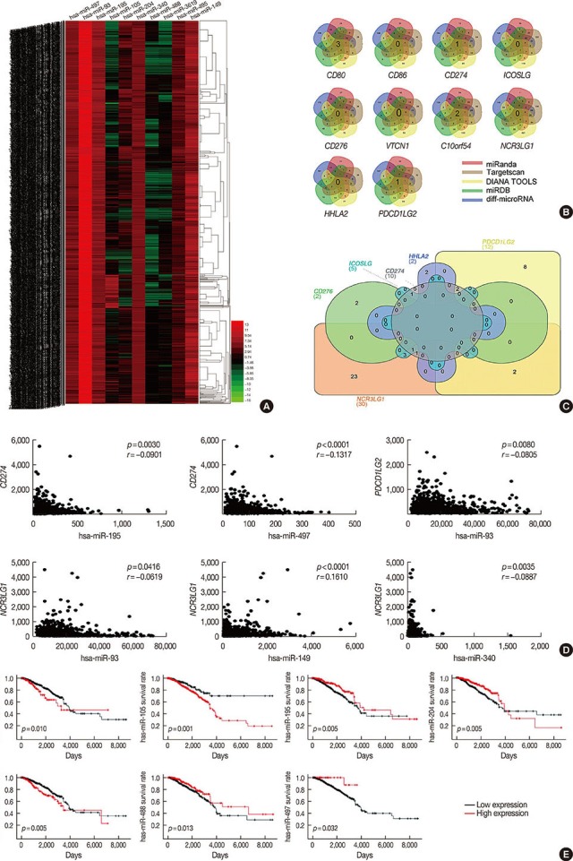
Figure 3 Identify the microRNA may target the B7 family in breast cancer. (A) Heat map of the log2-fold expression changes of 10 microRNAs in the The Cancer Genome Atlas (TCGA) BRCA database (n=1,092). The horizontal row represents different microRNAs, and the vertical column represents different patients. Green squares indicate increases, and red squares indicate decreases. (B) Venn diagrams depicting five sets of data: TargetScan, miRanda, DIANA TOOLS, miRDB and 256 differentially expressed microRNAs in the TCGA database of breast cancer. (C) Venn diagrams were used to select microRNAs that intersect with B7 family members, which have prognostic value in breast cancer. (D) We analyzed the correlation between target microRNAs and the gene expression of B7 family members. The horizontal axis represents microRNA, the vertical axis represents members of the B7 family (n=1,085) (p<0.05). (E) Relationship between microRNA and overall survival in breast cancer.CD80=cluster of differentiation 80; CD86=cluster of differentiation 86; CD274=cluster of differentiation 274; ICOSLG=inducible T-cell costimulator ligand; CD276=cluster of differentiation 276; VTCN1=V-set domain containing T cell activation inhibitor 1; C10orf54=chromosome 10 open reading frame 54; NCR3LG1=natural killer cell cytotoxicity receptor 3 ligand 1; HHLA2=HERV-H LTR-associating 2; PDCD1LG2=programmed cell death 1 ligand 2.
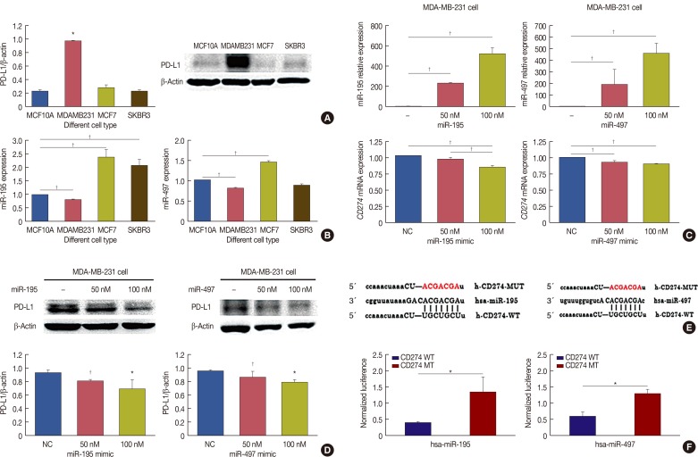
Figure 4 MicroRNA (miR)-195 and miR-497 directly targets CD274. (A) CD274 was detected in MCF7, MDA-MB-231, SK-BR-3 and MCF10A cells by Western blot. (B) Real-time polymerase chain reaction (RT-PCR) was performed to determine miR-195 and miR-497 expression in MCF7, MDA-MB-231, SK-BR-3 and MCF10A. (C) MDA-MB-231 was transfected with miR-195 or miR-497. miR-195, miR-497 and CD274 messenger RNA (mRNA) expression levels were determined via a quantitative RT-PCR assay. (D) MDA-MB-231 was transfected with miR-195 or miR-497. The expression levels of CD274 were determined by Western blotting. (E) CD274 are potential targets of miR-195 and miR-497. The miR-195 and miR-497 target sites and mutant sites in the 3′ untranslated region (3′UTR)s of CD274 are shown. (F) The luciferase vectors that contain the human wild-type (WT) and mutant (MT) CD274 3′UTR regions were co-transfected into MDA-MB-231 cells with miR-195/miR-497 mimic or negative control (NC) miR mimic. The normalized luciferase/Renilla activities were analyzed in the cells 48 hours after the transfection.CD274=cluster of differentiation; PD-L1=programmed death-ligand 1. *p<0.05; †p<0.01.
Reference
-
1. Ferlay J, Soerjomataram I, Dikshit R, Eser S, Mathers C, Rebelo M, et al. Cancer incidence and mortality worldwide: sources, methods and major patterns in GLOBOCAN 2012. Int J Cancer. 2015; 136:E359–E386. PMID: 25220842.
Article2. DeSantis CE, Fedewa SA, Goding Sauer A, Kramer JL, Smith RA, Jemal A. Breast cancer statistics, 2015: convergence of incidence rates between black and white women. CA Cancer J Clin. 2016; 66:31–42. PMID: 26513636.
Article3. Fan L, Strasser-Weippl K, Li JJ, St Louis J, Finkelstein DM, Yu KD, et al. Breast cancer in China. Lancet Oncol. 2014; 15:e279–e289. PMID: 24872111.
Article4. Lin S, Gregory RI. MicroRNA biogenesis pathways in cancer. Nat Rev Cancer. 2015; 15:321–333. PMID: 25998712.
Article5. Zou W, Chen L. Inhibitory B7-family molecules in the tumour microenvironment. Nat Rev Immunol. 2008; 8:467–477. PMID: 18500231.
Article6. Schildberg FA, Klein SR, Freeman GJ, Sharpe AH. Coinhibitory pathways in the B7-CD28 ligand-receptor family. Immunity. 2016; 44:955–972. PMID: 27192563.
Article7. Callahan MK, Postow MA, Wolchok JD. Targeting T cell co-receptors for cancer therapy. Immunity. 2016; 44:1069–1078. PMID: 27192570.
Article8. Zhao L, Yu H, Yi S, Peng X, Su P, Xiao Z, et al. The tumor suppressor miR-138-5p targets PD-L1 in colorectal cancer. Oncotarget. 2016; 7:45370–45384. PMID: 27248318.
Article9. Xu S, Tao Z, Hai B, Liang H, Shi Y, Wang T, et al. miR-424(322) reverses chemoresistance via T-cell immune response activation by blocking the PD-L1 immune checkpoint. Nat Commun. 2016; 7:11406. PMID: 27147225.
Article10. Robinson MD, McCarthy DJ, Smyth GK. edgeR: a Bioconductor package for differential expression analysis of digital gene expression data. Bioinformatics. 2010; 26:139–140. PMID: 19910308.
Article11. Budczies J, Klauschen F, Sinn BV, Győrffy B, Schmitt WD, Darb-Esfahani S, et al. Cutoff Finder: a comprehensive and straightforward Web application enabling rapid biomarker cutoff optimization. PLoS One. 2012; 7:e51862. PMID: 23251644.
Article12. Gibson J. Anti-PD-L1 for metastatic triple-negative breast cancer. Lancet Oncol. 2015; 16:e264.
Article13. Wu R, Li F, Zhu J, Tang R, Qi Q, Zhou X, et al. A functional variant at miR-132-3p, miR-212-3p, and miR-361-5p binding site in CD80 gene alters susceptibility to gastric cancer in a Chinese Han population. Med Oncol. 2014; 31:60. PMID: 24981235.
Article14. Yuan SM, Li H, Yang M, Zha H, Sun H, Li XR, et al. High intensity focused ultrasound enhances anti-tumor immunity by inhibiting the negative regulatory effect of miR-134 on CD86 in a murine melanoma model. Oncotarget. 2015; 6:37626–37637. PMID: 26485753.
Article15. Wu L, Chen Z, Zhang J, Xing Y. Effect of miR-513a-5p on etoposide-stimulating B7-H1 expression in retinoblastoma cells. J Huazhong Univ Sci Technolog Med Sci. 2012; 32:601–606. PMID: 22886978.
Article16. Guo W, Tan W, Liu S, Huang X, Lin J, Liang R, et al. MiR-570 inhibited the cell proliferation and invasion through directly targeting B7-H1 in hepatocellular carcinoma. Tumour Biol. 2015; 36:9049–9057. PMID: 26084609.
Article17. Zhu J, Chen L, Zou L, Yang P, Wu R, Mao Y, et al. MiR-20b, -21, and -130b inhibit PTEN expression resulting in B7-H1 over-expression in advanced colorectal cancer. Hum Immunol. 2014; 75:348–353. PMID: 24468585.
Article18. Chen L, Gibbons DL, Goswami S, Cortez MA, Ahn YH, Byers LA, et al. Metastasis is regulated via microRNA-200/ZEB1 axis control of tumour cell PD-L1 expression and intratumoral immunosuppression. Nat Commun. 2014; 5:5241. PMID: 25348003.
Article19. Wang X, Li J, Dong K, Lin F, Long M, Ouyang Y, et al. Tumor suppressor miR-34a targets PD-L1 and functions as a potential immunotherapeutic target in acute myeloid leukemia. Cell Signal. 2015; 27:443–452. PMID: 25499621.
Article20. Yang P, Tang R, Zhu J, Zou L, Wu R, Zhou H, et al. A functional variant at miR-24 binding site in B7-H2 alters susceptibility to gastric cancer in a Chinese Han population. Mol Immunol. 2013; 56:98–103. PMID: 23688438.
Article21. Xu H, Cheung IY, Guo HF, Cheung NK. MicroRNA miR-29 modulates expression of immunoinhibitory molecule B7-H3: potential implications for immune based therapy of human solid tumors. Cancer Res. 2009; 69:6275–6281. PMID: 19584290.
Article22. Wang ZS, Zhong M, Bian YH, Mu YF, Qin SL, Yu MH, et al. MicroRNA-187 inhibits tumor growth and invasion by directly targeting CD276 in colorectal cancer. Oncotarget. 2016; 7:44266–44276. PMID: 27329595.
Article23. Wang L, Kang FB, Sun N, Wang J, Chen W, Li D, et al. The tumor suppressor miR-124 inhibits cell proliferation and invasion by targeting B7-H3 in osteosarcoma. Tumour Biol. 2016; 37:14939–14947. PMID: 27644254.
Article24. Zhou X, Mao Y, Zhu J, Meng F, Chen Q, Tao L, et al. TGF-beta1 promotes colorectal cancer immune escape by elevating B7-H3 and B7-H4 via the miR-155/miR-143 axis. Oncotarget. 2016; 7:67196–67211. PMID: 27626488.25. Muenst S, Schaerli AR, Gao F, Däster S, Trella E, Droeser RA, et al. Expression of programmed death ligand 1 (PD-L1) is associated with poor prognosis in human breast cancer. Breast Cancer Res Treat. 2014; 146:15–24. PMID: 24842267.
Article26. Baptista MZ, Sarian LO, Derchain SF, Pinto GA, Vassallo J. Prognostic significance of PD-L1 and PD-L2 in breast cancer. Hum Pathol. 2016; 47:78–84. PMID: 26541326.
Article27. Faget J, Sisirak V, Blay JY, Caux C, Bendriss-Vermare N, Ménétrier-Caux C. ICOS is associated with poor prognosis in breast cancer as it promotes the amplification of immunosuppressive CD4(+) T cells by plasmacytoid dendritic cells. Oncoimmunology. 2013; 2:e23185. PMID: 23802069.28. Mao Y, Li W, Chen K, Xie Y, Liu Q, Yao M, et al. B7-H1 and B7-H3 are independent predictors of poor prognosis in patients with non-small cell lung cancer. Oncotarget. 2015; 6:3452–3461. PMID: 25609202.
Article
- Full Text Links
-
- Actions
-
Cited
- CITED
-
- Close
- Share
-
- Similar articles
-
- miR-205 and miR-200c: Predictive Micro RNAs for Lymph Node Metastasis in Triple Negative Breast Cancer
- LncRNA DLG1-AS1 Promotes Cancer Cell Proliferation in Triple Negative Breast Cancer by Downregulating miR-203
- The Significance of MicroRNA Let-7b, miR-30c, and miR-200c Expression in Breast Cancers
- Low Expression of Circulating MicroRNA-34c is Associated with Poor Prognosis in Triple-Negative Breast Cancer
- MicroRNA-222 Expression as a Predictive Marker for Tumor Progression in Hormone Receptor-Positive Breast Cancer
