Alkyl Cinnamates Induce Protein Kinase C Translocation and Anticancer Activity against Breast Cancer Cells through Induction of the Mitochondrial Pathway of Apoptosis
- Affiliations
-
- 1Malaria Research Group, Department of Biosciences and Bioengineering, Indian Institute of Technology Guwahati, Guwahati, India. vtrivedi@iitg.ernet.in vishalash_1999@yahoo.com
- 2Laboratory of Biological Chemistry, Department of Chemistry, Indian Institute of Technology Guwahati, Guwahati, India.
- KMID: 2362922
- DOI: http://doi.org/10.4048/jbc.2016.19.4.358
Abstract
- PURPOSE
The protein kinase C (PKC) family of serine-threonine kinases plays an important role in cancer cell progression. Thus, molecules that target PKC have potential as anticancer agents. The current study aims to understand the treatment of breast cancer cells with alkyl cinnamates. We have also explored the mechanistic details of their anticancer action and the underlying molecular signaling.
METHODS
3-(4,5-Dimethylthiazol-2-yl)-2,5-diphenyltetrazolium bromide (MTT) assay was performed to measure the viability of MDAMB-231 breast cancer cells to assess the anticancer activity of these compounds. In addition, flow cytometry was performed to study the effect of alkyl cinnamates on the cell cycle and apoptosis. Immunoblotting and immunofluorescence techniques were performed to study PKC translocation, cytochrome c release, and modulation of the mitochondrial membrane potential in breast cancer cells targeted with alkyl cinnamates.
RESULTS
The PKC agonist DM-2-8 translocated 16.6%±1.7% PKCα from cytosol to the plasma membrane and showed excellent anticancer activity with an half maximal inhibitory concentration (IC50) of 4.13±0.27 µg/mL against cancer cells. The treated cells had an abnormal morphology and exhibited cell cycle defects with G2/M arrest and reduced S phase. Cancer cells treated with DM-2-3, DM-2-4, or DM-2-8 underwent apoptosis as the major pathway of cell death, further confirmed by genomic DNA fragmentation. Furthermore, the mitochondrial membrane potential was perturbed, indicating involvement of the mitochondrial pathway of apoptosis. Immunolocalization studies revealed cytochrome c release from mitochondria to cytosol. Cancer cells treated with DM-2-8 and curcumin showed activation of caspase-9 and caspase-3 as downstream molecular components of the apoptotic pathway. Alkyl cinnamates also caused oxidative stress, which regulates the apoptotic machinery (DNA fragmentation), cell death, and morphological abnormalities in cancer cells.
CONCLUSION
Alkyl cinnamates specifically target cancer cells through induction of PKC translocation and the mitochondrial pathway of apoptosis, and could be promising anticancer drugs.
Keyword
MeSH Terms
-
Antineoplastic Agents
Apoptosis*
Breast Neoplasms*
Breast*
Caspase 3
Caspase 9
Caspases
Cell Cycle
Cell Death
Cell Membrane
Cinnamates*
Curcumin
Cytochromes c
Cytosol
DNA Fragmentation
Flow Cytometry
Fluorescent Antibody Technique
Humans
Immunoblotting
Membrane Potential, Mitochondrial
Mitochondria
Oxidative Stress
Protein Kinase C*
Protein Kinases*
Protein-Serine-Threonine Kinases
S Phase
Antineoplastic Agents
Caspase 3
Caspase 9
Caspases
Cinnamates
Curcumin
Cytochromes c
Protein Kinase C
Protein Kinases
Protein-Serine-Threonine Kinases
Figure
-
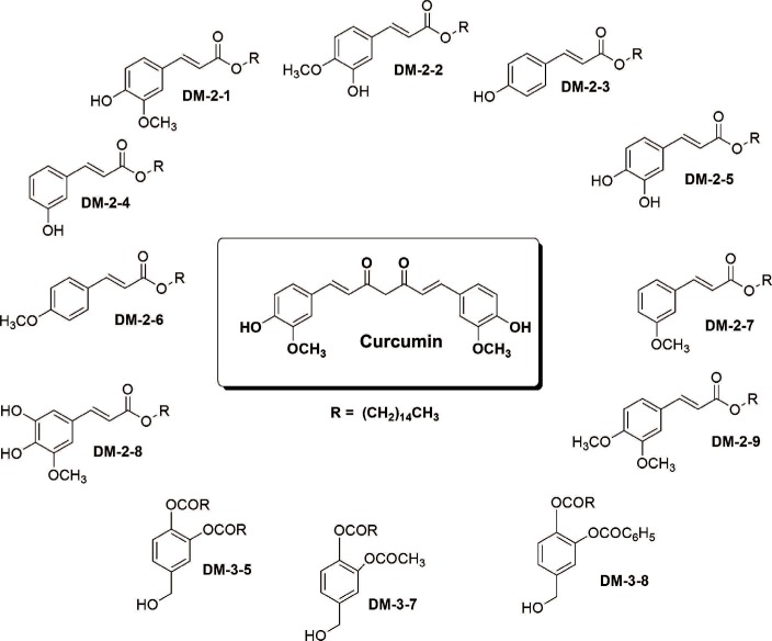
Figure 1 Chemical structure of different alkyl cinnamates with their respective compound codes.
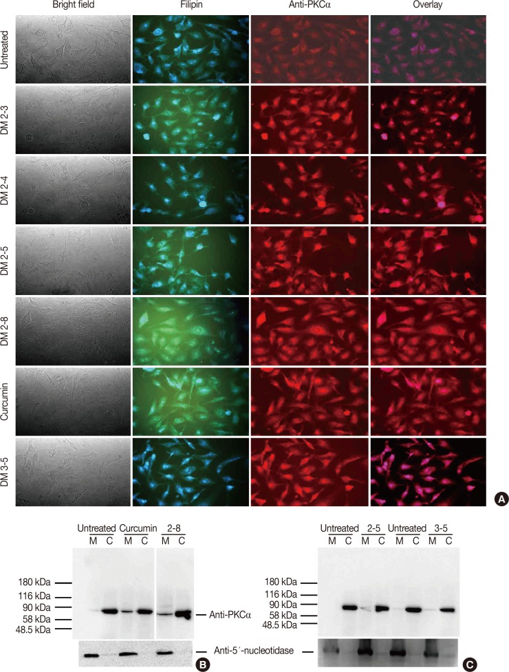
Figure 2 Alkyl cinnamates have potential to exhibits protein kinase C (PKC)-α translocation from cytosol to the plasma membrane in MDAMB-231 cells. (A) Immunolocalization of PKCα in MDAMB-231 treated with different alkyl cinnamates to monitor PKC translocation from cytosol to plasma membrane. MDAMB-231 cells were treated and PKCα immunolocalization was done as given in Methods. In each panel, bright field, PKCα (red), filipin to stain cell membrane (blue) and overlay. Untreated cells show a low level of PKCα at plasma membrane compared to significant high signal in alkyl cinnamate treated cells. (B, C) Immunoblotting of PKCα in MDAMB-231 treated with different alkyl cinnamates to monitor PKC translocation from cytosol to plasma membrane. MDAMB-231 cells were treated and PKCα immunolocalization was done as given in Methods. The purity of cytosolic or membrane fraction was asses by testing the presence of lactate dehydrogenase (LDH) and 5′-nucleotidase in these fractions. LDH activity assay was performed as described previously whereas presence of 5′-nucleotidase was done by immune-blotting with anti-5′-nucleotidase antibodies. Untreated cells show a basal level of PKCα at plasma membrane whereas a high level of PKCα was found in alkyl cinnamate treated cells.
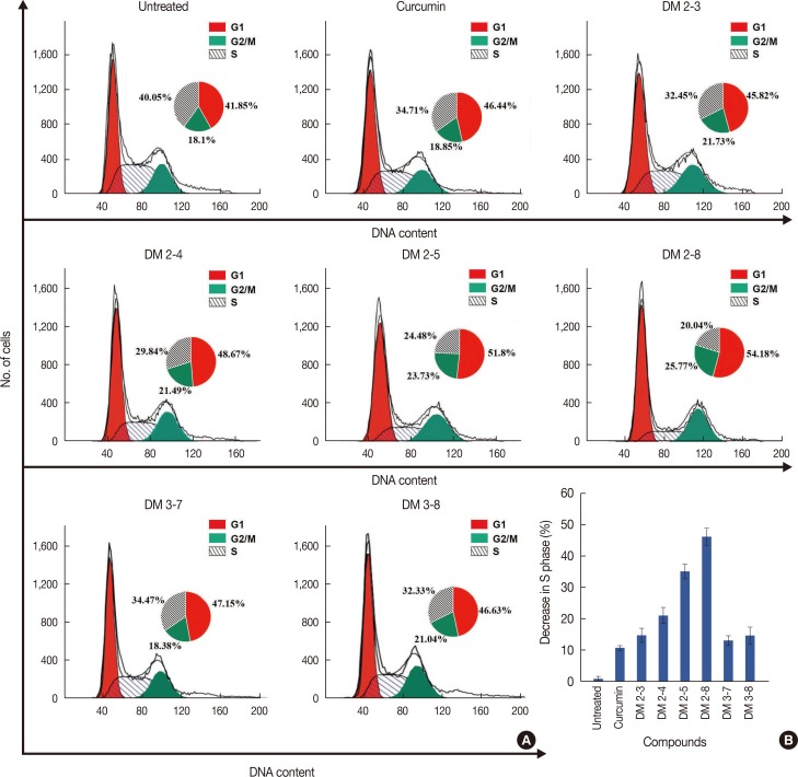
Figure 3 Alkyl cinnamates disturb cell-cycle in MDAMB-231w cells. (A) Cells were treated with different alkyl cinnamates (half maximal inhibitory concentrations [IC50]) for 24 hours in incomplete media, stained with propidium iodide as given in Methods, and immediately analyzed by flow cytometry. The cells treated with different compounds show G2/M arrest and reduction of S phase. (B) Plot of change in S phase of MDA-MB-231 cells treated with different alkyl cinnamates.
Figure 4 Alkyl cinnamates are causing death of MDAMB-231 following apoptosis. (A) MDAMB-231 treated with different alkyl cinnamates and distribution of healthy, dead, apoptotic and necrotic cells. Cells were treated alkyl cinnamates (IC50 for 24 hours), stained with acridine orange and propidium iodide and analyzed by flow cytometry. (B) MDAMB-231 treated with different alkyl cinnamates exhibits DNA fragmentation. Cells were treated with different alkyl cinnamates (the IC50 values of the compounds for 12 hours) in serum-free media and samples were processed for DNA laddering analysis. A laddering pattern of DNA is observed in alkyl cinnamate treated cells compared to the intact genomic DNA in untreated cells.
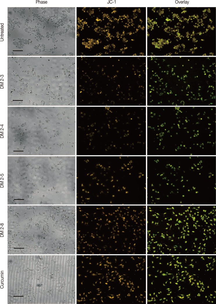
Figure 5 Loss of mitochondrial membrane potential in alkyl cinnamates treated MDAMB-231 breast cancer cells. MDAMB-231 breast cancer cells were treated with alkyl cinnamates (IC50) in serum-free media for 24 hours. After treatment, cells were stained with JC-1 dye for 20 minutes and were observed in Cytell Imaging System (GE Healthcare). Alkyl cinnamates treated cells show a decrease in mitochondrial membrane potential as indicated by loss of orange and appearance of green fluorescence (scale bar, 130 µm).
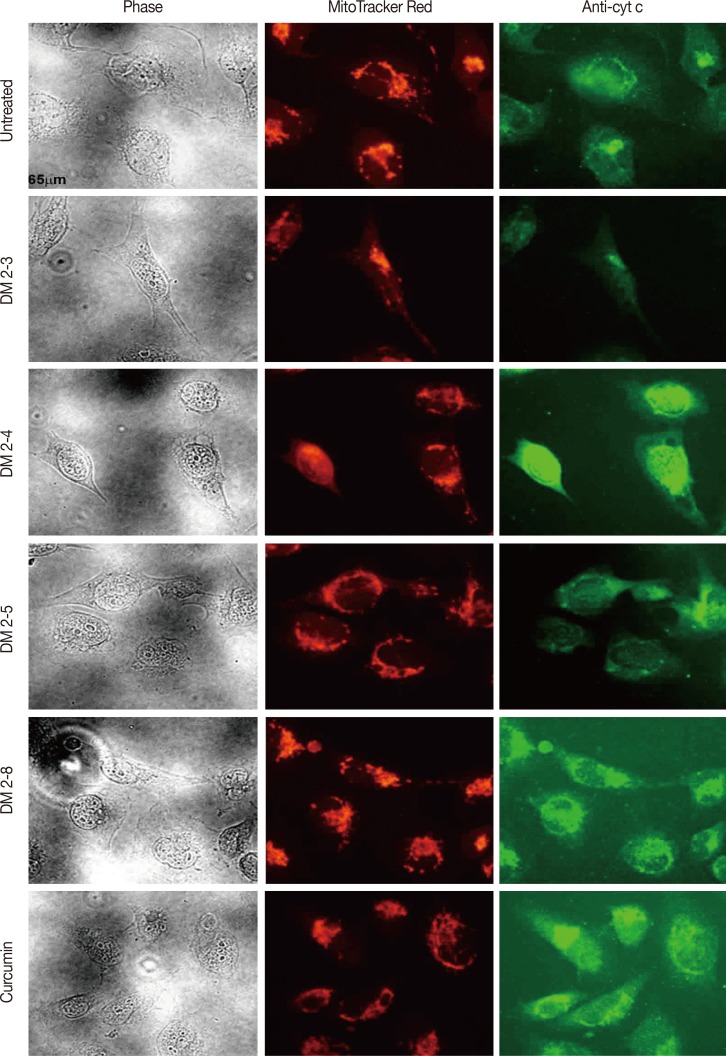
Figure 6 Release of cytochrome-c (cyt c) from alkyl cinnamate treated MDAMB-231 breast cancer cells. MDAMB-231 breast cancer cells were treated with alkyl cinnamates (IC50) in serum-free media for 24 hours. After treatment, cells were immune-stained with anti-cyt c antibodies and location of mitochondria in MDAMB-231 cells is determined by loading the cells with MitoTracker (BD-Biosciences; 200 nM for 45 minutes) as given in material and methods. Labelled cells were observed in a fluorescent microscope 80i (Nikon). In each panel, bright field, cyt c (green), MitoTracker Red (red) is given. High resolution fluorescence microscopy detected release of cyt c from DM2-3, DM2-4, DM2-5 and curcumin treated cells. Untreated cells seem healthy with the cyt c signal overlapping with MitoTracker signal.
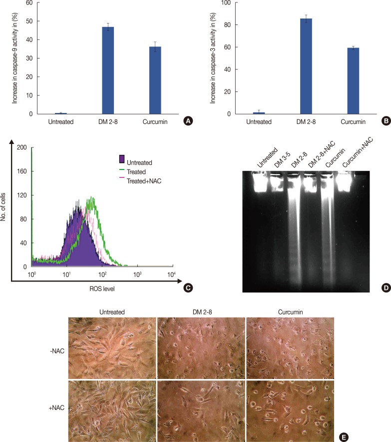
Figure 7 Cytochrome C release activates down-stream cytosolic caspases. Caspase 9 (A) and caspase 3 (B) activity in MDAMB-231 cells treated with alkyl cinnamates. Caspase-3 activity levels increased by 90% and 65% in DM2-8 and curcumin. (C) Flow cytometric measurement of reactive oxygen species (ROS) levels in alkyl cinnamate treated breast cancer cells. Cells were treated with alkyl cinnamates (IC50) for 3 hours and DCF-DHA was used as a ROS probe. DM-2-8 treated cells show an increased level of ROS compared to untreated cells. The preincubation of cells in N-acetylcysteine (NAC) reduces ROS level in treated cell. The preincubation of cells in N-acetylcysteine (NAC) reduces ROS level and apperance of DNA fragments (D) in treated cell. Cells were treated with DM 2-8 or curcumin in the absence or presence of NAC (5 mM) and presence of NAC give intact genomic DNA with no visible appearance of DNA fragments. (E) Microscopic observation of MDAMB-231 cells treated with DM-2-8 or curcumin for 48 hours in absence or presence of NAC. Images were taken with high resolution Nikon L22 camera. Cells preincubated with NAC gives an improved cellular morphology compared to DM-2-8 or curcumin treated cells.
Reference
-
2. Koivunen J, Aaltonen V, Peltonen J. Protein kinase C (PKC) family in cancer progression. Cancer Lett. 2006; 235:1–10. PMID: 15907369.
Article3. Griner EM, Kazanietz MG. Protein kinase C and other diacylglycerol effectors in cancer. Nat Rev Cancer. 2007; 7:281–294. PMID: 17384583.
Article4. O'Brian C, Vogel VG, Singletary SE, Ward NE. Elevated protein kinase C expression in human breast tumor biopsies relative to normal breast tissue. Cancer Res. 1989; 49:3215–3217. PMID: 2720675.5. Powell CT, Brittis NJ, Stec D, Hug H, Heston WD, Fair WR. Persistent membrane translocation of protein kinase C alpha during 12-0-tetradecanoylphorbol-13-acetate-induced apoptosis of LNCaP human prostate cancer cells. Cell Growth Differ. 1996; 7:419–428. PMID: 9052983.6. Wu TT, Hsieh YH, Hsieh YS, Liu JY. Reduction of PKC alpha decreases cell proliferation, migration, and invasion of human malignant hepatocellular carcinoma. J Cell Biochem. 2008; 103:9–20. PMID: 17486587.7. Tanaka Y, Gavrielides MV, Mitsuuchi Y, Fujii T, Kazanietz MG. Protein kinase C promotes apoptosis in LNCaP prostate cancer cells through activation of p38 MAPK and inhibition of the Akt survival pathway. J Biol Chem. 2003; 278:33753–33762. PMID: 12824193.
Article8. Chen ZF, Fang JY, Weng YR, Sun DF, Wang X, Lu R. The effect of PKC-delta inhibitor rottlerin on human colon cancer cell line SW1116 and its mechanism. Zhonghua Zhong Liu Za Zhi. 2006; 28:564–567. PMID: 17236547.9. Santiago-Walker AE, Fikaris AJ, Kao GD, Brown EJ, Kazanietz MG, Meinkoth JL. Protein kinase C delta stimulates apoptosis by initiating G1 phase cell cycle progression and S phase arrest. J Biol Chem. 2005; 280:32107–32114. PMID: 16051606.10. Ferriola PC, Cody V, Middleton E Jr. Protein kinase C inhibition by plant flavonoids. Kinetic mechanisms and structure-activity relationships. Biochem Pharmacol. 1989; 38:1617–1624. PMID: 2730676.11. Yang EB, Zhao YN, Zhang K, Mack P. Daphnetin, one of coumarin derivatives, is a protein kinase inhibitor. Biochem Biophys Res Commun. 1999; 260:682–685. PMID: 10403826.
Article12. Aitken A. The activation of protein kinase C by daphnane, ingenane and tigliane diterpenoid esters. Bot J Linn Soc. 1987; 94:247–263.
Article13. Mamidi N, Gorai S, Sahoo J, Manna D. Alkyl cinnamates as regulator for the C1 domain of protein kinase C isoforms. Chem Phys Lipids. 2012; 165:320–330. PMID: 22414757.
Article14. Trivedi V, Zhang SC, Castoreno AB, Stockinger W, Shieh EC, Vyas JM, et al. Immunoglobulin G signaling activates lysosome/phagosome docking. Proc Natl Acad Sci U S A. 2006; 103:18226–18231. PMID: 17110435.
Article15. Deshmukh R, Trivedi V. Phagocytic uptake of oxidized heme polymer is highly cytotoxic to macrophages. PLoS One. 2014; 9:e103706. PMID: 25078090.
Article16. Trivedi V, Chand P, Srivastava K, Puri SK, Maulik PR, Bandyopadhyay U. Clotrimazole inhibits hemoperoxidase of Plasmodium falciparum and induces oxidative stress: proposed antimalarial mechanism of clotrimazole. J Biol Chem. 2005; 280:41129–41136. PMID: 15863504.17. Black AR, Black JD. Protein kinase C signaling and cell cycle regulation. Front Immunol. 2013; 3:423. PMID: 23335926.
Article18. Xue X, Yu JL, Sun DQ, Kong F, Qu XJ, Zou W, et al. Curcumin induces apoptosis in SGC-7901 gastric adenocarcinoma cells via regulation of mitochondrial signaling pathways. Asian Pac J Cancer Prev. 2014; 15:3987–3992. PMID: 24935585.
Article19. Wender PA, Baryza JL, Brenner SE, DeChristopher BA, Loy BA, Schrier AJ, et al. Design, synthesis, and evaluation of potent bryostatin analogs that modulate PKC translocation selectivity. Proc Natl Acad Sci U S A. 2011; 108:6721–6726. PMID: 21415363.
Article20. Géczy T, Oláh A, Tóth BI, Czifra G, Szöllősi AG, Szabó T, et al. Protein kinase C isoforms have differential roles in the regulation of human sebocyte biology. J Invest Dermatol. 2012; 132:1988–1997. PMID: 22475757.
Article21. Frey MR, Saxon ML, Zhao X, Rollins A, Evans SS, Black JD. Protein kinase C isozyme-mediated cell cycle arrest involves induction of p21(waf1/cip1) and p27(kip1) and hypophosphorylation of the retinoblastoma protein in intestinal epithelial cells. J Biol Chem. 1997; 272:9424–9435. PMID: 9083081.22. Gutcher I, Webb PR, Anderson NG. The isoform-specific regulation of apoptosis by protein kinase C. Cell Mol Life Sci. 2003; 60:1061–1070. PMID: 12861375.
Article23. Garcia-Bermejo ML, Leskow FC, Fujii T, Wang Q, Blumberg PM, Ohba M, et al. Diacylglycerol (DAG)-lactones, a new class of protein kinase C (PKC) agonists, induce apoptosis in LNCaP prostate cancer cells by selective activation of PKCalpha. J Biol Chem. 2002; 277:645–655. PMID: 11584014.24. Clark JA, Black AR, Leontieva OV, Frey MR, Pysz MA, Kunneva L, et al. Involvement of the ERK signaling cascade in protein kinase C-mediated cell cycle arrest in intestinal epithelial cells. J Biol Chem. 2004; 279:9233–9247. PMID: 14670956.
Article25. Wong RS. Apoptosis in cancer: from pathogenesis to treatment. J Exp Clin Cancer Res. 2011; 30:87. PMID: 21943236.
Article26. Tong QS, Zheng LD, Lu P, Jiang FC, Chen FM, Zeng FQ, et al. Apoptosis-inducing effects of curcumin derivatives in human bladder cancer cells. Anticancer Drugs. 2006; 17:279–287. PMID: 16520656.
Article27. Subramaniam D, May R, Sureban SM, Lee KB, George R, Kuppusamy P, et al. Diphenyl difluoroketone: a curcumin derivative with potent in vivo anticancer activity. Cancer Res. 2008; 68:1962–1969. PMID: 18339878.
Article28. Sun A, Lu YJ, Hu H, Shoji M, Liotta DC, Snyder JP. Curcumin analog cytotoxicity against breast cancer cells: exploitation of a redox-dependent mechanism. Bioorg Med Chem Lett. 2009; 19:6627–6631. PMID: 19854644.
Article
- Full Text Links
-
- Actions
-
Cited
- CITED
-
- Close
- Share
-
- Similar articles
-
- Synthetic Chenodeoxycholic Acid Derivative HS-1200-induced Apoptosis of Human Melanoma Cells
- Activation of SAPK and increase in Bak levels during ceramide and indomethacin-induced apoptosis in HT29 cells
- Recombinant Rv0753c Protein of Mycobacterium tuberculosis Induces Apoptosis Through Reactive Oxygen Species-JNK Pathway in Macrophages
- Induction of cytoprotective autophagy by morusin via AMP-activated protein kinase activation in human non-small cell lung cancer cells
- Expression Status and Prognostic Value of bcl-2 Protein in Breast Cancer
