Inhibition of miR-128 Abates Aβ-Mediated Cytotoxicity by Targeting PPAR-γ via NF-κB Inactivation in Primary Mouse Cortical Neurons and Neuro2a Cells
- Affiliations
-
- 1Department of Rehabilitation Medicine, Huaihe Hospital of Henan University, Kaifeng, China. angelcindtg@yahoo.com
- 2Department of Neurology, Huaihe Hospital of Henan University, Kaifeng, China.
- KMID: 2422495
- DOI: http://doi.org/10.3349/ymj.2018.59.9.1096
Abstract
- PURPOSE
Alzheimer's disease (AD) is the sixth most common cause of death in the United States. MicroRNAs have been identified as vital players in neurodegenerative diseases, including AD. microRNA-128 (miR-128) has been shown to be dysregulated in AD. This study aimed to explore the roles and molecular mechanisms of miR-128 in AD progression.
MATERIALS AND METHODS
Expression patterns of miR-128 and peroxisome proliferator-activated receptor gamma (PPAR-γ) messenger RNA in clinical samples and cells were measured using RT-qPCR assay. PPAR-γ protein levels were determined by Western blot assay. Cell viability was determined by MTT assay. Cell apoptotic rate was detected by flow cytometry via double-staining of Annexin V-FITC/PI. Caspase 3 and NF-κB activity was determined by a Caspase 3 Activity Assay Kit or NF-κB p65 Transcription Factor Assay Kit, respectively. Bioinformatics prediction and luciferase reporter assay were used to investigate interactions between miR-128 and PPAR-γ 3"²UTR.
RESULTS
MiR-128 expression was upregulated and PPAR-γ expression was downregulated in plasma from AD patients and amyloid-β (Aβ)-treated primary mouse cortical neurons (MCN) and Neuro2a (N2a) cells. Inhibition of miR-128 decreased Aβ-mediated cytotoxicity through inactivation of NF-κB in MCN and N2a cells. Moreover, PPAR-γ was a target of miR-128. PPAR-γ upregulation attenuated Aβ-mediated cytotoxicity by inactivating NF-κB in MCN and N2a cells. Furthermore, PPAR-γ downregulation was able to abolish the effect of anti-miR-128 on cytotoxicity and NF-κB activity in MCN and N2a cells.
CONCLUSION
MiR-128 inhibitor decreased Aβ-mediated cytotoxicity by upregulating PPAR-γ via inactivation of NF-κB in MCN and N2a cells, providing a new potential target in AD treatment.
Keyword
MeSH Terms
-
Alzheimer Disease
Animals
Blotting, Western
Caspase 3
Cause of Death
Cell Survival
Computational Biology
Down-Regulation
Flow Cytometry
Humans
Luciferases
Mice*
MicroRNAs
Neurodegenerative Diseases
Neurons*
Plasma
PPAR gamma
RNA, Messenger
Transcription Factor RelA
United States
Up-Regulation
Caspase 3
Luciferases
MicroRNAs
PPAR gamma
RNA, Messenger
Transcription Factor RelA
Figure
-
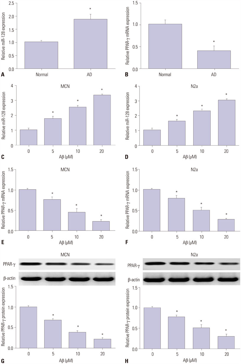
Fig. 1 MiR-128 expression is upregulated and PPAR-γ expression is downregulated in plasma from AD patients and Aβ-treated MCN and N2a cells. (A and B) Expression patterns of miR-128 and PPAR-γ in plasma from healthy volunteers (Normal) (n=20) and AD patients (n=20) were detected using RT-qPCR assay. (C–H) Primary MCN cells and N2a cells were treated with different concentrations of Aβ (0, 5, 10, 20 µM) for 24 h. Then, levels of miR-128 (C and D) and PPAR-γ mRNA (E and F) were determined by RT-qPCR assay, and PPAR-γ protein expression (G and H) was measured using Western blot assay. *p<0.05. PPAR-γ, proliferator-activated receptor gamma; AD, Alzheimer's disease; Aβ, amyloid-β; MCN, mouse cortical neurons; N2a, Neuro2a; mRNA, messenger RNA.
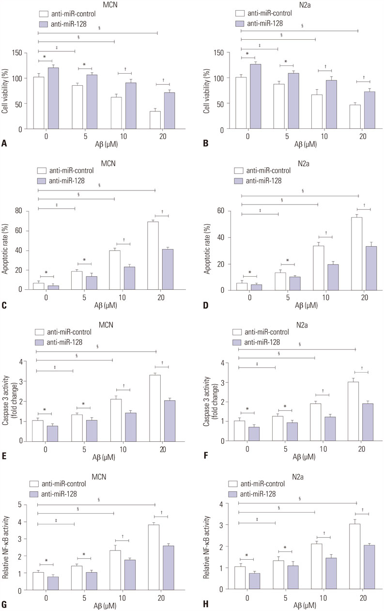
Fig. 2 Inhibition of miR-128 abates Aβ-mediated cytotoxicity by inactivating NF-κB in MCN and N2a cells. (A–H) MCN and N2a cells were transfected with antimiR-control or anti-miR-128 for 24 h and then treated with different concentrations of Aβ (0, 5, 10, 20 µM) for another 24 h, followed by detection of cell viability (A and B), apoptotic rate (C and D), caspase 3 activity (E and F), and NF-κB activity (G and H). *p<0.05, †p<0.01, ‡p<0.05, §p<0.01. Aβ, amyloid-β; MCN, mouse cortical neurons; N2a, Neuro2a.
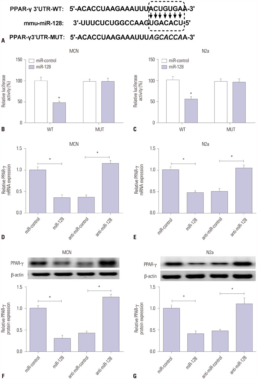
Fig. 3 PPAR-γ is a target of miR-128. (A) Putative binding sites between PPAR-γ 3′UTR and mouse miR-128, and mutant sites in MUT PPAR-γ reporter. (B and C) MCN and N2a cells were co-transfected with WT or MUT PPAR-γ reporter and miR-control or miR-128 mimic for 48 h, followed by measurement of luciferase activity via double luciferase reporter assay. (D–G) MCN and N2a cells were transfected with miR-control, miR-128 mimic, anti-miR-control, or anti-miR-128 for 48 h. Then, PPAR-γ expressions at mRNA (D and E) and protein (F and G) levels were determined by RT-qPCR and Western blot assays, respectively. *p<0.05. PPAR-γ, proliferator-activated receptor gamma; MCN, mouse cortical neurons; N2a, Neuro2a; mRNA, messenger RNA; WT, wild type; MUT, mutant type.
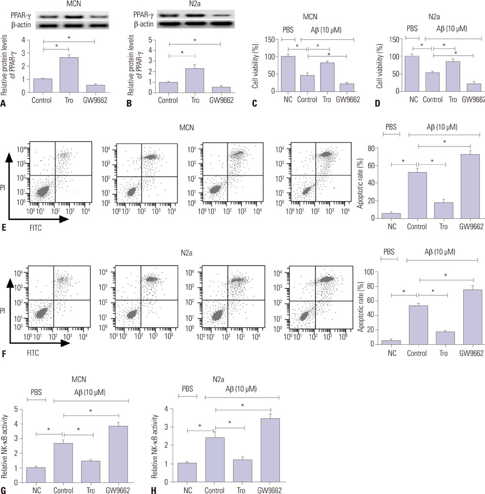
Fig. 4 PPAR-γ attenuates Aβ-mediated cytotoxicity by inactivating NF-κB in MCN and N2a cells. (A and B) MCN and N2a cells were treated with Control (DMSO), Tro (20 µM), or GW9662 (10 µM) for 24 h, followed by detection of PPAR-γ protein level via Western blot assay. (C–H) MCN and N2a cells were treated with Aβ (10 µM) for 24 h and stimulated with Control (DMSO), Tro (20 µM), or GW9662 (10 µM) for another 24 h. Next, at the indicated time point, cell viability (C and D), apoptotic rate (E and F), and NF-κB activity (G and H) were determined. *p<0.05. PPAR-γ, proliferator-activated receptor gamma; Aβ, amyloid-β; MCN, mouse cortical neurons; N2a, Neuro2a; Tro, troglitazone.

Fig. 5 miR-128 inhibitor decreases Aβ-mediated cytotoxicity by upregulating PPAR-γ via inactivation of NF-κB signaling in MCN and N2a cells. (A–F) MCN and N2a cells were treated with Aβ (10 µM) for 24 h, followed by stimulation of control (DMSO) or Tro (20 µM) for another 24 h. Aβ-treated cells were transfected with anti-miR-control or anti-miR-128 for another 24 h, together with or without the treatment of control (DMSO) or GW9662 (10 µM) for an additional 24 h. Following this, cell viability (A and B), apoptotic rate (C and D), and NF-κB activity (E and F) were determined in treated cells. *p<0.05. Aβ; amyloid-β PPAR-γ, proliferator-activated receptor gamma; MCN, mouse cortical neurons; N2a, Neuro2a; Tro, troglitazone.
Reference
-
1. Alzheimer's Association. 2016 Alzheimer's disease facts and figures. Alzheimers Dement. 2016; 12:459–509.2. Reitz C, Mayeux R. Alzheimer disease: epidemiology, diagnostic criteria, risk factors and biomarkers. Biochem Pharmacol. 2014; 88:640–651.
Article3. Kumar A, Singh A, Ekavali . A review on Alzheimer's disease pathophysiology and its management: an update. Pharmacol Rep. 2015; 67:195–203.
Article4. Kim DH, Yeo SH, Park JM, Choi JY, Lee TH, Park SY, et al. Genetic markers for diagnosis and pathogenesis of Alzheimer's disease. Gene. 2014; 545:185–193.
Article5. Selkoe DJ, Hardy J. The amyloid hypothesis of Alzheimer's disease at 25 years. EMBO Mol Med. 2016; 8:595–608.
Article6. Kang S, Lee YH, Lee JE. Metabolism-Centric Overview of the Pathogenesis of Alzheimer's Disease. Yonsei Med J. 2017; 58:479–488.
Article7. Mémet S. NF-kappaB functions in the nervous system: from development to disease. Biochem Pharmacol. 2006; 72:1180–1195.8. Shi ZM, Han YW, Han XH, Zhang K, Chang YN, Hu ZM, et al. Upstream regulators and downstream effectors of NF-κB in Alzheimer's disease. J Neurol Sci. 2016; 366:127–134.
Article9. Cai Y, Yu X, Hu S, Yu J. A brief review on the mechanisms of miRNA regulation. Genomics Proteomics Bioinformatics. 2009; 7:147–154.
Article10. Karnati HK, Panigrahi MK, Gutti RK, Greig NH, Tamargo IA. miRNAs: key players in neurodegenerative disorders and epilepsy. J Alzheimers Dis. 2015; 48:563–580.
Article11. Adlakha YK, Saini N. Brain microRNAs and insights into biological functions and therapeutic potential of brain enriched miRNA-128. Mol Cancer. 2014; 13:33.
Article12. Lukiw WJ. Micro-RNA speciation in fetal, adult and Alzheimer's disease hippocampus. Neuroreport. 2007; 18:297–300.
Article13. Müller M, Kuiperij HB, Claassen JA, Küsters B, Verbeek MM. MicroRNAs in Alzheimer's disease: differential expression in hippocampus and cell-free cerebrospinal fluid. Neurobiol Aging. 2014; 35:152–158.
Article14. Jiang Q, Heneka M, Landreth GE. The role of peroxisome proliferator-activated receptor-gamma (PPARgamma) in Alzheimer's disease: therapeutic implications. CNS Drugs. 2008; 22:1–14.
Article15. Sastre M, Dewachter I, Rossner S, Bogdanovic N, Rosen E, Borghgraef P, et al. Nonsteroidal anti-inflammatory drugs repress beta-secretase gene promoter activity by the activation of PPARgamma. Proc Natl Acad Sci U S A. 2006; 103:443–448.
Article16. Combs CK, Johnson DE, Karlo JC, Cannady SB, Landreth GE. Inflammatory mechanisms in Alzheimer's disease: inhibition of beta-amyloid-stimulated proinflammatory responses and neurotoxicity by PPARgamma agonists. J Neurosci. 2000; 20:558–567.
Article17. Fakhfouri G, Ahmadiani A, Rahimian R, Grolla AA, Moradi F, Haeri A. WIN55212-2 attenuates amyloid-beta-induced neuroinflammation in rats through activation of cannabinoid receptors and PPAR-γ pathway. Neuropharmacology. 2012; 63:653–666.
Article18. Dubois B, Feldman HH, Jacova C, Hampel H, Molinuevo JL, Blennow K, et al. Advancing research diagnostic criteria for Alzheimer's disease: the IWG-2 criteria. Lancet Neurol. 2014; 13:614–629.
Article19. Schmidt K. Clinical dementia rating scale. In : Michalos AC, editor. Encyclopedia of quality of life and well-being research. Dordrecht: Springer;2014. p. 957–960.20. Mitchell AJ. The Mini-Mental State Examination (MMSE): update on its diagnostic accuracy and clinical utility for cognitive disorders. In : Larner AJ, editor. Cognitive screening instruments. Cham: Springer;2017. p. 37–48.21. Krichevsky AM, Kosik KS. Neuronal RNA granules: a link between RNA localization and stimulation-dependent translation. Neuron. 2001; 32:683–696.22. Chen YC, Wu JS, Tsai HD, Huang CY, Chen JJ, Sun GY, et al. Peroxisome proliferator-activated receptor gamma (PPAR-γ) and neurodegenerative disorders. Mol Neurobiol. 2012; 46:114–124.
Article23. Reddy PH, Tonk S, Kumar S, Vijayan M, Kandimalla R, Kuruva CS, et al. A critical evaluation of neuroprotective and neurodegenerative MicroRNAs in Alzheimer’s disease. Biochem Biophys Res Commun. 2017; 483:1156–1165.
Article24. Femminella GD, Ferrara N, Rengo G. The emerging role of microRNAs in Alzheimer's disease. Front Physiol. 2015; 6:40.
Article25. Millan MJ. Linking deregulation of non-coding RNA to the core pathophysiology of Alzheimer's disease: an integrative review. Prog Neurobiol. 2017; 156:1–68.
Article26. Long JM, Ray B, Lahiri DK. MicroRNA-153 physiologically inhibits expression of amyloid-β precursor protein in cultured human fetal brain cells and is dysregulated in a subset of Alzheimer disease patients. J Biol Chem. 2012; 287:31298–31310.
Article27. Absalon S, Kochanek DM, Raghavan V, Krichevsky AM. MiR-26b, upregulated in Alzheimer’s disease, activates cell cycle entry, tauphosphorylation, and apoptosis in postmitotic neurons. J Neurosci. 2013; 33:14645–14659.
Article28. McSweeney KM, Gussow AB, Bradrick SS, Dugger SA, Gelfman S, Wang Q, et al. Inhibition of microRNA 128 promotes excitability of cultured cortical neuronal networks. Genome Res. 2016; 26:1411–1416.
Article29. Tiribuzi R, Crispoltoni L, Porcellati S, Di Lullo M, Florenzano F, Pirro M, et al. miR128 up-regulation correlates with impaired amyloid β(1-42) degradation in monocytes from patients with sporadic Alzheimer's disease. Neurobiol Aging. 2014; 35:345–356.
Article30. Guidi M, Muiños-Gimeno M, Kagerbauer B, Martí E, Estivill X, Espinosa-Parrilla Y. Overexpression of miR-128 specifically inhibits the truncated isoform of NTRK3 and upregulates BCL2 in SHSY5Y neuroblastoma cells. BMC Mol Biol. 2010; 11:95.
Article31. Kaltschmidt B, Uherek M, Volk B, Baeuerle PA, Kaltschmidt C. Transcription factor NF-kappaB is activated in primary neurons by amyloid beta peptides and in neurons surrounding early plaques from patients with Alzheimer disease. Proc Natl Acad Sci U S A. 1997; 94:2642–2647.
Article32. Valerio A, Boroni F, Benarese M, Sarnico I, Ghisi V, Bresciani LG, et al. NF-kappaB pathway: a target for preventing beta-amyloid (Abeta)-induced neuronal damage and Abeta42 production. Eur J Neurosci. 2006; 23:1711–1720.
Article33. Lin W, Ding M, Xue J, Leng W. The role of TLR2/JNK/NF-κB pathway in amyloid β peptide-induced inflammatory response in mouse NG108-15 neural cells. Int Immunopharmacol. 2013; 17:880–884.
Article34. Villapol S. Roles of peroxisome proliferator-activated receptor gamma on brain and peripheral inflammation. Cell Mol Neurobiol. 2018; 38:121–132.
Article35. Corona JC, Duchen MR. PPARγ as a therapeutic target to rescue mitochondrial function in neurological disease. Free Radic Biol Med. 2016; 100:153–163.
Article36. Lezana JP, Dagan SY, Robinson A, Goldstein RS, Fainzilber M, Bronfman FC, et al. Axonal PPARγ promotes neuronal regeneration after injury. Dev Neurobiol. 2016; 76:688–701.
Article37. Landreth G, Jiang Q, Mandrekar S, Heneka M. PPARgamma agonists as therapeutics for the treatment of Alzheimer's disease. Neurotherapeutics. 2008; 5:481–489.
Article38. Toba J, Nikkuni M, Ishizeki M, Yoshii A, Watamura N, Inoue T, et al. PPARγ agonist pioglitazone improves cerebellar dysfunction at pre-Aβ deposition stage in APPswe/PS1dE9 Alzheimer's disease model mice. Biochem Biophys Res Commun. 2016; 473:1039–1044.
Article39. de la Monte SM, Wands JR. Molecular indices of oxidative stress and mitochondrial dysfunction occur early and often progress with severity of Alzheimer's disease. J Alzheimers Dis. 2006; 9:167–181.
Article40. Kitamura Y, Shimohama S, Koike H, Kakimura Ji, Matsuoka Y, Nomura Y, et al. Increased expression of cyclooxygenases and peroxisome proliferator-activated receptor-gamma in Alzheimer's disease brains. Biochem Biophys Res Commun. 1999; 254:582–586.
Article41. Inestrosa NC, Godoy JA, Quintanilla RA, Koenig CS, Bronfman M. Peroxisome proliferator-activated receptor gamma is expressed in hippocampal neurons and its activation prevents beta-amyloid neurodegeneration: role of Wnt signaling. Exp Cell Res. 2005; 304:91–104.
Article42. Sastre M, Dewachter I, Landreth GE, Willson TM, Klockgether T, van Leuven F, et al. Nonsteroidal anti-inflammatory drugs and peroxisome proliferator-activated receptor-gamma agonists modulate immunostimulated processing of amyloid precursor protein through regulation of beta-secretase. J Neurosci. 2003; 23:9796–9804.
Article43. Camacho IE, Serneels L, Spittaels K, Merchiers P, Dominguez D, De Strooper B. Peroxisome-proliferator-activated receptor gamma induces a clearance mechanism for the amyloid-beta peptide. J Neurosci. 2004; 24:10908–10917.
Article44. d'Abramo C, Massone S, Zingg JM, Pizzuti A, Marambaud P, Dalla Piccola B, et al. Role of peroxisome proliferator-activated receptor gamma in amyloid precursor protein processing and amyloid beta-mediated cell death. Biochem J. 2005; 391(Pt 3):693–698.
- Full Text Links
-
- Actions
-
Cited
- CITED
-
- Close
- Share
-
- Similar articles
-
- miR-215 Enhances HCV Replication by Targeting TRIM22 and Inactivating NF-κB Signaling
- Galangin Suppresses Pro-Inflammatory Gene Expression in Polyinosinic-Polycytidylic Acid-Stimulated Microglial Cells
- MiR-506 Promotes Natural Killer Cell Cytotoxicity against Human Hepatocellular Carcinoma Cells by Targeting STAT3
- Long Noncoding RNA NEAT1 Aggravates Aβ-Induced Neuronal Damage by Targeting miR-107 in Alzheimer's Disease
- MiR-590 Inhibits Endothelial Cell Apoptosis by Inactivating the TLR4/NF-κB Pathway in Atherosclerosis
