Antitumor Effect of KX-01 through Inhibiting Src Family Kinases and Mitosis
- Affiliations
-
- 1Cancer Research Institute, Seoul National University, Seoul, Korea. moisa@snu.ac.kr
- 2Biomedical Research Institute, Seoul National University Hospital, Seoul, Korea.
- 3Department of Internal Medicine, Seoul National University College of Medicine, Seoul, Korea.
- 4Department of Internal Medicine, Seoul National University Bundang Hospital, Seoul National University College of Medicine, Seongnam, Korea.
- 5Kinex Pharmaceutical Corporation, New York State Center of Excellence in Bioinformartics and Life Sciences, NY, USA.
- KMID: 2388310
- DOI: http://doi.org/10.4143/crt.2016.168
Abstract
- PURPOSE
KX-01 is a novel dual inhibitor of Src and tubulin. Unlike previous Src inhibitors that failed to show clinical benefit during treatment of breast cancer, KX-01 can potentially overcome the therapeutic limitations of current Src inhibitors through inhibition of both Src and tubulin. The present study further evaluates the activity and mechanism of KX-01 in vitro and in vivo.
MATERIALS AND METHODS
The antitumor effect of KX-01 in triple negative breast cancer (TNBC) cell lines was determined by MTT assay. Wound healing and immunofluorescence assays were performed to evaluate the action mechanisms of KX-01. Changes in the cell cycle and molecular changes induced by KX-01 were also evaluated. A MDA-MB-231 mouse xenograft model was used to demonstrate the in vivo effects.
RESULTS
KX-01 effectively inhibited the growth of breast cancer cell lines. The expression of phospho-Src and proliferative-signaling molecules were down-regulated in KX-01-sensitive TNBC cell lines. In addition, migration inhibition was observed by wound healing assay. KX-01-induced G2/M cell cycle arrest and increased the aneuploid cell population in KX-01-sensitive cell lines. Multi-nucleated cells were significantly increased after KX-01 treatment. Furthermore, KX-01 effectively delayed tumor growth in a MDA-MB-231 mouse xenograft model.
CONCLUSION
KX-01 effectively inhibited cell growth and migration of TNBC cells. Moreover, this study demonstrated that KX-01 showed antitumor effects through the inhibition of Src signaling and the induction of mitotic catastrophe. The antitumor effects of KX-01 were also demonstrated in vivo using a mouse xenograft model.
Keyword
MeSH Terms
Figure
-
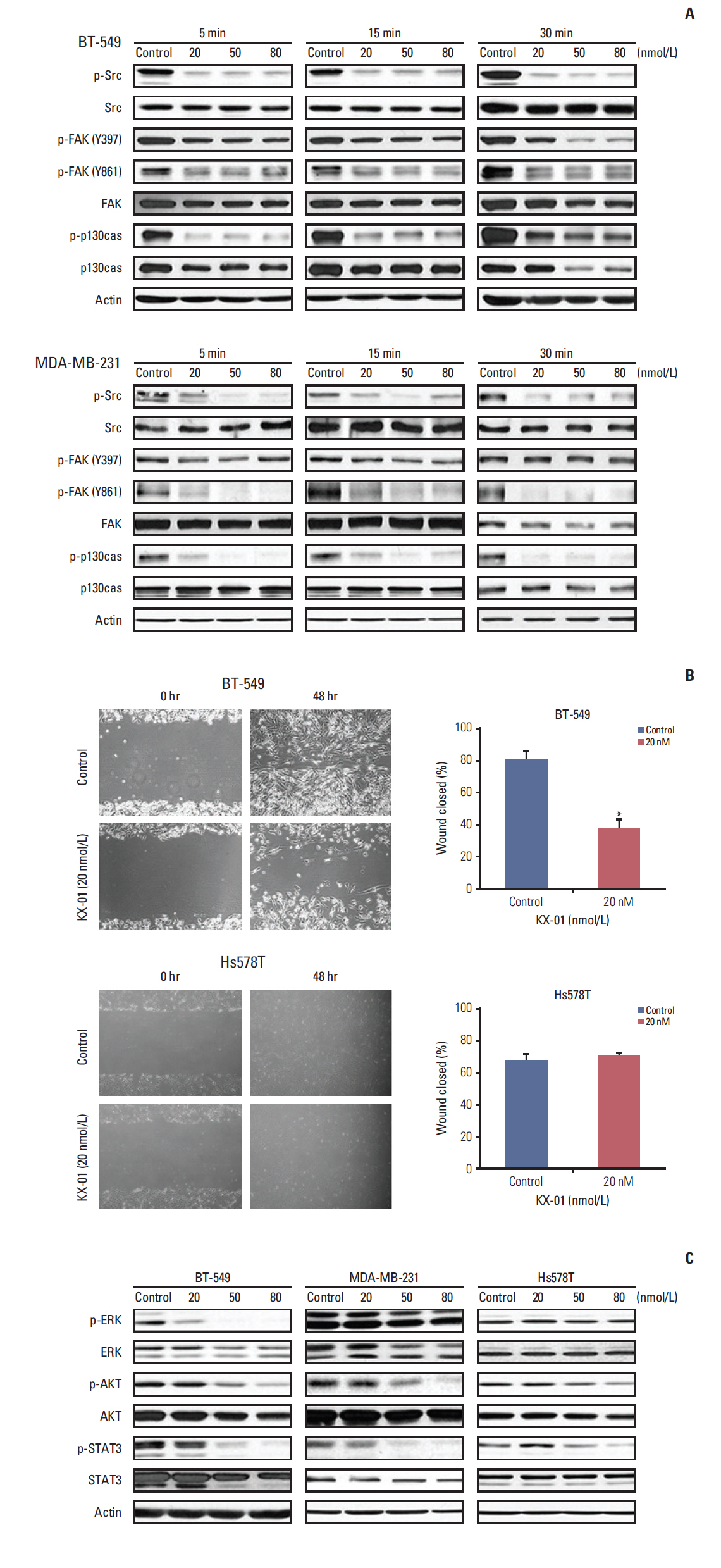
Fig. 1. KX-01 treatment in TNBC inhibits Src activity and the migration of cancer cells. (A) BT-549 and MDA-MB-231 cells were treated with KX-01 at the indicated time and dose. Western blot analysis showed molecular expression changes following KX-01 treatment. The active form of Src, FAK, and p130cas were all down-regulated by KX-01 treatment. (B) BT-549 and Hs578T cells were incubated with dimethyl sulfoxide (control) or KX-01 for 48 hours. Wound healing assay results demonstrate the migration inhibitory effect of KX-01. The columns are shown with error bars (±standard error). *p < 0.05. (C) BT-549, MDA-MB-231, and Hs578T cells were exposed to KX-01 for 24 hours. Western blot results show molecular expression changes, which are related to Src signaling.
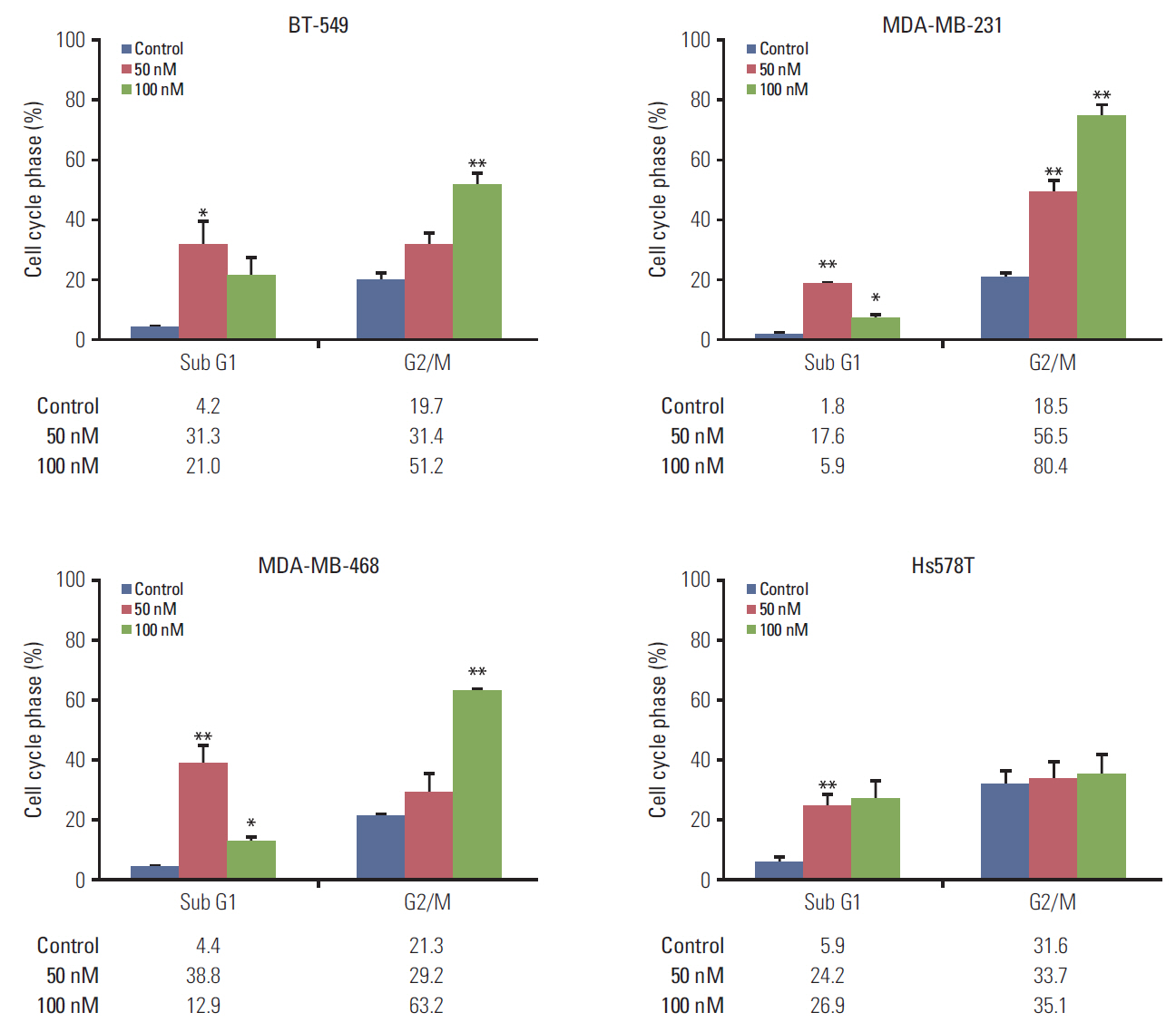
Fig. 2. KX-01 causes breast cancer cell death and G2/M cell cycle arrest. BT-549, MDA-MB-231, MDA-MB-468, and Hs578T cells were treated with the indicated concentrations of KX-01 for 48 hours. The percentages of cells in the G2/M or Sub G1 phase were determined by flow cytometry analysis. The columns represent the means of three independent experiments and are shown with error bars (±standard error). *p < 0.05, **p < 0.005.
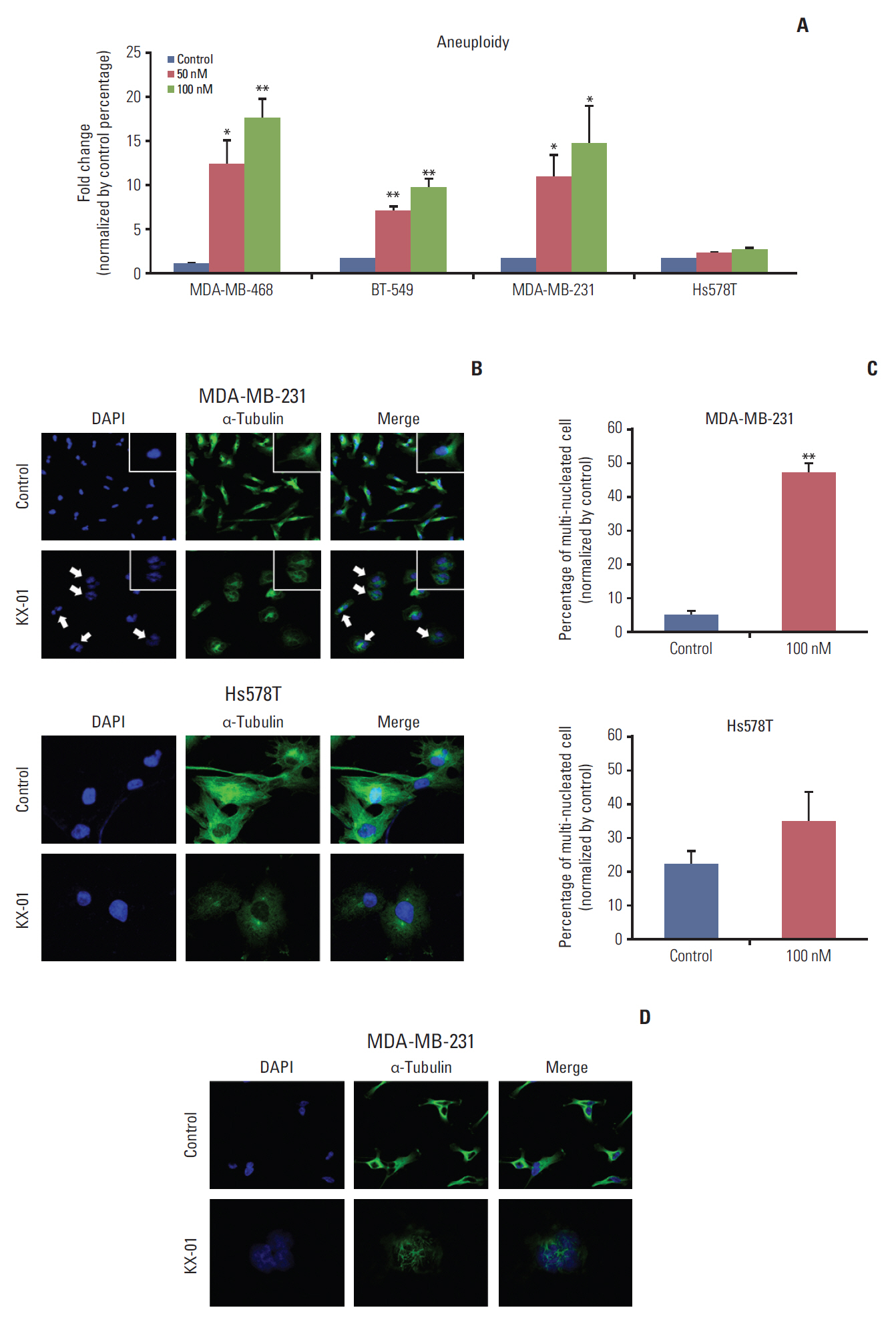
Fig. 3. KX-01 increases aneuploidy and induces mitotic catastrophe by inhibiting microtubule polymerization. (A) BT-549, MDA-MB-231, MDA-MB-468, and Hs578T cells were treated with indicated concentrations of KX-01 for 48 hours. The percentages of cells that contained more than 6N were determined by flow cytometry analysis and compared to the control values. Each column is shown with error bars (±standard error). *p < 0.05, **p < 0.005. (B) MDA-MB-231 and Hs578T cells were incubated with 100 nmol/L of KX-01 or dimethyl sulfoxide (DMSO, control) for 24 hours. Confocal microscopy was used to observe the signal corresponding to α-tubulin (green) and DNA was counterstained with DAPI (blue). Arrows indicate multinucleated cells. (C) One hundred cells in each KX-01 treatment level indicated were counted and the number of multinucleated cells were represented by a percentage. The columns represent the means of three independent experiments and are shown with error bars (±standard error). **p < 0.005. (D) Microtubule conformation was analyzed with 100 nmol/L of KX-01 or DMSO control for 48 hours. Confocal microscopy was used to observe the signal corresponding to α-tubulin (green) and DNA was counterstained with DAPI (blue).
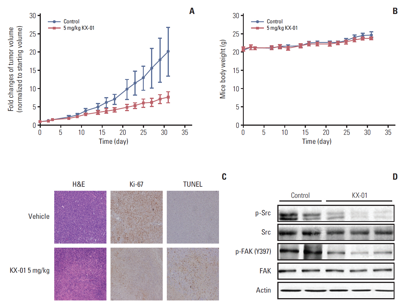
Fig. 4. KX-01 inhibits in vivo tumor growth in MDA-MB-231 mouse xenograft model. (A) BALB/c nude mice were injected with 5×10⁷ MDA-MB-231 cells. The vehicle group received 10% (2-hydroxypropyl)-β-cyclodextrin solution in phosphate buffered saline and the other group was treated with 5 mg/kg of KX-01 administered by oral gavage twice daily for 4 weeks. Tumor volumes were recorded as mm3 and compared to the starting tumor sizes values. (B) Mouse weights were measured three times weekly. Each dot indicates the mean mouse weight. No significant differences in body weight were detected. Mean values are shown ±standard error. (C) The tumors were removed from the mice after KX-01 treatment ended, and pathologic examination was conducted using H&E slides (×200). Immunohistochemical staining for Ki-67 and terminal deoxynucletidyltransferase-mediated dUTP nick end labeling (TUNEL) assays showed decreased Ki-67 with increased apoptosis in KX-01 treatment tumors. (D) On the final day of treatment, total cell protein was extracted from mouse tissues for immunoblotting with the indicated antibodies.
Reference
-
References
1. Cleator S, Heller W, Coombes RC. Triple-negative breast cancer: therapeutic options. Lancet Oncol. 2007; 8:235–44.
Article2. Tryfonopoulos D, Walsh S, Collins DM, Flanagan L, Quinn C, Corkery B, et al. Src: a potential target for the treatment of triple-negative breast cancer. Ann Oncol. 2011; 22:2234–40.
Article3. Hiscox S, Nicholson RI. Src inhibitors in breast cancer therapy. Expert Opin Ther Targets. 2008; 12:757–67.
Article4. Finn RS. Targeting Src in breast cancer. Ann Oncol. 2008; 19:1379–86.
Article5. Chen T, Pengetnze Y, Taylor CC. Src inhibition enhances paclitaxel cytotoxicity in ovarian cancer cells by caspase-9-independent activation of caspase-3. Mol Cancer Ther. 2005; 4:217–24.6. Finn RS, Bengala C, Ibrahim N, Roche H, Sparano J, Strauss LC, et al. Dasatinib as a single agent in triple-negative breast cancer: results of an open-label phase 2 study. Clin Cancer Res. 2011; 17:6905–13.
Article7. Gucalp A, Sparano JA, Caravelli J, Santamauro J, Patil S, Abbruzzi A, et al. Phase II trial of saracatinib (AZD0530), an oral SRC-inhibitor for the treatment of patients with hormone receptor-negative metastatic breast cancer. Clin Breast Cancer. 2011; 11:306–11.
Article8. Carey L, Winer E, Viale G, Cameron D, Gianni L. Triple-negative breast cancer: disease entity or title of convenience? Nat Rev Clin Oncol. 2010; 7:683–92.
Article9. Castedo M, Perfettini JL, Roumier T, Andreau K, Medema R, Kroemer G. Cell death by mitotic catastrophe: a molecular definition. Oncogene. 2004; 23:2825–37.
Article10. Gardner MK, Zanic M, Howard J. Microtubule catastrophe and rescue. Curr Opin Cell Biol. 2013; 25:14–22.
Article11. Topham CH, Taylor SS. Mitosis and apoptosis: how is the balance set? Curr Opin Cell Biol. 2013; 25:780–5.
Article12. Vakifahmetoglu H, Olsson M, Zhivotovsky B. Death through a tragedy: mitotic catastrophe. Cell Death Differ. 2008; 15:1153–62.
Article13. Vitale I, Galluzzi L, Castedo M, Kroemer G. Mitotic catastrophe: a mechanism for avoiding genomic instability. Nat Rev Mol Cell Biol. 2011; 12:385–92.
Article14. Anbalagan M, Ali A, Jones RK, Marsden CG, Sheng M, Carrier L, et al. Peptidomimetic Src/pretubulin inhibitor KX-01 alone and in combination with paclitaxel suppresses growth, metastasis in human ER/PR/HER2-negative tumor xenografts. Mol Cancer Ther. 2012; 11:1936–47.
Article15. Anbalagan M, Carrier L, Glodowski S, Hangauer D, Shan B, Rowan BG. KX-01, a novel Src kinase inhibitor directed toward the peptide substrate site, synergizes with tamoxifen in estrogen receptor alpha positive breast cancer. Breast Cancer Res Treat. 2012; 132:391–409.16. Antonarakis ES, Heath EI, Posadas EM, Yu EY, Harrison MR, Bruce JY, et al. A phase 2 study of KX2-391, an oral inhibitor of Src kinase and tubulin polymerization, in men with bonemetastatic castration-resistant prostate cancer. Cancer Chemother Pharmacol. 2013; 71:883–92.
Article17. Naing A, Cohen R, Dy GK, Hong DS, Dyster L, Hangauer DG, et al. A phase I trial of KX2-391, a novel non-ATP competitive substrate-pocket- directed SRC inhibitor, in patients with advanced malignancies. Invest New Drugs. 2013; 31:967–73.
Article18. Liu T, Hu W, Dalton HJ, Choi HJ, Huang J, Kang Y, et al. Targeting SRC and tubulin in mucinous ovarian carcinoma. Clin Cancer Res. 2013; 19:6532–43.
Article19. Kang S, Min A, Im SA, Song SH, Kim SG, Kim HA, et al. TGF-β suppresses COX-2 expression by tristetraprolin-mediated RNA destabilization in A549 human lung cancer cells. Cancer Res Treat. 2015; 47:101–9.
Article20. Min A, Im SA, Yoon YK, Song SH, Nam HJ, Hur HS, et al. RAD51C-deficient cancer cells are highly sensitive to the PARP inhibitor olaparib. Mol Cancer Ther. 2013; 12:865–77.
Article21. Nam HJ, Im SA, Oh DY, Elvin P, Kim HP, Yoon YK, et al. Antitumor activity of saracatinib (AZD0530), a c-Src/Abl kinase inhibitor, alone or in combination with chemotherapeutic agents in gastric cancer. Mol Cancer Ther. 2013; 12:16–26.
Article22. Macurek L, Draberova E, Richterova V, Sulimenko V, Sulimenko T, Draberova L, et al. Regulation of microtubule nucleation from membranes by complexes of membrane-bound gamma-tubulin with Fyn kinase and phosphoinositide 3-kinase. Biochem J. 2008; 416:421–30.23. Kasahara K, Nakayama Y, Nakazato Y, Ikeda K, Kuga T, Yamaguchi N. Src signaling regulates completion of abscission in cytokinesis through ERK/MAPK activation at the midbody. J Biol Chem. 2007; 282:5327–39.
Article24. Nakayama Y, Matsui Y, Takeda Y, Okamoto M, Abe K, Fukumoto Y, et al. c-Src but not Fyn promotes proper spindle orientation in early prometaphase. J Biol Chem. 2012; 287:24905–15.
Article25. Soeda S, Nakayama Y, Honda T, Aoki A, Tamura N, Abe K, et al. v-Src causes delocalization of Mklp1, Aurora B, and INCENP from the spindle midzone during cytokinesis failure. Exp Cell Res. 2013; 319:1382–97.
Article26. Park AY, Shen TL, Chien S, Guan JL. Role of focal adhesion kinase Ser-732 phosphorylation in centrosome function during mitosis. J Biol Chem. 2009; 284:9418–25.
Article27. Rea K, Sensi M, Anichini A, Canevari S, Tomassetti A. EGFR/MEK/ERK/CDK5-dependent integrin-independent FAK phosphorylated on serine 732 contributes to microtubule depolymerization and mitosis in tumor cells. Cell Death Dis. 2013; 4:e815.
Article28. Chee CE, Krishnamurthi S, Nock CJ, Meropol NJ, Gibbons J, Fu P, et al. Phase II study of dasatinib (BMS-354825) in patients with metastatic adenocarcinoma of the pancreas. Oncologist. 2013; 18:1091–2.
Article
- Full Text Links
-
- Actions
-
Cited
- CITED
-
- Close
- Share
-
- Similar articles
-
- The Regulatory Mechanism of Src Familly Kinase in Lipopolysaccharide (LPS) induced HF-kappaB Activation Pathway
- Involvement of Src Family Tyrosine Kinase in Apoptosis of Human Neutrophils Induced by Protozoan Parasite Entamoeba histolytica
- Clathrin and Lipid Raft-dependent Internalization of Porphyromonas gingivalis in Endothelial Cells
- Metastasis-suppressor KAI1/CD82 induces homotypic aggregation of human prostate cancer cells through Src-dependent pathway
- Differentially Expressed Genes by Inhibition of C-terminal Src Kinase by siRNA in Human Vascular Smooth Muscle Cells and Their Association with Blood Pressure
