Remifentanil induces autophagy and prevents hydrogen peroxide-induced apoptosis in Cos-7 cells
- Affiliations
-
- 1Department of Dental Anesthesia and Pain Medicine, Pusan National University Dental Hospital, Yangsan, Korea. wjdental@hanmail.net
- 2Department of Anesthesia and Pain Medicine, Pusan National University Yangsan Hospital, Yangsan, Korea.
- KMID: 2354652
- DOI: http://doi.org/10.17245/jdapm.2016.16.3.175
Abstract
- BACKGROUND
This study investigated the effect of remifentanil pretreatment on Cos-7 cells exposed to oxidative stress, and the influence of remifentanil on intracellular autophagy and apoptotic cell death.
METHODS
Cells were divided into 4 groups: (1) Control: non-pretreated cells were incubated in normoxia (5% COâ‚‚, 21% Oâ‚‚, and 74% Nâ‚‚). (2) Hâ‚‚Oâ‚‚: non-pretreated cells were exposed to Hâ‚‚Oâ‚‚ for 24 h. (3) RPC+Hâ‚‚Oâ‚‚: cells pretreated with remifentanil were exposed to Hâ‚‚Oâ‚‚ for 24 h. (4) 3-MA+RPC+Hâ‚‚Oâ‚‚: cells pretreated with 3-Methyladenine (3-MA) and remifentanil were exposed to Hâ‚‚Oâ‚‚ for 24 h. We determined the cell viability of each group using an MTT assay. Hoechst staining and FACS analysis of Cos-7 cells were performed to observe the effect of remifentanil on apoptosis. Autophagy activation was determined by fluorescence microscopy, MDC staining, and AO staining. The expression of autophagy-related proteins was observed using western blotting.
RESULTS
Remifentanil pretreatment increased the viability of Cos-7 cells exposed to oxidative stress. Hoechst staining and FACS analysis revealed that oxidative stress-dependent apoptosis was suppressed by the pretreatment. Additionally, fluorescence microscopy showed that remifentanil pretreatment led to autophagy-induction in Cos-7 cells, and the expression of autophagy-related proteins was increased in the RPC+Hâ‚‚Oâ‚‚ group.
CONCLUSIONS
The study showed that remifentanil pretreatment stimulated autophagy and increased viability in an oxidative stress model of Cos-7 cells. Therefore, we suggest that apoptosis was activated upon oxidative stress, and remifentanil preconditioning increased the survival rate of the cells by activating autophagy.
Keyword
MeSH Terms
Figure
-

Fig. 1 Cell viability of Cos-7 cells assessed by MTT assay. (A) The effect of remifentanil and 3-MA + remifentanil preconditioning on Cos-7 cell viability at various concentrations (0, 0.1, 0.5, 1, 2 ng/ml) without exposure to H2O2, (B) Cell viability of Cos-7 cells upon exposure to various concentrations (0, 50, 100, 200, 400) of H2O2.
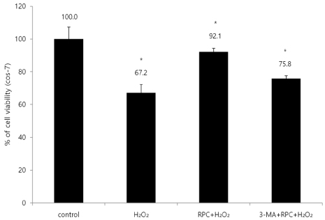
Fig. 2 The effect of remifentanil pretreatment on Cos-7 cell viability with or without autophagy inhibition, as examined by MTT assay. *P < 0.05 as compared with control group. Control: no H2O2, remifentanil treatment group, H2O2: hydrogen peroxide exposure only, RPC + H2O2: remifentanil pretreatment group plus hydrogen peroxide exposure, 3-MA + RPC + H2O2: pretreatment with 3-MA and remifentanil plus hydrogen peroxide exposure.
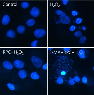
Fig. 3 Hoechst staining: Morphological changes in Cos-7 cells upon pretreatment with remifentanil alone or in combination with 3-MA before exposure to oxidative stress. Cos-7 cells were pretreated with remifentanil or remifentanil and 3-MA, then exposed to oxidative stress. Cells were stained with Hoechst stain and observed by fluorescence microscopy. Apoptotic bodies were seen in H2O2 and 3-MA + RPC + H2O2 group cells. In contrast, they were remarkably reduced in RPC + H2O2 group cells. Control: no H2O2, remifentanil treatment group, H2O2: hydrogen peroxide exposure only, RPC + H2O2: remifentanil pretreatment group plus hydrogen peroxide exposure, 3-MA + RPC + H2O2: pretreatment with 3-MA and remifentanil plus hydrogen peroxide exposure.

Fig. 4 Fluorescence activated cell sorting (FACS) analysis of Cos-7 cells to determine apoptotic cell populations of each experimental group. Control: no H2O2, remifentanil treatment group, H2O2: hydrogen peroxide exposure only, RPC + H2O2: remifentanil pretreatment group plus hydrogen peroxide exposure, 3-MA + RPC + H2O2: pretreatment with 3-MA and remifentanil plus hydrogen peroxide exposure.

Fig. 5 Fluorescence microscopic analysis of autophagosomes in Cos-7 cells under oxidative stress to observe autophagy activation upon different pretreatment conditions. (A) MDC staining of cytoplasmic vacuoles after preconditioning with remifentanil in Cos-7 cells followed by exposure to oxidative stress. Cellular oxidative stress resulted in greater accumulation of autophagosomes containing partially digested cytoplasmic contents compared to the control group. The remifentanil pretreatment group showed increased formation of autophagosomes. Compared with the RPC + H2O2 group, the autophagy pathway inhibitor, 3-MA, blocked formation of autophagosomes. Magnification: 400 ×, (B) AO staining of autophagosomes after remifentanil pretreatment in Cos-7 cells followed by exposure to oxidative stress. AO staining indicated by red fluorescent spots appeared in RPC + H2O2 group cells, while the control, H2O2, and 3-MA + RPC + H2O2 groups showed mainly green cytoplasmic fluorescence. The green shows where the dye has stained the nucleus and the red indicates autophagosome formation. Magnification: 400 ×. Control: no H2O2, remifentanil treatment group, H2O2: hydrogen peroxide exposure only, RPC + H2O2: remifentanil pretreatment group plus hydrogen peroxide exposure, 3-MA + RPC + H2O2: pretreatment with 3-MA and remifentanil plus hydrogen peroxide exposure.
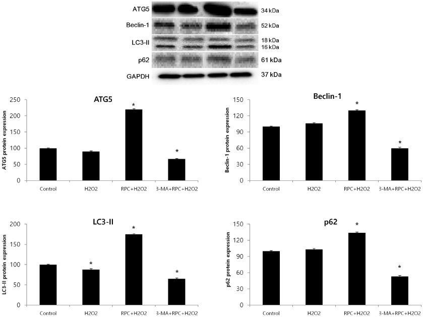
Fig. 6 Western blot analysis and densitometry to determine the effects of remifentanil treatment ATG5, Beclin-1, LC3-II, and p62 protein levels in each experimental group of Cos-7 cells. In RPC + H2O2 groups, cellular expression of ATG5, Beclin-1, LC3-II and p62 were upregulated, while these proteins were downregulated in 3-MA+RPC+H2O2 groups. Control: no H2O2, remifentanil treatment group, H2O2: hydrogen peroxide exposure only, RPC+H2O2: remifentanil pretreatment group plus hydrogen peroxide exposure, 3-MA+RPC+H2O2: pretreatment with 3-MA and remifentanil plus hydrogen peroxide exposure. *P < 0.05 as compared with the control group.
Reference
-
1. Elahi MM, Kong YX, Matata BM. Oxidative stress as a mediator of cardiovascular disease. Oxid Med Cell Longev. 2009; 2:259–269.
Article2. Droge W. Free radicals in the physiological control of cell function. Physiol Rev. 2002; 82:47–95.
Article3. Halliwell B. Reactive species and antioxidants. Redox biology is a fundamental theme of aerobic life. Plant Physiol. 2006; 141:312–322.
Article4. Valko M, Rhodes C, Moncol J, Izakovic M, Mazur M. Free radicals, metals and antioxidants in oxidative stress-induced cancer. Chem Biol Interact. 2006; 160:1–40.
Article5. Zhao Y, Zhao B. Oxidative stress and the pathogenesis of alzheimer's disease. Oxid Med Cell Longev. 2013; 2013:316523.
Article6. Singh DK, Winocour P, Farrington K. Oxidative stress in early diabetic nephropathy: fueling the fire. Nat Rev Endocrinol. 2011; 7:176–184.
Article7. Giordano FJ. Oxygen, oxidative stress, hypoxia, and heart failure. J Clin Invest. 2005; 115:500–508.
Article8. Ryter SW, Kim HP, Hoetzel A, Park JW, Nakahira K, Wang X, et al. Mechanisms of cell death in oxidative stress. Antioxid Redox Signal. 2007; 9:49–89.9. Mizushima N. The pleiotropic role of autophagy: From protein metabolism to bactericide. Cell Death Differ. 2005; 12:1535–1541.10. Guan X, Qian Y, Shen Y, Zhang L, Du Y, Dai H, et al. Autophagy protects renal tubular cells against ischemia/reperfusion injury in a time-dependent manner. Cell Physiol Biochem. 2015; 36:285–298.
Article11. Kim YH, Kang JM, Kim IR, Lee BY, Yoon JY, Kim CH, et al. Protective effect of propofol against hypoxia-reoxygenation injury in HaCaT human keratinocytes. Int J Oral Biol. 2014; 39:97–105.
Article12. Scherz-Shouval R, Elazar Z. Regulation of autophagy by ROS: Physiology and pathology. Trends Biochem Sci. 2011; 36:30–38.
Article13. Kwon JY, Park BS, Kim YH, Kim YD, Kim CH, Yoon JY, et al. Remifentanil protects human keratinocytes against hypoxia-reoxygenation injury through activation of autophagy. PloS One. 2015; 10:e0116982.
Article14. Scott LJ, Perry CM. Spotlight on remifentanil for general anaesthesia. CNS drugs. 2005; 19:1069–1074.
Article15. Komatsu R, Turan A, Orhan-Sungur M, McGuire J, Radke O, Apfel C. Remifentanil for general anaesthesia: A systematic review. Anaesthesia. 2007; 62:1266–1280.
Article16. Marsh D, Hodkinson B. Remifentanil in paediatric anaesthetic practice. Anaesthesia. 2009; 64:301–308.
Article17. Cho SS, Rudloff I, Berger PJ, Irwin MG, Nold MF, Cheng W, et al. Remifentanil ameliorates intestinal ischemia-reperfusion injury. BMC Gastroenterol. 2013; 13:69.
Article18. Zhang Y, Irwin MG, Wong TM, Chen M, Cao CM. Remifentanil preconditioning confers cardioprotection via cardiac kappa- and delta-opioid receptors. Anesthesiology. 2005; 102:371–378.
Article19. Kim H, Cho J, Hong S, Kim S, Shim J, Kwak Y. Remifentanil protects myocardium through activation of anti-apoptotic pathways of survival in ischemia-reperfused rat heart. Physiol Res. 2010; 59:347–356.
Article20. Zhang TZ, Zhou J, Jin Q, Sun YJ, Diao YG, Zhang YN, et al. Protective effects of remifentanil preconditioning on cerebral injury during pump-assisted coronary artery bypass graft. Genet Mol Res. 2014; 13:7658–7665.
Article21. Chang C, Chen P, Lu S, Hsieh M, Lin C, Lee H, et al. Propofol-enhanced autophagy increases motility and angiogenic capacity of cultured human umbilical vascular endothelial cells. Life Sci. 2015; 142:49–59.
Article22. Anding AL, Baehrecke EH. Chapter three-autophagy in cell life and cell death. Curr Top Dev Biol. 2015; 114:67–91.
Article23. Baehrecke EH. Autophagy: Dual roles in life and death? Nat Rev Mol Cell Biol. 2005; 6:505–510.
Article24. Fitzwalter BE, Thorburn A. Recent insights into cell death and autophagy. FEBS J. 2015; 282:4279–4288.
Article25. Levine B, Yuan J. Autophagy in cell death: An innocent convict? J Clin Invest. 2005; 115:2679–2688.
Article26. Huang C, Lee C, Lin H, Chen M, Lin C, Chang J. Autophagy-regulated ROS from xanthine oxidase acts as an early effector for triggering late mitochondria-dependent apoptosis in cathepsin S-targeted tumor cells. PloS one. 2015; 10:e0128045.
Article27. Elahi M, Matata B. Free radicals in blood: Evolving concepts in the mechanism of ischemic heart disease. Arch Biochem Biophys. 2006; 450:78–88.
Article28. Finkel T, Holbrook NJ. Oxidants, oxidative stress and the biology of ageing. Nature. 2000; 408:239–247.
Article29. Yue Z, Friedman L, Komatsu M, Tanaka K. The cellular pathways of neuronal autophagy and their implication in neurodegenerative diseases. Biochim Biophys Acta. 2009; 1793:1496–1507.
Article30. Yan L, Sadoshima J, Vatner DE, Vatner SF. Autophagy: A novel protective mechanism in chronic ischemia. Cell Cycle. 2006; 5:1175–1177.
Article31. Scherz-Shouval R, Elazar Z. Regulation of autophagy by ROS: Physiology and pathology. Trends Biochem Sci. 2011; 36:30–38.
Article32. Zhao G, Shen X, Nan H, Yan L, Zhao H, Yu J, et al. Remifentanil protects liver against ischemia/reperfusion injury through activation of anti-apoptotic pathways. J Surg Res. 2013; 183:827–834.
Article33. Peart JN, Gross ER, Gross GJ. Opioid-induced preconditioning: Recent advances and future perspectives. Vascul Pharmacol. 2005; 42:211–218.
Article34. Headrick JP, See Hoe LE, Du Toit EF, Peart JN. Opioid receptors and cardioprotection–'opioidergic conditioning' of the heart. Br J Pharmacol. 2015; 172:2026–2050.
Article35. Costa-Malaquias A, Almeida MB, Monteiro JRS, de Matos Macchi B, do Nascimento JLM, Crespo-Lopez ME. Morphine protects against methylmercury intoxication: A role for opioid receptors in oxidative stress? PloS One. 2014; 9:e110815.
Article
- Full Text Links
-
- Actions
-
Cited
- CITED
-
- Close
- Share
-
- Similar articles
-
- Propofol protects against oxidative-stress-induced COS-7 cell apoptosis by inducing autophagy
- Effects of Remifentanil Preconditioning Attenuating Oxidative Stress in Human Dermal Fibroblast
- Activation of Caspase-3 in Hydrogen Peroxide-Induced Apoptosis of Human Leukemia HL 60 Cells
- Induction of Apoptosis in Synovial Cells from Patients with Rheumatoid Arthritis
- Propofol protects human keratinocytes from oxidative stress via autophagy expression
