Methylglyoxal Induces Apoptosis Mediated by Reactive Oxygen Species in Bovine Retinal Pericytes
- Affiliations
-
- 1Division of Endocrinology and Metabolism, Department of Internal Medicine, College of Medicine, Chung-Ang University, Seoul, Korea. jtkim@cau.ac.kr
- KMID: 1785701
- DOI: http://doi.org/10.3346/jkms.2004.19.1.95
Abstract
- One of the histopathologic hallmarks of early diabetic retinopathy is the loss of pericytes. Evidences suggest that the pericyte loss in vivo is mediated by apoptosis. However, the underlying cause of pericyte apoptosis is not fully understood. This study investigated the influence of methylglyoxal (MGO), a reactive -dicarbonyl compound of glucose metabolism, on apoptotic cell death in bovine retinal pericytes. Analysis of internucleosomal DNA fragmentation by ELISA showed that MGO (200 to 800 micrometer) induced apoptosis in a concentration-dependent manner. Intracellular reactive oxygen species were generated earlier and the antioxidant, N-acetyl cysteine, inhibited the MGO-induced apoptosis. NF-kB activation and increased caspase- 3 activity were detected. Apoptosis was also inhibited by the caspase-3 inhibitor, Z-DEVD-fmk, or the NF- kB inhibitor, pyrrolidine dithiocarbamate. These data suggest that elevated MGO levels observed in diabetes may cause apoptosis in bovine retinal pericytes through an oxidative stress mechanism and suggests that the nuclear activation of NF-kB are involved in the apoptotic process.
Keyword
MeSH Terms
-
Acetylcysteine/pharmacology
Animals
*Apoptosis
Caspases/metabolism
Cattle
Cell Death
Cell Survival
DNA Fragmentation
Dose-Response Relationship, Drug
Enzyme-Linked Immunosorbent Assay
Flow Cytometry
Glucose/metabolism
NF-kappa B/metabolism
Nucleosomes/metabolism
Oxidative Stress
Pericytes/*drug effects
Pyruvaldehyde/*pharmacology
*Reactive Oxygen Species
Retina/cytology/*drug effects
Figure
-

Fig. 1 Dose-dependent cytotoxic effects of MGO in retinal pericytes. Cytotoxicity was measured by MTT assay after 6 hr. Data are means±SD of triplicate experiments. *p<0.05, **p<0.01.

Fig. 2 (A) Effect of MGO on intracellular nucleosome enrichment. Cells were treated with 200, 400, 600, 800 µM MGO for 6 hr and the nucleosome concentration within the cell and in the cell culture supernatant was measured by ELISA. (B) Caspase-3 activity in retinal pericytes. Data are means±SD of triplicate experiments. *p<0.05, **p<0.01 compared to the value of the control.

Fig. 3 Measurement of intracellular ROS production by flow cytometry using DCF-DA. (A) Cells incubated with or without MGO (control, purplish; 400 µM, green; 800 µM, red) for 2 hr. (B) Cells co-treated with 2 mM NAC were incubated with or without MGO for 2 hr. (control, purplish; 400 µM+NAC, green; 800 µM+NAC, red). Data are representative results from three separate experiments.
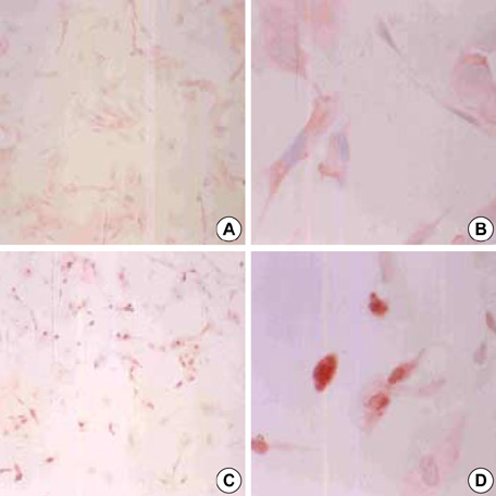
Fig. 4 Subcellular localization of NF-κB p65 subunits. (A, B) In control cells, NF-κB is located in the cytoplasm. (C, D) In cells treated 800 µM MGO for 6 hr, NF-κB is translocated into the nuclei (red). Magnification, (A, C) ×100; (B, D)×400.

Fig. 5 Effects of MGO on NF-κB binding. Pericytes were treated with MGO (800 µM) for 6 hr. Nuclear extracts from the treated and untreated control cells were isolated and used in an EMSA with 32P-labeled NF-κB oligonucleotide as a probe. The arrow indicates the NF-κB binding complex. Competitor, 100-fold molar excess of unlabeled NF-κB probe. Data are representative results from three separate experiments.

Fig. 6 Inhibitory effects of NAC, PDTC, and Z-DEVD-fmk on MGO-induced pericyte apoptosis. Apoptosis was measured by ELISA after 400 or 800 µM MGO treatment for 6 hr. Data are means±SD of triplicate experiments. *p<0.05, **p<0.01 vs without NAC or inhibitor.
Reference
-
1. Mizutani M, Kern TS, Lorenzi M. Accelerated death of retinal microvascular cells in human and experimental diabetic retinopathy. J Clin Invest. 1996. 97:2883–2890.
Article2. Engerman RL, Pfaffenbach D, Davis MD. Cell turnover of capillaries. Lab Invest. 1967. 17:738–743.3. Engerman RL. Pathogenesis of diabetic retinopathy. Diabetes. 1989. 38:1203–1206.
Article4. Naruse K, Nakamura J, Hamada Y, Nakayama M, Chaya S, Komori T, Kato K, Kasuya Y, Miwa K, Hotta N. Aldose reductase inhibition prevents glucose-induced apoptosis in cultured bovine retinal microvascular pericytes. Exp Eye Res. 2000. 71:309–315.
Article5. Denis U, Lecomte M, Paget C, Ruggiero D, Wiernsperger N, Lagarde M. Advanced glycation end-products induce apoptosis of bovine retinal pericytes in culture: involvement of diacylglycerol/ceramide production and oxidative stress induction. Free Radic Biol Med. 2002. 33:236–247.
Article6. Yamagishi S, Amano S, Inagaki Y, Okamoto T, Koga K, Sasaki N, Yamamoto H, Takeuchi M, Makita Z. Advanced glycation end products-induced apoptosis and overexpression of vascular endothelial growth factor in bovine retinal pericytes. Biochem Biophys Res Commun. 2002. 290:973–978.
Article7. Kim J, Kim KS, Shinn JW, Oh YS, Kim HT, Jo I, Shinn SH. The effect of antioxidants on glycated albumin-induced cytotoxicity in bovine retinal pericytes. Biochem Biophys Res Commun. 2002. 292:1010–1016.
Article8. Du J, Suzuki H, Nagase F, Akhand AA, Ma XY, Yokoyama T, Miyata T, Nakashima I. Superoxide-mediated early oxidation and activation of ASK1 are important for initiating methylglyoxal-induced apoptosis process. Free Radic Biol Med. 2001. 31:469–478.
Article9. Murata-Kamiya N, Kamiya H. Methylglyoxal, an endogenous aldehyde, crosslinks DNA polymerase and the substrate DNA. Nucleic Acids Res. 2001. 29:3433–3438.
Article10. Baynes JW, Thorpe SR. Role of oxidative stress in diabetic complications: a new perspective on an old paradigm. Diabetes. 1999. 48:1–9.
Article11. Vander Jagt DL, Hunsaker LA, Vander Jagt TJ, Gomez MS, Gonzales DM, Deck LM, Royer RE. Inactivation of glutathione reductase by 4-hydroxynonenal and other endogenous aldehydes. Biochem Pharmacol. 1997. 53:1133–1140.
Article12. Okado A, Kawasaki Y, Hasuike Y, Takahashi M, Teshima T, Fujii J, Taniguchi N. Induction of apoptotic cell death by methylglyoxal and 3-deoxyglucosone in macrophage-derived cell lines. Biochem Biophys Res Commun. 1996. 225:219–224.
Article13. Wu L, Juurlink BH. Increased methylglyoxal and oxidative stress in hypertensive rat vascular smooth muscle cells. Hypertension. 2002. 39:809–814.
Article14. Thornalley PJ, Hooper NI, Jennings PE, Florkowski CM, Jones AF, Lunec J, Barnett AH. The human red blood cell glyoxalase system in diabetes mellitus. Diabetes Res Clin Pract. 1989. 7:115–120.
Article15. McLellan AC, Thornalley PJ, Benn J, Sonksen PH. Glyoxalase system in clinical diabetes mellitus and correlation with diabetic complications. Clin Sci (Lond). 1994. 87:21–29.
Article16. Beisswenger PJ, Howell SK, Touchette AD, Lal S, Szwergold BS. Metformin reduces systemic methylglyoxal levels in type 2 diabetes. Diabetes. 1999. 48:198–202.
Article17. Bass DA, Parce JW, Dechatelet LR, Szejda P, Seeds MC, Thomas M. Flow cytometric studies of oxidative product formation by neutrophils: a graded response to membrane stimulation. J Immunol. 1983. 130:1910–1917.18. Schreiber E, Matthias P, Muller MM, Schaffner W. Rapid detection of octamer binding proteins with 'mini-extracts' prepared from a small number of cells. Nucleic Acids Res. 1989. 17:6419.19. Thornalley PJ. Pharmacology of methylglyoxal. Gen Pharmacol. 1996. 27:565–573.20. Kikuchi S, Shinpo K, Moriwaka F, Makita Z, Miyata T, Tashiro KJ. Neurotoxicity of methylglyoxal and 3-deoxyglucosone on cultured cortical neurons: synergism between glycation and oxidative stress, possibly involved in neurodegenerative diseases. J Neurosci Res. 1999. 57:280–289.
Article21. Chaplen FW, Fahl WE, Cameron DC. Evidence of high levels of methylglyoxal in cultured Chinese hamster ovary cells. Proc Natl Acad Sci USA. 1998. 95:5533–5538.
Article22. Li W, Chan LS, Khatami M, Rockey JH. Characterization of glucose transport by bovine retinal capillary pericytes in culture. Exp Eye Res. 1985. 41:191–199.
Article23. Dalton TP, Shertzer HG, Puga A. Regulation of gene expression by reactive oxygen. Annu Rev Pharmacol Toxicol. 1999. 39:67–101.
Article24. Meister A. Glutathione metabolism and its selective modification. J Biol Chem. 1988. 263:17205–17208.
Article25. Ferrari G, Yan CY, Greene LA. N-acetylcysteine (D- and L-stereoisomers) prevents apoptotic death of neuronal cells. J Neurosci. 1995. 15:2857–2866.
Article26. DiPietrantonio AM, Hsieh T, Wu JM. Activation of caspase-3 in HL-60 cells exposed to hydrogen peroxide. Biochem Biophys Res Commun. 1999. 255:477–482.27. Matsura T, Kai M, Fujii Y, Ito H, Yamada K. Hydrogen peroxide-induced apoptosis in HL-60 cells requires caspase-3 activation. Free Radic Res. 1999. 30:73–83.
Article28. Siebenlist U, Franzoso G, Brown K. Structure, regulation and function of NF-kappa B. Annu Rev Cell Biol. 1994. 10:405–455.29. Barkett M, Gilmore TD. Control of apoptosis by Rel/NF-kappaB transcription factors. Oncogene. 1999. 18:6910–6924.30. Droge W. Free radicals in the physiological control of cell function. Physiol Rev. 2002. 82:47–95.31. Römeo G, Liu WH, Asnaghi V, Kern TS, Lorenzi M. Activation of nuclear factor-kappaB induced by diabetes and high glucose regulates a proapoptotic program in retinal pericytes. Diabetes. 2002. 51:2241–2248.32. Bourcier T, Sukhova G, Libby P. The nuclear factor-κB signaling pathway participates in dysregulation of vascular smooth muscle cells in vitro and in human atherosclerosis. J Biol Chem. 1997. 272:15817–15824.33. Herrmann JL, Beham AW, Sarkiss M, Chiao PJ, Rands MT, Bruckheimer EM, Brisbay S, McDonnell TJ. Bcl-2 suppresses apoptosis resulting from disruption of the NF-kappa B survival pathway. Exp Cell Res. 1997. 237:101–109.
- Full Text Links
-
- Actions
-
Cited
- CITED
-
- Close
- Share
-
- Similar articles
-
- Study on the Methylglyoxal-induced Apoptosis in Bovine Retinal Pericytes
- Eugenol Induces a Reactive Oxygen Species-mediated Apoptosis in HL-60 Human Promyelocytic Leukemia Cells
- Effect of Methylglyoxal on the Oxidative Stress in Trabecular Meshwork Cells
- Hypericin, a Naphthodianthrone Derivative, Prevents Methylglyoxal-Induced Human Endothelial Cell Dysfunction
- Sesamin induces A549 cell mitophagy and mitochondrial apoptosis via a reactive oxygen species-mediated reduction in mitochondrial membrane potential
