Asiatic Acid Induces Apoptosis and Autophagy and Reduces MiR-17 and MiR-21 Expression in Pancreatic Cancer Cell Lines
- Affiliations
-
- 1College of Pharmacy and Wonkwang Oriental Medicines Research Institute, Wonkwang University, Iksan, Republic of Korea. ejlee7@wku.ac.kr
- 2College of Pharmacy, Korea University, Sejong, Republic of Korea. kylee11@korea.ac.kr
- KMID: 2468062
- DOI: http://doi.org/10.20307/nps.2019.25.4.298
Abstract
- This study investigated the cytotoxic effects and mechanism of action of asiatic acid in pancreatic cancer cell lines. First, we confirmed the cell viability of MIA PaCa-2 and PANC-1 cells after asiatic acid administration for 48 and 72 h. The viability of MIA PaCa-2 and PANC-1 cells decreased in a dose-dependent manner following asiatic acid administration. To investigate the underlying mechanism, we performed a terminal deoxynucleotidyl transferase dUTP nick end labeling (TUNEL) assay, annexin V assay, and western blotting. Asiatic acid induced apoptosis and autophagy through activation of AMP-activated protein kinase (AMPK) and inhibition of mammalian target of rapamycin (mTOR) in MIA PaCa-2 cells. Finally, the expression of miR-17 and miR-21, known as oncogenes in pancreatic cancer, was decreased by asiatic acid. These results indicate that asiatic acid has potential as a new therapeutic agent against pancreatic cancer.
Keyword
MeSH Terms
Figure
-
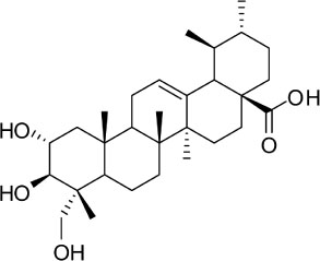
Fig. 1 The chemical structure of asiatic acid.
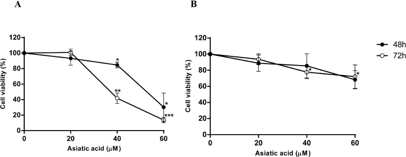
Fig. 2 Asiatic acid inhibits the growth of pancreatic cancer cells. MIA PaCa-2 cells (A) and PANC-1 cells (B) were seeded onto a 96-well plate and incubated with 0, 20, 40, and 60 µM of asiatic acid for 48 (filled circle) and 72 h (unfilled circle). Cell viability was analyzed using the WST-1 assay and mean values were calculated from triplicate wells. This experiment was repeated three times (*p ≤ 0.05; **p ≤ 0.01; ***p ≤ 0.001).
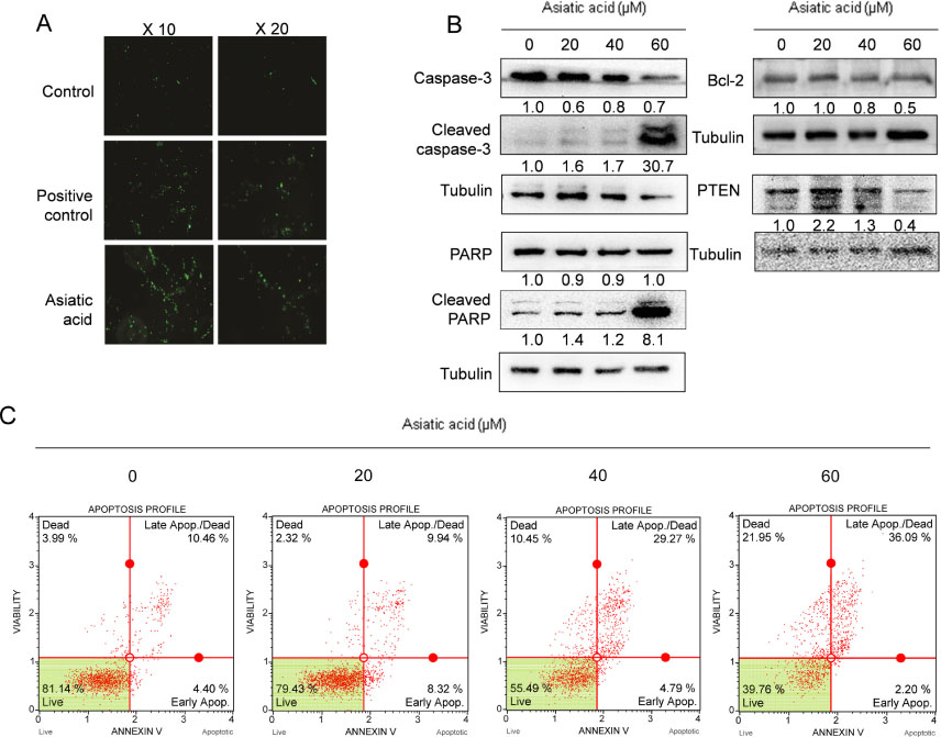
Fig. 3 Asiatic acid induces apoptosis in MIA PaCa-2 cells. (A) The effect of asiatic acid in MIA PaCa-2 cells using the TUNEL assay. Positive control was HSW345. Cells were photographed at magnifications of 10 and 20X. (B) The effect of asiatic acid on the expression of apoptosis-related proteins in MIA PaCa-2 cells. (C) The effect of asiatic acid on apoptosis in MIA PaCa-2 cells was estimated by annexin V assay.
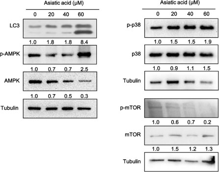
Fig. 4 The effect of asiatic acid on the expression of autophagy-related proteins in MIA PaCa-2 cells. Western blotting was performed to detect the protein expression of LC-3II, p-AMPK, AMPK, p-p38, p38, p-mTOR, mTOR, and tubulin. Asiatic acid was dissolved in DMSO. MIA PaCa-2 cells were treated with different concentrations of asiatic acid (0, 20, 40 and 60 µM) for 48 h. Tubulin was used as a loading control and band density was calculated using Image J.

Fig. 5 Expression level of microRNAs in MIA PaCa-2 cells treated with asiatic acid. Cells were treated with 60 µM of asiatic acid for 48 and 72 h. The CT mean was calculated from three wells and RNU6B was used as a control to determine the relative miRNA expression. The 2−ΔΔCT method was used for the relative quantitation of miR-17 (A) and miR-21 (B).
Reference
-
1. Campbell PJ, Yachida S, Mudie LJ, Stephens PJ, Pleasance ED, Stebbings LA, Morsberger LA, Latimer C, McLaren S, Lin ML, McBride DJ, Varela I, Nik-Zainal SA, Leroy C, Jia M, Menzies A, Butler AP, Teague JW, Griffin CA, Burton J, Swerdlow H, Quail MA, Stratton MR, Iacobuzio-Donahue C, Futreal PA. Nature. 2010; 467:1109–1113.2. Lee HJ. Nat Prod Sci. 2014; 20:170–175.3. Silverman DT, Schiffman M, Everhart J, Goldstein A, Lillemoe KD, Swanson GM, Schwartz AG, Brown LM, Greenberg RS, Schoenberg JB, Pottern LM, Hoover RN, Fraumeni JF Jr. Br J Cancer. 1999; 80:1830–1837.4. Strimpakos A, Saif MW, Syrigos KN. Cancer Metastasis Rev. 2008; 27:495–522.5. Kokubo M, Nishimura Y, Shibamoto Y, Sasai K, Kanamori S, Hosotani R, Imamura M, Hiraoka M. Int J Radiat Oncol Biol Phys. 2000; 48:1081–1087.6. Burris HA 3rd, Moore MJ, Andersen J, Green MR, Rothenberg ML, Modiano MR, Cripps MC, Portenoy RK, Storniolo AM, Tarassoff P, Nelson R, Dorr FA, Stephens CD, Von Hoff DD. J Clin Oncol. 1997; 15:2403–2413.7. Howes MJ, Houghton PJ. Pharmacol Biochem Behav. 2003; 75:513–527.8. Huang FX, Lin XH, He WN, Song W, Ye M, Yang WZ, Guo DA. J Asian Nat Prod Res. 2012; 14:1039–1045.9. Guo FF, Feng X, Chu ZY, Li DP, Zhang L, Zhang ZS. J Asian Nat Prod Res. 2013; 15:15–21.10. Lee YS, Jin DQ, Kwon EJ, Park SH, Lee ES, Jeong TC, Nam DH, Huh K, Kim JA. Cancer Lett. 2002; 186:83–91.11. Bunpo P, Kataoka K, Arimochi H, Nakayama H, Kuwahara T, Vinitketkumnuen U, Ohnishi Y. T. J Med Invest. 2005; 52:65–73.12. Kavitha CV, Agarwal C, Agarwal R, Deep G. PloS one. 2011; 6:e22745.13. Seo JH, Jung KH, Son MK, Yan HH, Ryu YL, Kim J, Lee JK, Hong S, Hong SS. Cancer Lett. 2013; 338:271–281.14. Tang XL, Yang XY, Jung HJ, Kim SY, Jung SY, Choi DY, Park WC, Park H. Biol Pharm Bull. 2009; 32:1399–1405.15. Wu Q, Lv T, Chen Y, Wen L, Zhang J, Jiang X, Liu F. Mol Med Rep. 2015; 12:1429–1434.16. Lozano GM, Bejarano I, Espino J, González D, Ortiz A, García JF, Rodriguez AB, Pariente JA. J Reprod Dev. 2009; 55:615–621.17. Kaufmann SH, Desnoyers S, Ottaviano Y, Davidson NE, Poirier GG. Cancer Res. 1993; 53:3976–3985.18. Ogier-Denis E, Codogno P. Biochim Biophys Acta. 2003; 1603:113–128.19. Hippert MM, O'Toole PS, Thorburn A. Cancer Res. 2006; 66:9349–9351.20. Qu X, Yu J, Bhagat G, Furuya N, Hibshoosh H, Troxel A, Rosen J, Eskelinen EL, Mizushima N, Ohsumi Y, Cattoretti G, Levine B. J Clin Invest. 2003; 112:1809–1820.21. Apel A, Herr I, Schwarz H, Rodemann HP, Mayer A. Cancer Res. 2008; 68:1485–1494.22. Degenhardt K, Mathew R, Beaudoin B, Bray K, Anderson D, Chen G, Mukherjee C, Shi Y, Gélinas C, Fan Y, Nelson DA, Jin S, White E. Cancer cell. 2006; 10:51–64.23. Chresta CM, Davies BR, Hickson I, Harding T, Cosulich S, Critchlow SE, Vincent JP, Ellston R, Jones D, Sini P, James D, Howard Z, Dudley P, Hughes G, Smith L, Maguire S, Hummersone M, Malagu K, Menear K, Jenkins R, Jacobsen M, Smith GC, Guichard S, Pass M. Cancer Res. 2010; 70:288–298.24. Comes F, Matrone A, Lastella P, Nico B, Susca FC, Bagnulo R, Ingravallo G, Modica S, Lo Sasso G, Moschetta A, Guanti G, Simone C. Cell Death Differ. 2007; 14:693–702.25. Casas-Terradellas E, Tato I, Bartrons R, Ventura F, Rosa JL. Biochim Biophys Acta. 2008; 1783:2241–2254.26. Choi CH, Jung YK, Oh SH. Mol Pharmacol. 2010; 78:114–125.27. Tavazoie SF, Alvarez VA, Ridenour DA, Kwiatkowski DJ, Sabatini BL. Nat Neurosci. 2005; 8:1727–1734.28. Fitzwalter BE, Thorburn A. FEBS J. 2015; 282:4279–4288.29. Crino PB. Nat Rev Neurol. 2016; 12:379–392.30. Cheng AM, Byrom MW, Shelton J, Ford LP. Nucleic Acids Res. 2005; 33:1290–1297.31. du Rieu MC, Torrisani J, Selves J, Al Saati T, Souque A, Dufresne M, Tsongalis GJ, Suriawinata AA, Carrère N, Buscail L, Cordelier P. Clin Chem. 2010; 56:603–612.32. O'Donnell KA, Wentzel EA, Zeller KI, Dang CV, Mendell JT. Nature. 2005; 435:839–843.33. Volinia S, Calin GA, Liu CG, Ambs S, Cimmino A, Petrocca F, Visone R, Iorio M, Roldo C, Ferracin M, Prueitt RL, Yanaihara N, Lanza G, Scarpa A, Vecchione A, Negrini M, Harris CC, Croce CM. Proc Natl Acad Sci U S A. 2006; 103:2257–2261.34. Yu J, Ohuchida K, Mizumoto K, Fujita H, Nakata K, Tanaka M. Cancer Biol Ther. 2010; 10:748–757.
- Full Text Links
-
- Actions
-
Cited
- CITED
-
- Close
- Share
-
- Similar articles
-
- Upregulation of MicroRNA-1246 Is Associated with BRAF Inhibitor Resistance in Melanoma Cells with Mutant BRAF
- α, γ-Mangostins Induce Autophagy and Show Synergistic Effect with Gemcitabine in Pancreatic Cancer Cell Lines
- MicroRNA-765 is upregulated in myelodysplastic syndromes and induces apoptosis via PLP2 inhibition in leukemia cells
- MicroRNA expression profiling of diagnostic needle aspirates from surgical pancreatic cancer specimens
- STM2457 Inhibits METTL3-Mediated m6A Modification of miR-30c to Alleviate Spinal Cord Injury by Inducing the ATG5-Mediated Autophagy
