Modulation of Renal Parenchyma in Response to Allogeneic Adipose-Derived Mesenchymal Stem Cells Transplantation in Acute Kidney Injury
- Affiliations
-
- 1Stem Cells Research Laboratory (SCRL), Karachi, Pakistan. sumreenbegum@gmail.com
- 2Clinical Laboratory Sciences (CLS), Karachi, Pakistan.
- 3Department of Urology, Sindh Institute of Urology and Transplantation (SIUT), Karachi, Pakistan.
- KMID: 2447231
- DOI: http://doi.org/10.15283/ijsc18091
Abstract
- BACKGROUND AND OBJECTIVES
In regenerative medicine, mesenchymal stem cells derived from adipose tissues (Ad-MSCs) are a very attractive target to treat many diseases. In relation to nephrology, the aim of the current study is to investigate the effects of Ad-MSCs for the amelioration of acute kidney injury and to explore the mechanism of renal parenchymal changes in response to allogeneic transplantation of Ad-MSCs.
METHODS AND RESULTS
The nephrotoxicity was induced by cisplatin (CP) in balb/c mice according to RIFLE Class and AKIN Stage 3. PCR, qRT-PCR and fluorescent labeled cells infusion, histopathology, immunohistochemistry, functional analyses were used for genes and proteins expressions data acquisition respectively. We demonstrated that single intravenous infusion of 2.5×10⁷/kg mAd-MSCs in mice pre-injected with CP recruited to the kidney, restored the renal structure, and function, which resulted in progressive survival of mice. The renal tissue morphology was recovered in terms of diminished necrosis or epithelial cells damage, protein casts formation, infiltration of inflammatory cells, tubular dilatation, and restoration of brush border protein; Megalin and decreased Kim-1 expressions in mAd-MSCs transplanted mice. Significant reduction in serum creatinine with slashed urea and urinary protein levels were observed. Anti-BrdU staining displayed enhanced tubular cells proliferation. Predominantly, downgrade expressions of TNF-α and TGF-β1 were observed post seven days in mAd-MSCs transplanted mice.
CONCLUSIONS
Ad-MSCs exerts pro-proliferative, anti-inflammatory, and anti-fibrotic effects. Ad-MSCs transplantation without any chemical or genetic manipulation can provide the evidence of therapeutic strategy for the origin of regeneration and overall an improved survival of the system in functionally deprived failed kidneys.
Keyword
MeSH Terms
-
Acute Kidney Injury*
Animals
Cisplatin
Creatinine
Dilatation
Epithelial Cells
Immunohistochemistry
Infusions, Intravenous
Kidney
Low Density Lipoprotein Receptor-Related Protein-2
Mesenchymal Stromal Cells*
Mice
Microvilli
Necrosis
Nephrology
Polymerase Chain Reaction
Regeneration
Regenerative Medicine
Transplantation, Homologous
Urea
Cisplatin
Creatinine
Low Density Lipoprotein Receptor-Related Protein-2
Urea
Figure
-
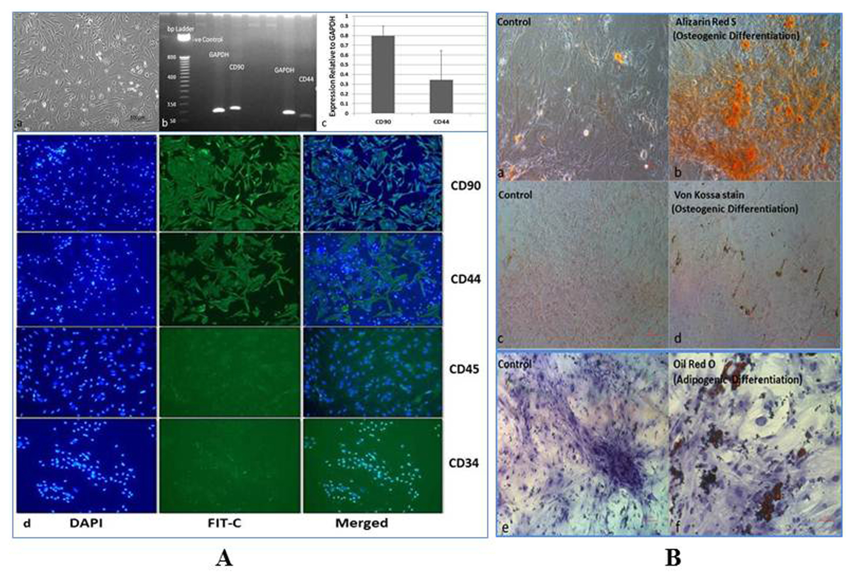
Fig. 1 Morphometry and characterization of mAd-MSCs from Balb/c mice. (A) (a) mAD-MSCs with spindle-shaped morphology (b) The transcriptomic expressions of stem cells markers CD90 and CD44 on 2% agarose gel electrophoresis and (c) Semi-quantification of these markers relative to Glyceraldehyde-3-Phosphate Dehydrogenase (GAPDH) (d) Immunocytochemistry for positive expressions of stem cells markers CD90 and CD44 (raw1 & 2), and negative expressions of CD45 and CD34 (raw 3 & 4). All proteins are shown with 4′, 6-diamidino-2-phenylindole (DAPI), fluorescein isothiocyanate (FIT-C) conjugated secondary antibody and merged images (20× Magnification). (B) (a~d) In vitro differentiation of mAd-MSCs into osteogenic lineage by the formation of calcium hydroxyapatite mineral positive cells, which were visualized by Alizarin Red S (orange-red) (b) and Von Kossa stain (brown to black pigments) (d) both are shown with respective controls (a & c). (f) Adipogenic lineage differentiation of mAd-MSCs for the formation of intracellular lipid vacuoles, which were visualized by Oil Red O stain and is shown with its control (e) (40× Magnification).
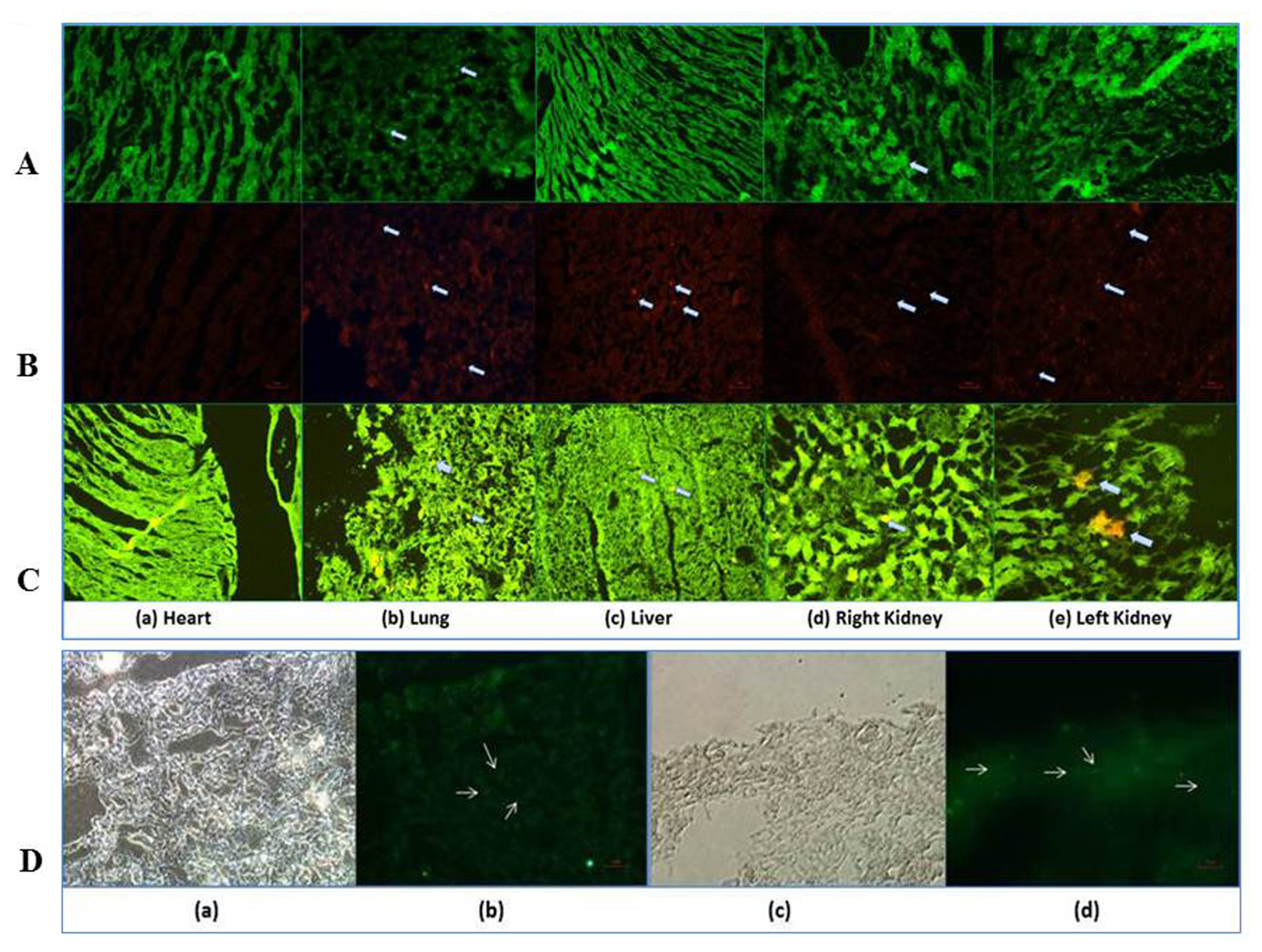
Fig. 2 mAd-MSCs localized to the kidney within 24~72 hours, which facilitate the regeneration of renal tubular cells by excessive proliferation rate. Fluorescence microscopic examinations of heart, lung, liver, and kidney are demonstrated for in vivo tracking or homing of mAd-MSCs within 24~72 hours in AKI mice and proliferation of injured kidney tissues post seven days of intravenous mAd-MSCs infusion in CP-treated mice. The viability and proliferation of cells were not affected by the dyes like CMFDA and Dil. In vivo transplanted mAd-MSCs were observed in kidneys of AKI produced by CP (18 mg/kg, s.c.). Localization of cells is shown by arrows. (A) CMFDA labeled mAd-MSCs in CP-induced AKI (B) Dil labeled mAd-MSCs in CP-induced AKI. (C) Merged images of Co-stained Dil labeled mAd-MSCs and fluorescein isothiocyanate (FIT-C) labeled anti-BrdU staining following injection of BrdU reagent to assess proliferation of cells in injured kidney slices. In each panel (a) Heart (b) Lung (c) Liver (d) Right Kidney (e) Left Kidney (20× Magnification). (D) Micrograph of the kidney slices of FIT-C labeled anti-BrdU staining. (a & b) CP-treated mice (96 hrs CP), (c & d) CP+mAd-MSCs transplanted kidney sections (20× Magnification).

Fig. 3 mAd-MSCs transplantation restores the kidney architecture. (A) Histopathological improvement in AKI model of balb/c mice in response to cellular therapy by mAd-MSCs. The model was developed by CP nephrotoxicity (18 mg/kg b.w. s.c.). Time course representative kidney sections were stained with Hematoxylin & Eosin (H&E) (raw1), Periodic Acid Schiff’s (PAS) (raw2), and Masson’s trichrome staining (TRI) (raw3), and examined by light microscope. Control (saline) showed the normal architecture of tubules and glomeruli. All changes are shown by arrows. Thick arrows show tubular protein casts formation; thin arrows show degeneration of tubules while double arrows show dilatation of tubules. Congestion (erythrocyte trapping) was displayed in the H&E stain of 24~48 hrs. Some rectangular features of tubules were also observed after 72~120 hrs of CP infusion. Shrinkage of glomeruli was observed in 96 hrs of CP infusion. Both are shown in H&E and PAS stains respectively. Accumulation of nuclei is shown in H&E and PAS stains of 72 hr. PAS stains displayed some sign of fibrosis post 72~120 hrs. All stains exhibited structural improvement post mAd-MSCs transplantation, as well as some clear areas (PAS), were also observed. a=control (saline); b=24 hr; c=48 hr; d=72 hr; e=96 hr; f=120 hr; g=CP+mAd-MSCs. CP: cisplatin, G: Glomerulus, PT: Proximal tubules, DT: Distal tubules (40× Magnification). (B) Representatives graphs of histopathological scoring through H&E and PAS stains for the presence of (a) Tubular epithelial cells’s necrosis or degeneration, (b) Tubular protein casts formation, (c) Infiltration of inflammatory cells, and (d) Renal tubular dilatation. The graphical results are presented as mean±s.d. ANOVA followed by Tucky-Kramer test. *p<0.05, **p<0.01 and ***p<0.001 were the levels of significance for all groups. [1=24 hr; 2=48 hr; 3=72 hr; 4=96 hr; 5=120 hr; 6=CP+mAd-MSCs transplanted]. (C) Immuno changes in Megalin (raw1) and Kim-1 (raw2) expressions, which were enhanced and reduced respectively in mAd-MSCs transplanted mice. These changes exposed the recovery of the renal tubular brush border and reduced necrosis in the renal tissues post cellular therapy. Protein expressions in these sections are represented by arrows. (a) Control (saline); (b) 72 hr; (c) 96 hr; (d) CP+mAd-MSCs transplant (40× Magnification).
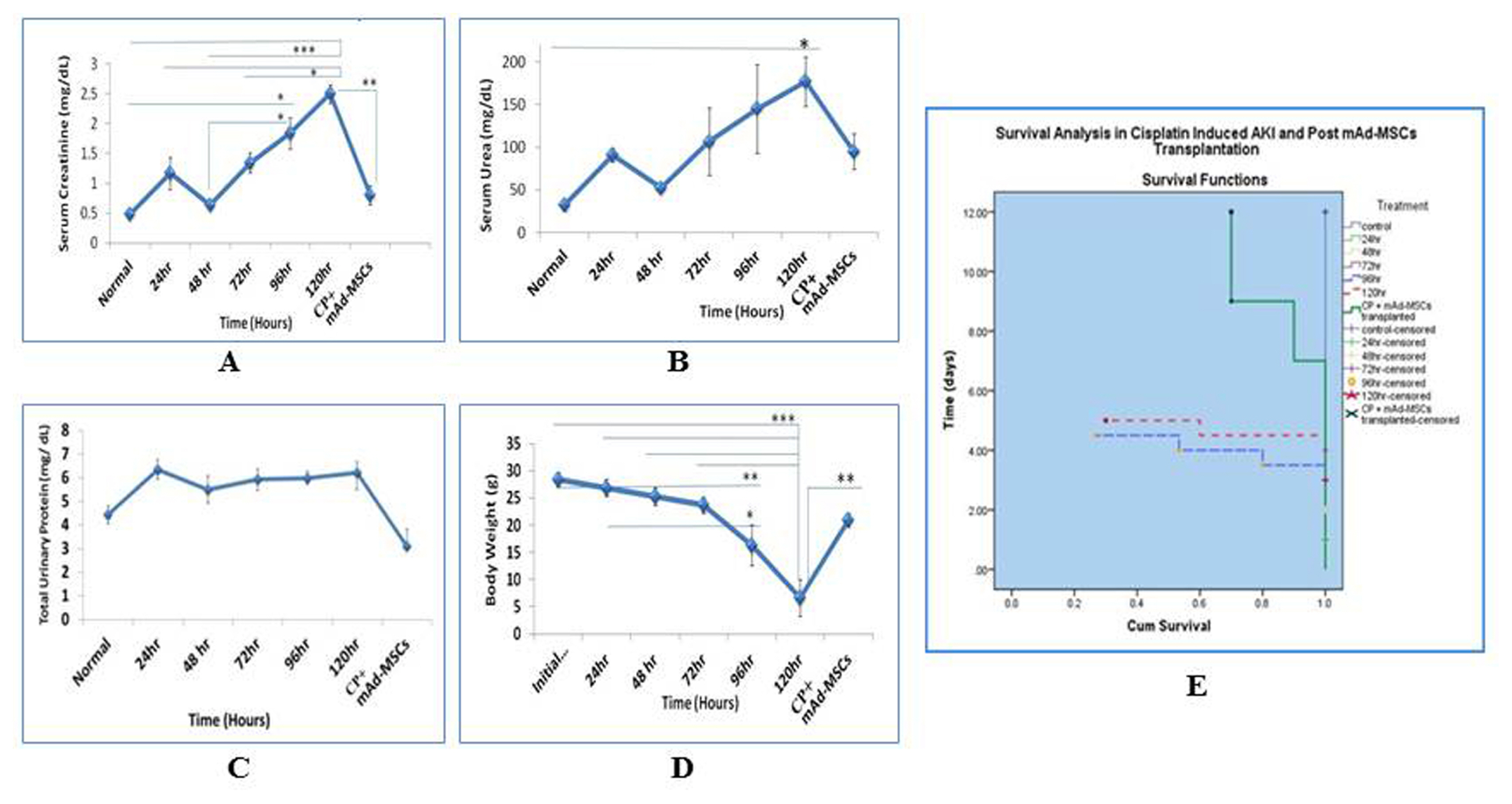
Fig. 4 mAd-MSCs ameliorate kidney failure by restoring its functions, which improved the survival of mice. Functional improvement of kidneys with the survival of balb/c mice was observed in AKI model by CP (18 mg/kg, s.c.) in post transplanted mAd-MSCs. (A) Serum Creatinine, (B) Serum Urea, (C) Total Urinary Protein, (D) Weight dynamics, (E) Survival function for a cumulative survival rate of balb/c mice in all groups was recorded from day 0~12. Data is presented as mean±s.e.m. ANOVA followed by Post-Hoc Tucky’s test for comparison was used. *p<0.5, **p<0.1, and ***p<0.001 were the levels of significance for all groups.
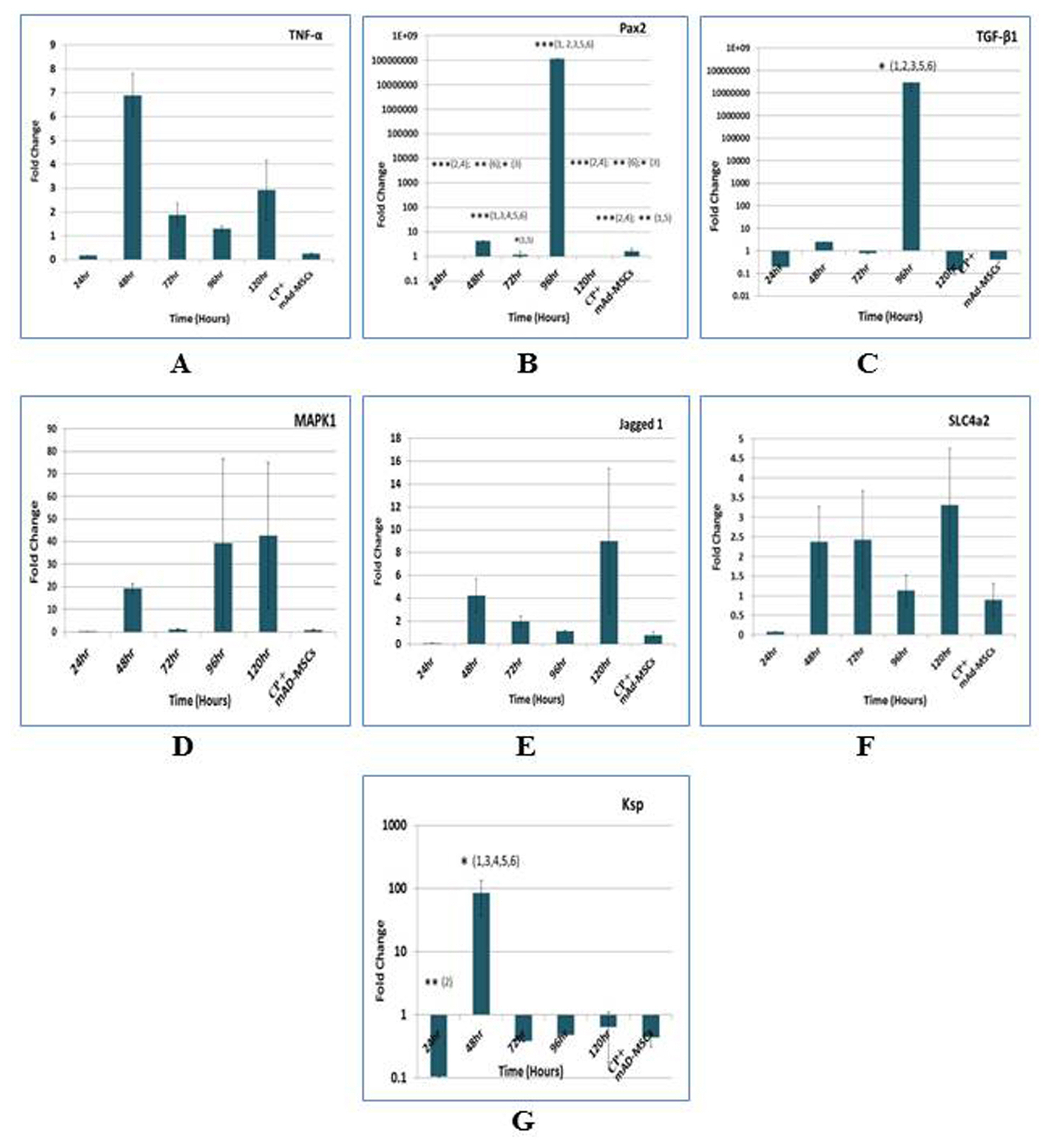
Fig. 5 Transcriptomic features demonstrate the anti-fibrotic and anti-inflammatory effects of mAd-MSCs. Quantitative transcriptomic expressions profiling of the genes in renal tissues of sacrificed balb/c mice by TaqMan qRT-PCR was evaluated in pre (24-120 hours) and post-transplantation of mAd-MSCs. The data was evaluated by the 2^−ΔΔCt method for fold change in genes expressions, which were used for multiple comparisons in ANOVA followed by Tucky’s test. The levels of significance *p<0.05, **p<0.01, and ***p<0.001 were used [log10 scale for Pax2, TGFβ-1, and Ksp]. (A) Tumor necrosis factor alpha (TNF-α). (B) Paired box gene 2 (Pax2). (C) Transforming growth factor beta 1 (TGFβ1). (D) Mitogen-activated protein kinase 1 (MAPK1). (E) Jagged1. (F) Solute carrier family 4 member 2 (SLC4a2). (G) Kidney-specific protein (Ksp-cadherin). [1=24 hr; 2=48 hr; 3=72 hr; 4=96 hr; 5=120 hr; 6=CP+mAd-MSCs transplanted].
Reference
-
References
1. Kanbay M, Covic A. Bone morphogenic protein-7: a new prognostic marker for acute kidney injury? NDT Plus. 2010; 3:106–107. DOI: 10.1093/ndtplus/sfp165. PMID: 25949422. PMCID: 4421545.
Article2. Yao W, Hu Q, Ma Y, Xiong W, Wu T, Cao J, Wu D. Human adipose-derived mesenchymal stem cells repair cisplatin-induced acute kidney injury through antiapoptotic pathways. Exp Ther Med. 2015; 10:468–476. DOI: 10.3892/etm.2015.2505. PMID: 26622339. PMCID: 4509364.
Article3. Morigi M, Imberti B, Zoja C, Corna D, Tomasoni S, Abbate M, Rottoli D, Angioletti S, Benigni A, Perico N, Alison M, Remuzzi G. Mesenchymal stem cells are renotropic, helping to repair the kidney and improve function in acute renal failure. J Am Soc Nephrol. 2004; 15:1794–1804. DOI: 10.1097/01.ASN.0000128974.07460.34. PMID: 15213267.
Article4. Barnes CJ, Distaso CT, Spitz KM, Verdun VA, Haramati A. Comparison of stem cell therapies for acute kidney injury. Am J Stem Cells. 2016; 5:1–10. PMID: 27335697. PMCID: 4913292.5. Patschan D, Buschmann I, Ritter O, Kribben A. Cell-based therapies in Acute Kidney Injury (AKI). Kidney Blood Press Res. 2018; 43:673–681. DOI: 10.1159/000489624. PMID: 29734169.
Article6. Ricci Z, Cruz DN, Ronco C. Classification and staging of acute kidney injury: beyond the RIFLE and AKIN criteria. Nat Rev Nephrol. 2011; 7:201–208. DOI: 10.1038/nrneph.2011.14. PMID: 21364520.
Article7. Mohsenin V. Practical approach to detection and management of acute kidney injury in critically ill patient. J Intensive Care. 2017; 5:57. DOI: 10.1186/s40560-017-0251-y. PMID: 28932401. PMCID: 5603084.
Article8. Luna AC, Madeira ME, Conceição TO, Moreira JA, Laiso RA, Maria DA. Characterization of adipose-derived stem cells of anatomical region from mice. BMC Res Notes. 2014; 7:552. DOI: 10.1186/1756-0500-7-552. PMID: 25138545. PMCID: 4156637.
Article9. Večerić-Haler Ž, Erman A, Cerar A, Motaln H, Kološa K, Lah Turnšek T, Sodin Šemrl S, Lakota K, Mrak-Poljšak K, Škrajnar Š, Kranjc S, Arnol M, Perše M. Improved protective effect of umbilical cord stem cell transplantation on cisplatin-induced kidney injury in mice pretreated with antithymocyte globulin. Stem Cells Int. 2016; 2016:3585362. DOI: 10.1155/2016/3585362. PMID: 26880955. PMCID: 4736416.
Article10. Lindoso RS, Verdoorn KS, Einicker-Lamas M. Renal recovery after injury: the role of Pax-2. Nephrol Dial Transplant. 2009; 24:2628–2633. DOI: 10.1093/ndt/gfp307. PMID: 19556301.
Article11. Ozkok A, Edelstein CL. Pathophysiology of cisplatin-induced acute kidney injury. Biomed Res Int. 2014; 2014:967826. DOI: 10.1155/2014/967826. PMID: 25165721. PMCID: 4140112.
Article12. Cheng K, Rai P, Plagov A, Lan X, Kumar D, Salhan D, Rehman S, Malhotra A, Bhargava K, Palestro CJ, Gupta S, Singhal PC. Transplantation of bone marrow-derived MSCs improves cisplatinum-induced renal injury through paracrine mechanisms. Exp Mol Pathol. 2013; 94:466–473. DOI: 10.1016/j.yexmp.2013.03.002. PMID: 23534987. PMCID: 3647016.
Article13. Park MS, De Leon M, Devarajan P. Cisplatin induces apoptosis in LLC-PK1 cells via activation of mitochondrial pathways. J Am Soc Nephrol. 2002; 13:858–865. PMID: 11912244.
Article14. Christensen EI, Verroust PJ. Megalin and cubilin, role in proximal tubule function and during development. Pediatr Nephrol. 2002; 17:993–999. DOI: 10.1007/s00467-002-0956-5. PMID: 12478347.
Article15. Gekle M, Knaus P, Nielsen R, Mildenberger S, Freudinger R, Wohlfarth V, Sauvant C, Christensen EI. Transforming growth factor-beta1 reduces megalin- and cubilin-mediated endocytosis of albumin in proximal-tubule-derived opossum kidney cells. J Physiol. 2003; 552:471–481. DOI: 10.1113/jphysiol.2003.048074. PMID: 14561830. PMCID: 2343374.
Article16. Ichimura T, Hung CC, Yang SA, Stevens JL, Bonventre JV. Kidney injury molecule-1: a tissue and urinary biomarker for nephrotoxicant-induced renal injury. Am J Physiol Renal Physiol. 2004; 286:F552–563. DOI: 10.1152/ajprenal.00285.2002. PMID: 14600030.
Article17. Vaidya VS, Ramirez V, Ichimura T, Bobadilla NA, Bonventre JV. Urinary kidney injury molecule-1: a sensitive quantitative biomarker for early detection of kidney tubular injury. Am J Physiol Renal Physiol. 2006; 290:F517–529. DOI: 10.1152/ajprenal.00291.2005. PMID: 16174863.
Article18. Bonventre JV. Kidney injury molecule-1 (KIM-1): a urinary biomarker and much more. Nephrol Dial Transplant. 2009; 24:3265–3268. DOI: 10.1093/ndt/gfp010. PMID: 19318357.
Article19. Ghaly EN, Gergis SW, Aziz JN, Yassa HD, Hassan HA. Role of mesenchymal stem cell therapy in cisplatin induced nephrotoxicity in adult albino rats: ultrastructural & biochemical study. Acta Medica Int. 2014; 1:57–66. DOI: 10.5530/ami.2014.2.3.
Article20. Zhang JB, Wang XQ, Lu GL, Huang HS, Xu SY. Adiposederived mesenchymal stem cells therapy for acute kidney injury induced by ischemia-reperfusion in a rat model. Clin Exp Pharmacol Physiol. 2017; 44:1232–1240. DOI: 10.1111/1440-1681.12811. PMID: 28688148.
Article21. Schubert R, Sann J, Frueh JT, Ullrich E, Geiger H, Baer PC. Tracking of adipose-derived mesenchymal stromal/stem cells in a model of cisplatin-induced acute kidney injury: comparison of bioluminescence imaging versus qRT-PCR. Int J Mol Sci. 2018; 19. pii: E2564. DOI: 10.3390/ijms19092564. PMID: 30158455. PMCID: 6165020.
Article22. Tsuruya K, Ninomiya T, Tokumoto M, Hirakawa M, Masutani K, Taniguchi M, Fukuda K, Kanai H, Kishihara K, Hirakata H, Iida M. Direct involvement of the receptor-mediated apoptotic pathways in cisplatin-induced renal tubular cell death. Kidney Int. 2003; 63:72–82. DOI: 10.1046/j.1523-1755.2003.00709.x. PMID: 12472770.
Article23. Ramesh G, Reeves WB. p38 MAP kinase inhibition ameliorates cisplatin nephrotoxicity in mice. Am J Physiol Renal Physiol. 2005; 289:F166–174. DOI: 10.1152/ajprenal.00401.2004. PMID: 15701814.
Article24. Luo J, Tsuji T, Yasuda H, Sun Y, Fujigaki Y, Hishida A. The molecular mechanisms of the attenuation of cisplatin-induced acute renal failure by N-acetylcysteine in rats. Nephrol Dial Transplant. 2008; 23:2198–2205. DOI: 10.1093/ndt/gfn090. PMID: 18385389.
Article25. Francescato HD, Costa RS, Silva CG, Coimbra TM. Treatment with a p38 MAPK inhibitor attenuates cisplatin nephrotoxicity starting after the beginning of renal damage. Life Sci. 2009; 84:590–597. DOI: 10.1016/j.lfs.2009.02.004. PMID: 26324989.
Article26. Malik S, Suchal K, Bhatia J, Gamad N, Dinda AK, Gupta YK, Arya DS. Molecular mechanisms underlying attenuation of cisplatin-induced acute kidney injury by epicate-chin gallate. Lab Invest. 2016; 96:853–861. DOI: 10.1038/labinvest.2016.60. PMID: 27239733.
Article27. Imgrund M, Gröne E, Gröne HJ, Kretzler M, Holzman L, Schlöndorff D, Rothenpieler UW. Re-expression of the developmental gene Pax-2 during experimental acute tubular necrosis in mice 1. Kidney Int. 1999; 56:1423–1431. DOI: 10.1046/j.1523-1755.1999.00663.x. PMID: 10504494.
Article28. Humphreys BD, Czerniak S, DiRocco DP, Hasnain W, Cheema R, Bonventre JV. Repair of injured proximal tubule does not involve specialized progenitors. Proc Natl Acad Sci U S A. 2011; 108:9226–9231. DOI: 10.1073/pnas.1100629108. PMID: 21576461. PMCID: 3107336.
Article29. Jiang YS, Jiang T, Huang B, Chen PS, Ouyang J. Epithelial-mesenchymal transition of renal tubules: divergent processes of repairing in acute or chronic injury? Med Hypotheses. 2013; 81:73–75. DOI: 10.1016/j.mehy.2013.03.020. PMID: 23601763.
Article30. Kobayashi T, Terada Y, Kuwana H, Tanaka H, Okado T, Kuwahara M, Tohda S, Sakano S, Sasaki S. Expression and function of the Delta-1/Notch-2/Hes-1 pathway during experimental acute kidney injury. Kidney Int. 2008; 73:1240–1250. DOI: 10.1038/ki.2008.74. PMID: 18418349.
Article31. Bielesz B, Sirin Y, Si H, Niranjan T, Gruenwald A, Ahn S, Kato H, Pullman J, Gessler M, Haase VH, Susztak K. Epithelial Notch signaling regulates interstitial fibrosis development in the kidneys of mice and humans. J Clin Invest. 2010; 120:4040–4054. DOI: 10.1172/JCI43025. PMID: 20978353. PMCID: 2964979.
Article32. Nyhan KC, Faherty N, Murray G, Cooey LB, Godson C, Crean JK, Brazil DP. Jagged/Notch signalling is required for a subset of TGFβ1 responses in human kidney epithelial cells. Biochim Biophys Acta. 2010; 1803:1386–1395. DOI: 10.1016/j.bbamcr.2010.09.001. PMID: 20833210.
Article33. Djudjaj S, Chatziantoniou C, Raffetseder U, Guerrot D, Dussaule JC, Boor P, Kerroch M, Hanssen L, Brandt S, Dittrich A, Ostendorf T, Floege J, Zhu C, Lindenmeyer M, Cohen CD, Mertens PR. Notch-3 receptor activation drives inflammation and fibrosis following tubulointerstitial kidney injury. J Pathol. 2012; 228:286–299. DOI: 10.1002/path.4076. PMID: 22806125.
Article34. Wang Z, Schultheis PJ, Shull GE. Three N-terminal variants of the AE2 Cl-/HCO3− exchanger are encoded by mRNAs transcribed from alternative promoters. J Biol Chem. 1996; 271:7835–7843. DOI: 10.1074/jbc.271.13.7835. PMID: 8631828.
Article35. Medina JF, Lecanda J, Acín A, Ciesielczyk P, Prieto J. Tissue-specific N-terminal isoforms from overlapping alternate promoters of the human AE2 anion exchanger gene. Biochem Biophys Res Commun. 2000; 267:228–235. DOI: 10.1006/bbrc.1999.1951. PMID: 10623603.
Article36. Alper SL, Darman RB, Chernova MN, Dahl NK. The AE gene family of Cl/HCO3− exchangers. J Nephrol. 2002; 15(Suppl 5):S41–53.37. Romero MF, Fulton CM, Boron WF. The SLC4 family of HCO 3 - transporters. Pflugers Arch. 2004; 447:495–509. DOI: 10.1007/s00424-003-1180-2. PMID: 14722772.38. Rodríguez-Soriano J. New insights into the pathogenesis of renal tubular acidosis--from functional to molecular studies. Pediatr Nephrol. 2000; 14:1121–1136. DOI: 10.1007/s004670000407. PMID: 11045400.
Article39. Stewart AK, Kurschat CE, Vaughan-Jones RD, Alper SL. Putative re-entrant loop 1 of AE2 transmembrane domain has a major role in acute regulation of anion exchange by pH. J Biol Chem. 2009; 284:6126–6139. DOI: 10.1074/jbc.M802051200. PMID: 19103596. PMCID: 2649077.
Article40. Jiang J, Dean D, Burghardt RC, Parrish AR. Disruption of cadherin/catenin expression, localization, and interactions during HgCl2-induced nephrotoxicity. Toxicol Sci. 2004; 80:170–182. DOI: 10.1093/toxsci/kfh143. PMID: 15084754.
Article
- Full Text Links
-
- Actions
-
Cited
- CITED
-
- Close
- Share
-
- Similar articles
-
- Transplantation of adipose derived mesenchymal stem cells for acute thoracolumbar disc disease with no deep pain perception in dogs
- Rapid deterioration of preexisting renal insufficiency after autologous mesenchymal stem cell therapy
- The Role of Adipose Tissue-Derived Stem Cells in Ischemic Acute Renal Injury in Rats
- Adipose-derived stem cells: characterization and clinical application
- Therapeutic potential of adipose tissue-derived stem cells for liver failure according to the transplantation routes
