Diarylbutane-type Lignans from Myristica fragrans (Nutmeg) show the Cytotoxicity against Breast Cancer Cells through Activation of AMP-activated Protein Kinase
- Affiliations
-
- 1College of Pharmacy, Chosun University, Gwangju 501-759, Republic of Korea.
- 2Korea Bioactive Natural Material Bank, Research Institute of Pharmaceutical Sciences, College of Pharmacy, Seoul National University, Seoul 151-742, Republic of Korea. wkoh1@snu.ac.kr
- 3Department of Pathology, College of Dentistry, Chosun University, Gwangju 501-759 Republic of Korea.
- KMID: 2376495
- DOI: http://doi.org/10.20307/nps.2017.23.1.21
Abstract
- In our program to search for new AMP-activated protein kinase (AMPK) activators from plants that exert potential anticancer property, we found that an EtOAc extract of Myristica fragrans (nutmeg) activated AMPK enzyme in human breast cancer MCF-7 cells. Two major diarylbutane-type lignans, macelignan and meso-dihydroguaiaretic acid (MDGA), were isolated as active principles from this extract. Treatment of breast cancer cells with two compounds induced cellular apoptosis, evidenced by cleavage of poly-(ADP-ribose) polymerase (PARP) and Ser 15 phosphorylation of p53. Moreover, macelignan and MDGA significantly inhibited the colony formation of MCF-7 breast cancer cells on soft agar. Intraperitoneal injection of macelignan and MDGA (20 mg/kg) suppressed the tumor growth of 4T1 mammary cancer cells. These results indicate that the chemopreventive effects of two major diarylbutane-type lignans from Myristica fragrans (nutmeg) may be associated with induction of apoptosis presumably through AMPK activation.
MeSH Terms
Figure
-
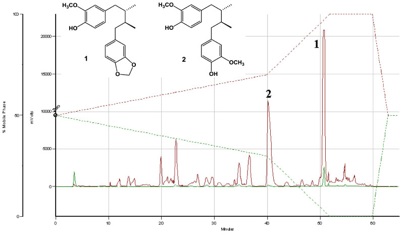
Fig. 1 A representative HPLC profile of major compounds from the total EtOAc layer of Myristica fragrans with detections at 205 and 280 nm. Key to peak identity: (1) macelignan, (2) MDGA. Chromatographic method used: 0 - 40 min (50 - 70% MeOH), 40 - 52 min (70 - 100% MeOH), 52 - 60 min (100% MeOH).
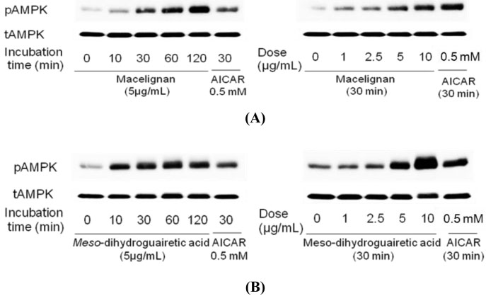
Fig. 2 Phosphorylation of AMPK by macelignan and MDGA in MCF-7 cells. Cells were seeded using DMEM supplemented with 10% heat-inactivated fetal bovine serum without antibiotic and cultured for 24 hrs at 37℃ in humidified air condition containing 5% CO2. The cells were then starved with serum free DMEM media for 24 hrs and treated with 5 µg/ml of macelignan and MDGA for 10 min, 30 min, 1 hr, and 2 hrs (the left panels of A and B) or treated with 0, 1, 2.5, 5, and 10 µg/ml of macelignan and MDGA (the right panels of A and B), respectively. The cells were harvested with cold PBS and lysed with 1 × NP40 lysis buffer. Proteins in whole cell lysates were separated by SDS-PAGE and immunoblotted with antibodies against phospho-AMPK and total AMPK. AICAR, an AMPK activator, was used as a positive control in this experiment. Data are representative of three independent experiments that gave similar results.
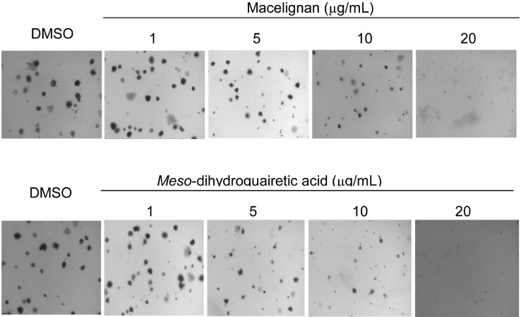
Fig. 3 Inhibitory effects of macelignan and MDGA on colony formation of MCF-7cells. MCF-7 cells were subjected to soft agar assays in the presence of macelignan and MDGA. Cells (8 × 10³/mL) were exposed to 1, 5, 10, and 20 µg/ml of macelignan and MDGA, respectively, in 1 ml of 0.3% basal medium Eagle (BME) agar containing 10% FBS, 2 mM L-glutamine, and 25 µg/ml gentamicin. The cultures were maintained at 37℃, in a 5% CO2 incubator for 10-14 days, and the cell colonies were scored using a microscope and the Image-Pro PLUS computer software program (Media Cybernetics, Silver Spring, MD).
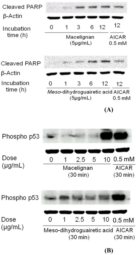
Fig. 4 Effects of macelignan and MDGA on PARP cleavage and p53 phosphorylation on MCF-7 cells. Cells were cultured using DMEM supplemented with 10% heat-inactivated fetal bovine serum for 24 hrs at 37℃. The cells were then starved with serum free DMEM media for 24 hrs and treated with 5 µg/ml of macelignan and MDGA for 1, 3, 6, and 12 hrs (the upper panels of A and B) for cleaved PARP and treated with 0, 1, 2.5, 5, and 10 µg/ml of macelignan and MDGA for 30 min for phospho-P53 (the lower panels of A and B), respectively. AICAR was used as a positive control. Data are representative of three independent experiments that gave similar results.
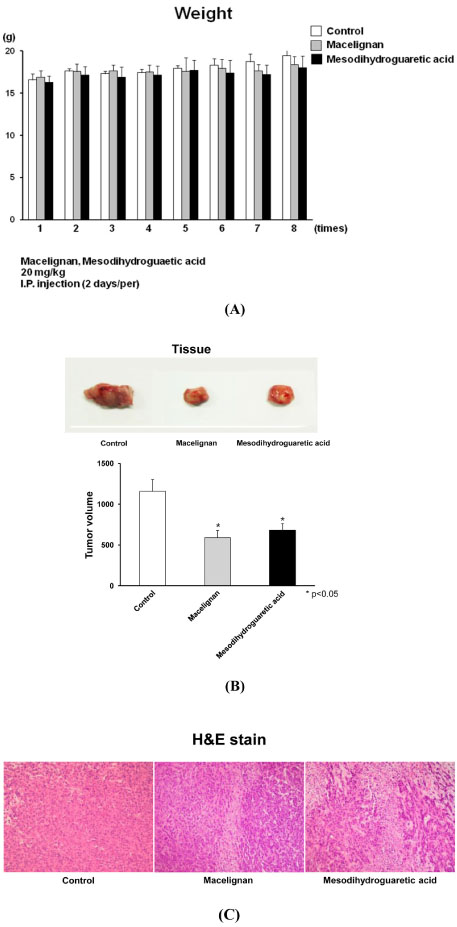
Fig. 5 In vivo anti-cancer effect on 4T1 mammary cancer cells. Six-week-old female Balb/c mice were anesthesized with 50 mg/kg pentobarbital and the rudimentary mammary ducts was cleared. 4T1 mammary cancer cells were harvested by trypsinization and centrifuged and resuspended in DMEM at a density of 3 × 10⁶/100 µl. 100 µl cell mixtures were injected into the cleared fat pad. Macelignan and MDGA (20 mg/kg) or an equal volume of the vehicle (n = 5 - 6 mice each group) were intraperitoneally injected by every two days for a total of 8 times. At the end of the treatment period, animals were sacrificed and solid tumors were excised for further studies. Body weights were recorded daily.
Reference
-
1. Evans DGR, Howell A. Breast Cancer Res. 2007; 9:213–220.2. Zhou W, Guan X, Wang L, Liao Y, Huang J. J Cancer Res Clin Oncol. 2012; 138:2085–2093.3. Nagalingam A, Arbiser JL, Bonner MY, Saxena NK, Sharma D. Breast Cancer Res. 2012; 14:R35.4. Guppy A, Jamal-Hanjani M, Pickering L. Future Oncol. 2011; 7:727–736.5. Li BX, Yamanaka K, Xiao X. Bioorg Med Chem. 2012; 20:6811–6820.6. Fortes C, Forastiere F, Farchi S, Mallone S, Trequattrinni T, Anatra F, Schmid G, Perucci CA. Nutr Cancer. 2003; 46:30–37.7. Tran TP, Kim HG, Choi JH, Na MK, Jeong HG. Phytomedicine. 2013; 20:622–631.8. Nguyen HB, Babcock JT, Wells CD, Quilliam LA. Oncogene. 2013; 32:4100–4109.9. Hadad SM, Baker L, Quinlan PR, Robertson KE, Bray SE, Thomson G, Kellock D, Jordan LB, Purdie CA, Hardie DG, Fleming S, Thompson AM. BMC Cancer. 2009; 9:307–315.10. Lee KE, Mun S, Pyun HB, Kim MS, Hwang JK. Biol Pharm Bull. 2012; 35:1669–1675.11. Pan JY, Chen SL, Yang MH, Wu J, Sinkkonen J, Zou K. Nat Prod Rep. 2009; 26:1251–1292.12. Nguyen PH, Le TVT, Kang HW, Chae J, Kim SK, Kwon KI, Seo DB, Lee SJ, Oh WK. Bioorg Med Chem Lett. 2010; 20:4128–4131.13. Woo WS, Shin KH, Wagner H, Lotter H. Phytochemistry. 1987; 26:1542–1543.14. Nakatani N, Ikeda K, Kikuzaki H, Kido M, Yamaguchi Y. Phytochemistry. 1988; 27:3127–3129.15. Hashimura T, Yoshida O. Jpn J Cancer Res. 1985; 76:321–323.16. Li Q, Ling Y, Yu L. J Cancer Res Clin Oncol. 2012; 138:1073–1079.17. Zhang X, Zhang S, Liu Y, Liu J, Ma Y, Zhu Y, Zhang J. Eur J Cancer. 2012; 48:1581–1592.18. Dumaz N, Meek DW. EMBO J. 1999; 18:7002–7010.19. Hardie DG. Curr Opin Cell Biol. 2005; 17:167–173.20. Kim MS, Park JY, Namkoong C, Jang PG, Ryu JW, Song HS, Yun JY, Namgoong IS, Ha J, Park IS, Lee IK, Viollet B, Youn JH, Lee HK, Lee KU. Nat Med. 2004; 10:727–733.21. Atherton PJ, Babraj J, Smith K, Singh J, Rennie MJ, Wackerhage H. FASEB J. 2005; 19:786–788.22. Lee DH, Lee TH, Jung CH, Kim YH. Cell Signal. 2012; 24:2216–2225.23. Choi EJ, Kang YG, Kim J, Hwang JK. Biol Pharm Bull. 2011; 34:748–754.
- Full Text Links
-
- Actions
-
Cited
- CITED
-
- Close
- Share
-
- Similar articles
-
- Cytotoxic Activities on Human Ovarian Cancer Cell Lines by Neolignans and Diarylnonanoids from the Seed of Myristica fragrans Houtt.
- Letter to the Editor: 17Beta-estradiol Stimulates Glucose Uptake Through Estrogen Receptor and AMP-activated Protein Kinase Activation in C2C12 Myotubes (Korean J Obes 2016;25:190-6)
- Effects of AMP-activated Protein Kinase Activating Compounds and Its Mechanism
- 5-aminoimidazole-4-carboxamide Riboside Induces Apoptosis Through AMP-activated Protein Kinase-independent and NADPH Oxidase-dependent Pathways
- Humanin suppresses receptor activator of nuclear factor-κB ligand-induced osteoclast differentiation via AMP-activated protein kinase activation
