Humanin suppresses receptor activator of nuclear factor-κB ligand-induced osteoclast differentiation via AMP-activated protein kinase activation
- Affiliations
-
- 1Department of Oral Biology, BK21 PLUS Project, Yonsei University College of Dentistry, Seoul 03722, Korea. dmshin@yuhs.ac
- KMID: 2455817
- DOI: http://doi.org/10.4196/kjpp.2019.23.5.411
Abstract
- Humanin (HN) is a mitochondrial peptide that exhibits cytoprotective actions against various stresses and diseases. HN has been shown to induce the phosphorylation of AMP-activated protein kinase (AMPK), which is a negative regulator of receptor activator of nuclear factor-κB ligand (RANKL). However, the role of HN in osteoclastogenesis or other skeletal disorders remains unknown. Here, we examined whether HN regulates osteoclastogenesis via AMPK activation using bone marrow-derived macrophage (BMM) cultures. Our results show that HN inhibited RANKL-induced osteoclast formation and reduced the expression of genes involved in osteoclastogenesis, including nuclear factor of activated T-cells cytoplasmic 1, osteoclast-associated receptor, cathepsin K, and tartrate-resistant acid phosphatase. Moreover, HN increased the levels of phosphorylated AMPK protein; compound C, an AMPK inhibitor, recovered HN-induced osteoclast differentiation. In addition, we found that HN significantly decreased the levels of RANKL-induced reactive oxygen species in BMMs. Therefore, these results indicate that HN plays an important role in osteoclastogenesis and may function as an inhibitor of bone disorders via AMPK activation.
Keyword
MeSH Terms
Figure
-
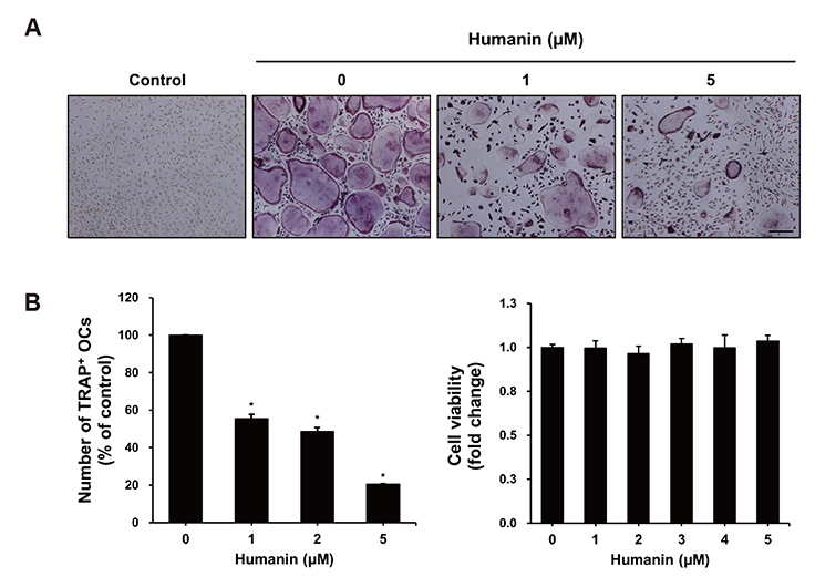
Fig. 1 Effects of humanin (HN) on receptor activator of nuclear factor-κB ligand (RANKL)-induced osteoclast differentiation. (A) Bone marrow-derived macrophages (BMMs) were cultured in the presence of macrophage colony-stimulating factor (M-CSF) (30 ng/ml), RANKL (50 ng/ml), and the indicated concentrations of HN for 6 days. Cells were stained for tartrate-resistant acid phosphatase (TRAP). Scale bar, 500 µm. (B) Numbers of TRAP+ multinucleated cells were counted (≥ 3 nuclei). BMMs were seeded into 96-well plates and incubated with various concentrations of HN. Cell viability was measured by the MTS assay. OCs, osteoclasts.*p < 0.05 compared with control.
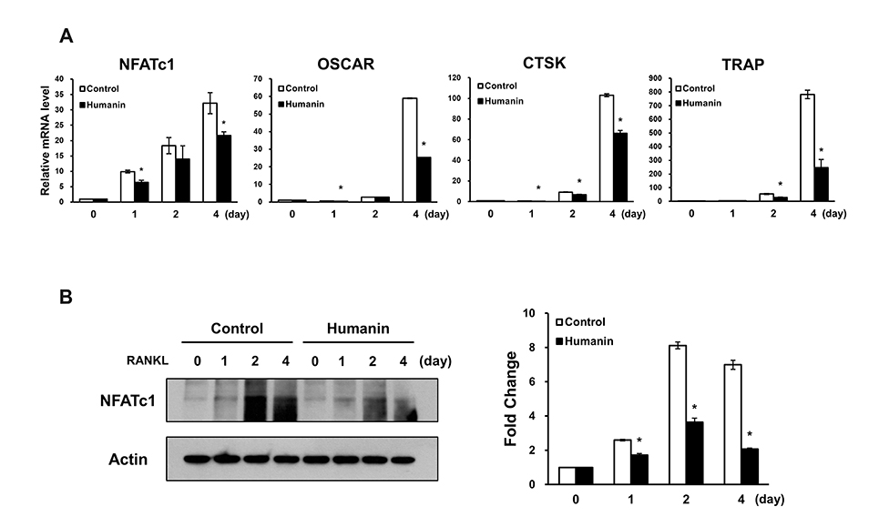
Fig. 2 Effects of humanin on the mRNA and protein levels of osteoclastogenesis-related genes. (A) The mRNA levels of receptor activator of nuclear factor-κB ligand (RANKL)-induced osteoclast differentiation markers were evaluated by quantitative RT-PCR. (B) NFATc1 protein levels were examined by Western blot analyses. OSCAR, osteoclast-associated receptor; CTSK, cathepsin K; TRAP, tartrate-resistant acid phosphatase. *p < 0.05 compared with control.
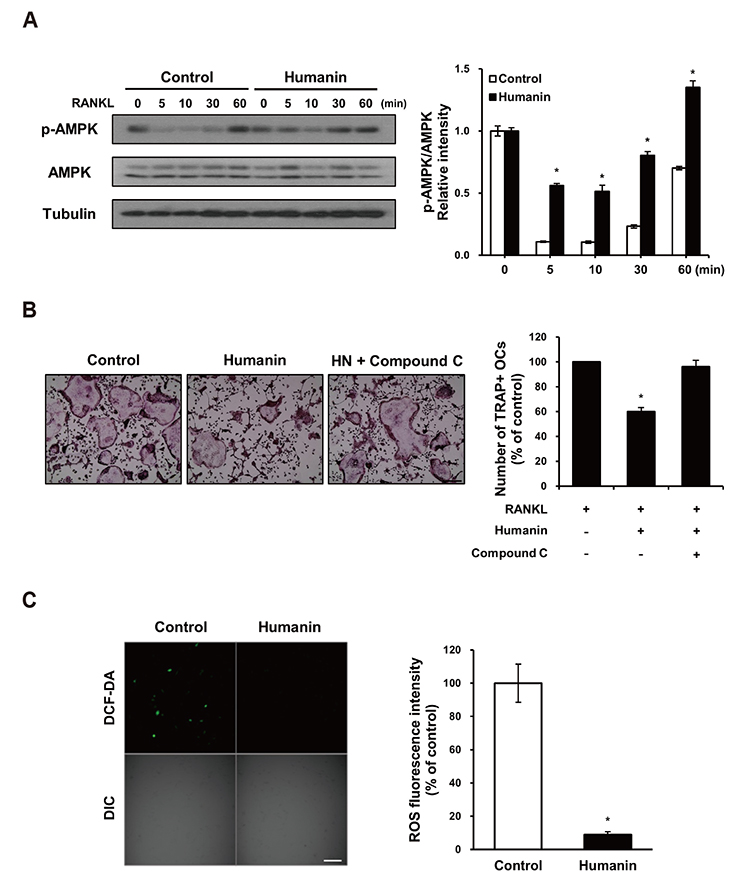
Fig. 3 Effects of humanin (HN) on AMP-activated protein kinase (AMPK) activation during osteoclast differentiation and receptor activator of nuclear factor-κB ligand (RANKL)-induced reactive oxygen species production in bone marrow-derived macrophages (BMMs). (A) Western blot analysis of phosphor (p)-AMPK and AMPK during RANKL-induced osteoclastogenesis in untreated or HN-treated BMMs. The p-AMPK:AMPK ratio was calculated. (B) BMMs were treated with or without HN, and compound C, and stained for tartrate-resistant acid phosphatase (TRAP). Numbers of TRAP+ multinucleated cells were counted (≥ 3 nuclei). Scale bar, 500 µm. (C) BMMs were treated with RANKL (50 ng/ml) for 10 min and loaded with 10 µM dichlorofluorescein diacetate (DCF-DA). Data are expressed relative to the value for control BMMs. Scale bar, 100 µm. OCs, osteoclasts. *p < 0.05 compared with control.
Reference
-
1. Datta HK, Ng WF, Walker JA, Tuck SP, Varanasi SS. The cell biology of bone metabolism. J Clin Pathol. 2008; 61:577–587.
Article2. Charles JF, Aliprantis AO. Osteoclasts: more than ‘bone eaters’. Trends Mol Med. 2014; 20:449–459.
Article3. Erkhembaatar M, Gu DR, Lee SH, Yang YM, Park S, Muallem S, Shin DM, Kim MS. Lysosomal Ca2+ signaling is essential for osteoclastogenesis and bone remodeling. J Bone Miner Res. 2017; 32:385–396.4. Florencio-Silva R, Sasso GR, Sasso-Cerri E, Simões MJ, Cerri PS. Biology of bone tissue: structure, function, and factors that influence bone cells. Biomed Res Int. 2015; 2015:421746.
Article5. Teitelbaum SL. Bone resorption by osteoclasts. Science. 2000; 289:1504–1508.
Article6. Mihaylova MM, Shaw RJ. The AMPK signalling pathway coordinates cell growth, autophagy and metabolism. Nat Cell Biol. 2011; 13:1016–1023.
Article7. Jeyabalan J, Shah M, Viollet B, Chenu C. AMP-activated protein kinase pathway and bone metabolism. J Endocrinol. 2012; 212:277–290.
Article8. Lee YS, Kim YS, Lee SY, Kim GH, Kim BJ, Lee SH, Lee KU, Kim GS, Kim SW, Koh JM. AMP kinase acts as a negative regulator of RANKL in the differentiation of osteoclasts. Bone. 2010; 47:926–937.
Article9. Ming W, Lu G, Xin S, Huanyu L, Yinghao J, Xiaoying L, Chengming X, Banjun R, Li W, Zifan L. Mitochondria related peptide MOTS-c suppresses ovariectomy-induced bone loss via AMPK activation. Biochem Biophys Res Commun. 2016; 476:412–419.
Article10. Hashimoto Y, Niikura T, Tajima H, Yasukawa T, Sudo H, Ito Y, Kita Y, Kawasumi M, Kouyama K, Doyu M, Sobue G, Koide T, Tsuji S, Lang J, Kurokawa K, Nishimoto I. A rescue factor abolishing neuronal cell death by a wide spectrum of familial Alzheimer's disease genes and Abeta. Proc Natl Acad Sci U S A. 2001; 98:6336–6341.11. Qin Q, Jin J, He F, Zheng Y, Li T, Zhang Y, He J. Humanin promotes mitochondrial biogenesis in pancreatic MIN6 β-cells. Biochem Biophys Res Commun. 2018; 497:292–297.
Article12. Lee C, Yen K, Cohen P. Humanin: a harbinger of mitochondrial-derived peptides? Trends Endocrinol Metab. 2013; 24:222–228.
Article13. Zhang Z, Welte T, Troiano N, Maher SE, Fu XY, Bothwell AL. Osteoporosis with increased osteoclastogenesis in hematopoietic cell-specific STAT3-deficient mice. Biochem Biophys Res Commun. 2005; 328:800–807.
Article14. Gidlund EK, von Walden F, Venojärvi M, Risérus U, Heinonen OJ, Norrbom J, Sundberg CJ. Humanin skeletal muscle protein levels increase after resistance training in men with impaired glucose metabolism. Physiol Rep. 2016; 4:e13063.
Article15. Widmer RJ, Flammer AJ, Herrmann J, Rodriguez-Porcel M, Wan J, Cohen P, Lerman LO, Lerman A. Circulating humanin levels are associated with preserved coronary endothelial function. Am J Physiol Heart Circ Physiol. 2013; 304:H393–H397.
Article16. Lee NK, Choi YG, Baik JY, Han SY, Jeong DW, Bae YS, Kim N, Lee SY. A crucial role for reactive oxygen species in RANKL-induced osteoclast differentiation. Blood. 2005; 106:852–859.
Article17. Kim MS, Yang YM, Son A, Tian YS, Lee SI, Kang SW, Muallem S, Shin DM. RANKL-mediated reactive oxygen species pathway that induces long lasting Ca2+ oscillations essential for osteoclastogenesis. J Biol Chem. 2010; 285:6913–6921.18. Sreekumar PG, Hinton DR, Kannan R. Endoplasmic reticulum-mitochondrial crosstalk: a novel role for the mitochondrial peptide humanin. Neural Regen Res. 2017; 12:35–38.
Article19. Liu X, Chhipa RR, Nakano I, Dasgupta B. The AMPK inhibitor compound C is a potent AMPK-independent antiglioma agent. Mol Cancer Ther. 2014; 13:596–605.
Article20. Matsuoka M, Hashimoto Y. Humanin and the receptors for humanin. Mol Neurobiol. 2010; 41:22–28.
Article21. Ikonen M, Liu B, Hashimoto Y, Ma L, Lee KW, Niikura T, Nishimoto I, Cohen P. Interaction between the Alzheimer's survival peptide humanin and insulin-like growth factor-binding protein 3 regulates cell survival and apoptosis. Proc Natl Acad Sci U S A. 2003; 100:13042–13047.
Article22. Yang YM, Kim MS, Son A, Hong JH, Kim KH, Seo JT, Lee SI, Shin DM. Alteration of RANKL-induced osteoclastogenesis in primary cultured osteoclasts from SERCA2+/− mice. J Bone Miner Res. 2009; 24:1763–1769.23. Boyle WJ, Simonet WS, Lacey DL. Osteoclast differentiation and activation. Nature. 2003; 423:337–342.
Article24. Feng X, Teitelbaum SL. Osteoclasts: new insights. Bone Res. 2013; 1:11–26.25. Teitelbaum SL. Osteoclasts, integrins, and osteoporosis. J Bone Miner Metab. 2000; 18:344–349.
Article26. Boyce BF, Xing L. Functions of RANKL/RANK/OPG in bone modeling and remodeling. Arch Biochem Biophys. 2008; 473:139–146.
Article27. Son A, Kim MS, Jo H, Byun HM, Shin DM. Effects of inositol 1,4,5-triphosphate on osteoclast differentiation in RANKL-induced osteoclastogenesis. Korean J Physiol Pharmacol. 2012; 16:31–36.
Article28. Takayanagi H, Kim S, Koga T, Nishina H, Isshiki M, Yoshida H, Saiura A, Isobe M, Yokochi T, Inoue J, Wagner EF, Mak TW, Kodama T, Taniguchi T. Induction and activation of the transcription factor NFATc1 (NFAT2) integrate RANKL signaling in terminal differentiation of osteoclasts. Dev Cell. 2002; 3:889–901.
Article29. Park B, Yang YM, Choi BJ, Kim MS, Shin DM. Activation of G proteins by aluminum fluoride enhances RANKL-mediated osteoclastogenesis. Korean J Physiol Pharmacol. 2013; 17:427–433.
Article30. Kim JH, Kim N. Regulation of NFATc1 in osteoclast differentiation. J Bone Metab. 2014; 21:233–241.
Article31. Sharma SM, Bronisz A, Hu R, Patel K, Mansky KC, Sif S, Ostrowski MC. MITF and PU.1 recruit p38 MAPK and NFATc1 to target genes during osteoclast differentiation. J Biol Chem. 2007; 282:15921–15929.
Article32. Takayanagi H. The role of NFAT in osteoclast formation. Ann N Y Acad Sci. 2007; 1116:227–237.
Article33. Saito Y, Chapple RH, Lin A, Kitano A, Nakada D. AMPK protects leukemia-initiating cells in myeloid leukemias from metabolic stress in the bone marrow. Cell Stem Cell. 2015; 17:585–596.
Article34. Kim J, Yang G, Kim Y, Kim J, Ha J. AMPK activators: mechanisms of action and physiological activities. Exp Mol Med. 2016; 48:e224.
Article35. Shackelford DB, Shaw RJ. The LKB1-AMPK pathway: metabolism and growth control in tumour suppression. Nat Rev Cancer. 2009; 9:563–575.
Article36. Hardie DG. AMP-activated/SNF1 protein kinases: conserved guardians of cellular energy. Nat Rev Mol Cell Biol. 2007; 8:774–785.
Article37. Kanazawa I, Yamaguchi T, Yano S, Yamauchi M, Yamamoto M, Sugimoto T. Adiponectin and AMP kinase activator stimulate proliferation, differentiation, and mineralization of osteoblastic MC3T3-E1 cells. BMC Cell Biol. 2007; 8:51.
Article38. Jang WG, Kim EJ, Bae IH, Lee KN, Kim YD, Kim DK, Kim SH, Lee CH, Franceschi RT, Choi HS, Koh JT. Metformin induces osteoblast differentiation via orphan nuclear receptor SHP-mediated transactivation of Runx2. Bone. 2011; 48:885–893.
Article
- Full Text Links
-
- Actions
-
Cited
- CITED
-
- Close
- Share
-
- Similar articles
-
- Effect of Cornus Officinalis on Receptor Activator of Nuclear Factor-kappaB Ligand (RANKL)-induced Osteoclast Differentiation
- Regulation of NFATc1 in Osteoclast Differentiation
- Fexaramine Inhibits Receptor Activator of Nuclear Factor-κB Ligand-induced Osteoclast Formation via Nuclear Factor of Activated T Cells Signaling Pathways
- Inhibitory Effect of Rosae Multiflorae Fructus Extracts on the Receptor Activator of NF-κB Ligand-Induced Osteoclastogenesis through Modulation of P38- and Ca2âº-Mediated Nuclear Factor of Activated T-Cells Cytoplasmic 1 Expression
- NF-kappaB-Mediated Regulation of Osteoclastogenesis
