Gene expression pattern during osteogenic differentiation of human periodontal ligament cells in vitro
- Affiliations
-
- 1Department of Periodontology, Kyungpook National University School of Dentistry, Daegu, Korea. jysuh@knu.ac.kr
- 2Institute for Hard tissue and Bio-tooth Regeneration, Kyungpook National University School of Dentistry, Daegu, Korea.
- KMID: 2094716
- DOI: http://doi.org/10.5051/jpis.2011.41.4.167
Abstract
- PURPOSE
Periodontal ligament (PDL) cell differentiation into osteoblasts is important in bone formation. Bone formation is a complex biological process and involves several tightly regulated gene expression patterns of bone-related proteins. The expression patterns of bone related proteins are regulated in a temporal manner both in vivo and in vitro. The aim of this study was to observe the gene expression profile in PDL cell proliferation, differentiation, and mineralization in vitro.
METHODS
PDL cells were grown until confluence, which were then designated as day 0, and nodule formation was induced by the addition of 50 microg/mL ascorbic acid, 10 mM beta-glycerophosphate, and 100 nM dexamethasone to the medium. The dishes were stained with Alizarin Red S on days 1, 7, 14, and 21. Real-time polymerase chain reaction was performed for the detection of various genes on days 0, 1, 7, 14, and 21.
RESULTS
On day 0 with a confluent monolayer, in the active proliferative stage, c-myc gene expression was observed at its maximal level. On day 7 with a multilayer, alkaline phosphatase, bone morphogenetic protein (BMP)-2, and BMP-4 gene expression had increased and this was followed by maximal expression of osteocalcin on day 14 with the initiation of nodule mineralization. In relationship to apoptosis, c-fos gene expression peaked on day 21 and was characterized by the post-mineralization stage. Here, various genes were regulated in a temporal manner during PDL fibroblast proliferation, extracellular matrix maturation, and mineralization. The gene expression pattern was similar.
CONCLUSIONS
We can speculate that the gene expression pattern occurs during PDL cell proliferation, differentiation, and mineralization. On the basis of these results, it might be possible to understand the various factors that influence PDL cell proliferation, extracellular matrix maturation, and mineralization with regard to gene expression patterns.
MeSH Terms
-
Alkaline Phosphatase
Anthraquinones
Apoptosis
Ascorbic Acid
Biological Processes
Bone Morphogenetic Proteins
Cell Differentiation
Cell Proliferation
Dexamethasone
Durapatite
Extracellular Matrix
Fibroblasts
Gene Expression
Genes, fos
Genes, myc
Glycerophosphates
Humans
Osteoblasts
Osteocalcin
Osteogenesis
Periodontal Ligament
Proteins
Real-Time Polymerase Chain Reaction
Transcriptome
Alkaline Phosphatase
Anthraquinones
Ascorbic Acid
Bone Morphogenetic Proteins
Dexamethasone
Durapatite
Glycerophosphates
Osteocalcin
Proteins
Figure
-

Figure 1 Morphology of the various stages and percentage of calcified areas during the formation of mineralized nodule by human periodontal ligament (PDL) cells. (A) shows the visual field at ×200 magnification on days 1(a), 7(b), 14(c), and 21(d). Developed mineralization is observed and size and number of nodules are increased compared to PDL cells on day 14. (B) Measurement of calcified areas by Alizarin Red S stain. Data are shown as the mean±SD of two patients. Percentages of the calcified areas are 5% and 33% of the culture dish on day 14 and 21, respectively.
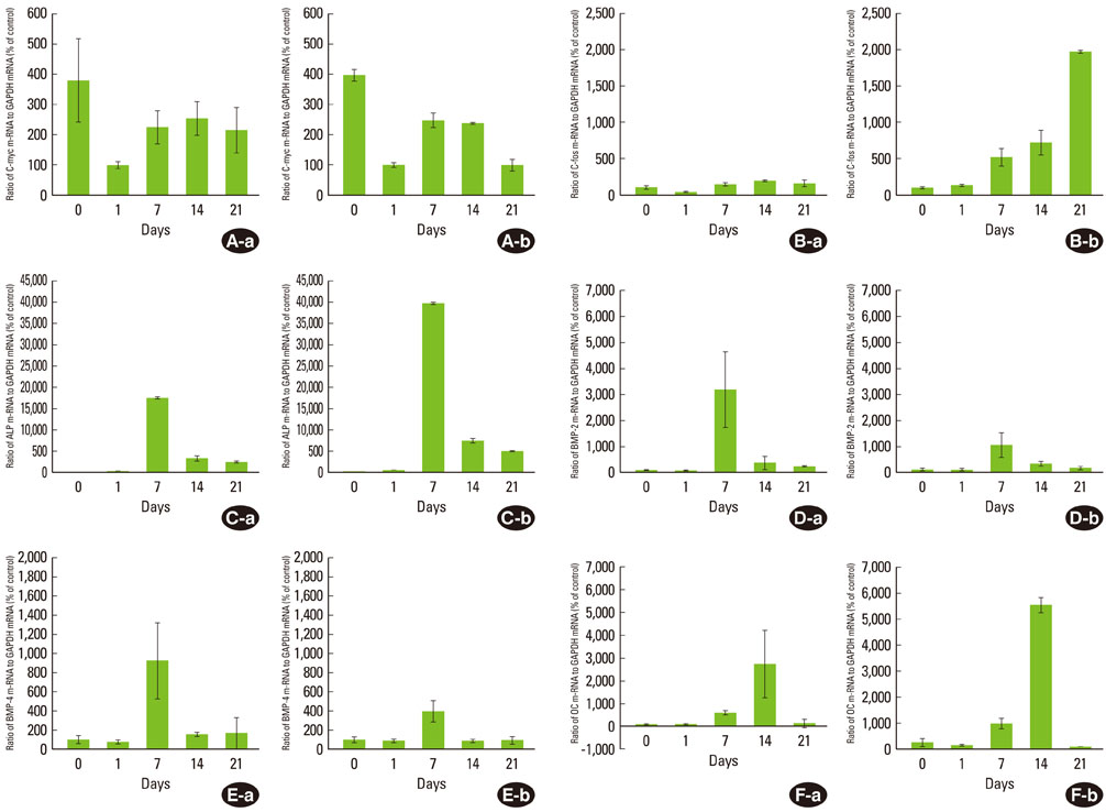
Figure 2 c-myc (A), c-fos (B), ALP (C), BMP-2 (D), BMP-4 (E), and OC (F) mRNA expression during mineralization of human periodontal ligament (PDL) cells. The expression of each gene in PDL1 (a) and PDL2 (b) is shown. The graphs show the ratio of mRNA to GAPDH mRNA from the real-time polymerase chain reaction results. Control means the minimal gene expression level during 21 days. Values are means±SD of two cultures.

Figure 3 Gene expression pattern during mineralization of periodontal ligament cells. Expression of genes was analyzed by real-time PCR and normalized to the levels of GAPDH mRNA. Values are presented as the percent of the maximal expression for each transcript. a)c-myc, b)c-fos, c)ALP, d)BMP-2, e)BMP-4, f)OC. a),c)-f)Statistically significant difference as compared with day 0 (P<0.05). b)(P>0/05).
Cited by 1 articles
-
The effects of dexamethasone on the apoptosis and osteogenic differentiation of human periodontal ligament cells
Sung-Mi Kim, Yong-Gun Kim, Jin-Woo Park, Jae-Mok Lee, Jo-Young Suh
J Periodontal Implant Sci. 2013;43(4):168-176. doi: 10.5051/jpis.2013.43.4.168.
Reference
-
1. Strutz F, Okada H, Lo CW, Danoff T, Carone RL, Tomaszewski JE, et al. Identification and characterization of a fibroblast marker: FSP1. J Cell Biol. 1995. 130:393–405.
Article2. Lackler KP, Cochran DL, Hoang AM, Takacs V, Oates TW. Development of an in vitro wound healing model for periodontal cells. J Periodontol. 2000. 71:226–237.
Article3. Nojima N, Kobayashi M, Shionome M, Takahashi N, Suda T, Hasegawa K. Fibroblastic cells derived from bovine periodontal ligaments have the phenotypes of osteoblasts. J Periodontal Res. 1990. 25:179–185.
Article4. Somerman MJ, Young MF, Foster RA, Moehring JM, Imm G, Sauk JJ. Characteristics of human periodontal ligament cells in vitro. Arch Oral Biol. 1990. 35:241–247.
Article5. Cho MI, Matsuda N, Lin WL, Moshier A, Ramakrishnan PR. In vitro formation of mineralized nodules by periodontal ligament cells from the rat. Calcif Tissue Int. 1992. 50:459–467.
Article6. Mukai M, Yoshimine Y, Akamine A, Maeda K. Bone-like nodules formed in vitro by rat periodontal ligament cells. Cell Tissue Res. 1993. 271:453–460.
Article7. Chung HB, Park JW, Suh JY. The effect of dexamethasone on the gene expression of the bone matrix protein in the periodontal ligament cells. J Korean Acad Periodontol. 2002. 32:445–456.
Article8. Stein GS, Lian JB. Molecular mechanisms mediating proliferation/differentiation interrelationships during progressive development of the osteoblast phenotype. Endocr Rev. 1993. 14:424–442.
Article9. Choi JY, Lee BH, Song KB, Park RW, Kim IS, Sohn KY, et al. Expression patterns of bone-related proteins during osteoblastic differentiation in MC3T3-E1 cells. J Cell Biochem. 1996. 61:609–618.
Article10. Zhumabayeva BD, Lin WL, Choung PH, Chien HH, Sodek J, Sampath KT, et al. Differential induction of bone sialoprotein by dexamethasone and osteogenic protein-1 (OP-1, BMP-7) in rat periodontal ligament cells in vitro: relationship to the mineralization of tissue nodules. Int J Oral Biol. 1998. 23:91–101.11. Strayhorn CL, Garrett JS, Dunn RL, Benedict JJ, Somerman MJ. Growth factors regulate expression of osteoblast-associated genes. J Periodontol. 1999. 70:1345–1354.
Article12. Lian JB, Stein GS. Concepts of osteoblast growth and differentiation: basis for modulation of bone cell development and tissue formation. Crit Rev Oral Biol Med. 1992. 3:269–305.
Article13. Ripamonti U. Lindholm TS, editor. Induction of cementogenesis and periodontal ligament regeneration by bone morphogenetic proteins. Bone morphogenetic proteins: biology, biochemistry and reconstructive surgery. 1996. San Diego: Academic Press;189–198.14. King GN, King N, Cruchley AT, Wozney JM, Hughes FJ. Recombinant human bone morphogenetic protein-2 promotes wound healing in rat periodontal fenestration defects. J Dent Res. 1997. 76:1460–1470.
Article15. Ivanovski S, Li H, Haase HR, Bartold PM. Expression of bone associated macromolecules by gingival and periodontal ligament fibroblasts. J Periodontal Res. 2001. 36:131–141.
Article16. Shalhoub V, Gerstenfeld LC, Collart D, Lian JB, Stein GS. Downregulation of cell growth and cell cycle regulated genes during chick osteoblast differentiation with the reciprocal expression of histone gene variants. Biochemistry. 1989. 28:5318–5322.
Article17. Preston GA, Lyon TT, Yin Y, Lang JE, Solomon G, Annab L, et al. Induction of apoptosis by c-Fos protein. Mol Cell Biol. 1996. 16:211–218.
Article18. McCabe LR, Banerjee C, Kundu R, Harrison RJ, Dobner PR, Stein JL, et al. Developmental expression and activities of specific fos and jun proteins are functionally related to osteoblast maturation: role of Fra-2 and Jun D during differentiation. Endocrinology. 1996. 137:4398–4408.
Article19. Aubin JE, Turken K, Heersche JNM. Noda M, editor. Osteoblastic cell lineage. Cellular and molecular biology of bone. 1993. San Diego: Academic Press;1–44.
Article20. Onyia JE, Hale LV, Miles RR, Cain RL, Tu Y, Hulman JF, et al. Molecular characterization of gene expression changes in ROS 17/2.8 cells cultured in diffusion chambers in vivo. Calcif Tissue Int. 1999. 65:133–138.
Article21. Lynch MP, Capparelli C, Stein JL, Stein GS, Lian JB. Apoptosis during bone-like tissue development in vitro. J Cell Biochem. 1998. 68:31–49.
Article22. Hughes FJ, Collyer J, Stanfield M, Goodman SA. The effects of bone morphogenetic protein-2, -4, and -6 on differentiation of rat osteoblast cells in vitro. Endocrinology. 1995. 136:2671–2677.
Article23. Zegzula HD, Buck DC, Brekke J, Wozney JM, Hollinger JO. Bone formation with use of rhBMP-2 (recombinant human bone morphogenetic protein-2). J Bone Joint Surg Am. 1997. 79:1778–1790.
Article24. Thesleff I, Vaahtokari A, Kettunen P, Aberg T. Epithelial-mesenchymal signaling during tooth development. Connect Tissue Res. 1995. 32:9–15.25. Boden SD, McCuaig K, Hair G, Racine M, Titus L, Wozney JM, et al. Differential effects and glucocorticoid potentiation of bone morphogenetic protein action during rat osteoblast differentiation in vitro. Endocrinology. 1996. 137:3401–3407.
Article26. Kobayashi M, Takiguchi T, Suzuki R, Yamaguchi A, Deguchi K, Shionome M, et al. Recombinant human bone morphogenetic protein-2 stimulates osteoblastic differentiation in cells isolated from human periodontal ligament. J Dent Res. 1999. 78:1624–1633.
Article27. Zaman KU, Sugaya T, Kato H. Effect of recombinant human platelet-derived growth factor-BB and bone morphogenetic protein-2 application to demineralized dentin on early periodontal ligament cell response. J Periodontal Res. 1999. 34:244–250.
Article28. Chen D, Harris MA, Rossini G, Dunstan CR, Dallas SL, Feng JQ, et al. Bone morphogenetic protein 2 (BMP-2) enhances BMP-3, BMP-4, and bone cell differentiation marker gene expression during the induction of mineralized bone matrix formation in cultures of fetal rat calvarial osteoblasts. Calcif Tissue Int. 1997. 60:283–290.
Article29. Xu WP, Shiba H, Mizuno N, Uchida Y, Mouri Y, Kawaguchi H, et al. Effect of bone morphogenetic proteins-4, -5 and -6 on DNA synthesis and expression of bone-related proteins in cultured human periodontal ligament cells. Cell Biol Int. 2004. 28:675–682.
Article30. Nakashima M, Nagasawa H, Yamada Y, Reddi AH. Regulatory role of transforming growth factor-beta, bone morphogenetic protein-2, and protein-4 on gene expression of extracellular matrix proteins and differentiation of dental pulp cells. Dev Biol. 1994. 162:18–28.
Article31. Franceschi RT, Iyer BS. Relationship between collagen synthesis and expression of the osteoblast phenotype in MC3T3-E1 cells. J Bone Miner Res. 1992. 7:235–246.
Article32. Gerstenfeld LC, Chipman SD, Glowacki J, Lian JB. Expression of differentiated function by mineralizing cultures of chicken osteoblasts. Dev Biol. 1987. 122:49–60.
Article33. Romberg RW, Werness PG, Riggs BL, Mann KG. Inhibition of hydroxyapatite crystal growth by bone-specific and other calcium-binding proteins. Biochemistry. 1986. 25:1176–1180.
Article34. Glowacki J, Lian JB. Impaired recruitment and differentiation of osteoclast progenitors by osteocalcin-deplete bone implants. Cell Differ. 1987. 21:247–254.
Article35. Prigodich RV, Vesely MR. Characterization of the complex between bovine osteocalcin and type I collagen. Arch Biochem Biophys. 1997. 345:339–341.
Article36. Ritter NM, Farach-Carson MC, Butler WT. Evidence for the formation of a complex between osteopontin and osteocalcin. J Bone Miner Res. 1992. 7:877–885.
Article37. Aronow MA, Gerstenfeld LC, Owen TA, Tassinari MS, Stein GS, Lian JB. Factors that promote progressive development of the osteoblast phenotype in cultured fetal rat calvaria cells. J Cell Physiol. 1990. 143:213–221.
Article38. Malaval L, Modrowski D, Gupta AK, Aubin JE. Cellular expression of bone-related proteins during in vitro osteogenesis in rat bone marrow stromal cell cultures. J Cell Physiol. 1994. 158:555–572.
Article39. Shin JH, Park JW, Yeo SI, Noh WC, Kim MK, Kim JC, et al. Identification of matrix mineralization-related genes in human periodontal ligament cells using cDNA microarray. J Korean Acad Periodontol. 2007. 37:Suppl. 447–463.
Article40. Smeyne RJ, Vendrell M, Hayward M, Baker SJ, Miao GG, Schilling K, et al. Continuous c-fos expression precedes programmed cell death in vivo. Nature. 1993. 363:166–169.
Article
- Full Text Links
-
- Actions
-
Cited
- CITED
-
- Close
- Share
-
- Similar articles
-
- Analysis of gene expression during mineralization of cultured human periodontal ligament cells
- Indirect Co-Culture of Stem Cells from Human Exfoliated Deciduous Teeth and Oral Cells in a Microfluidic Platform
- Effects of nitric oxide on the proliferation and differentiation of human periodontal ligament cells
- Comparison of Gene Expression from Supernumerary Dental Pulp and Periodontal Ligament Stem Cells
- A study on differentiation potency of adult stem cells from pulp, periodontal ligament, and dental follicle to osteoblast
