Effect of Diquafosol on Hyperosmotic Stress-induced Tumor Necrosis Factor-α and Interleukin-6 Expression in Human Corneal Epithelial Cells
- Affiliations
-
- 1Myungmoon Bio, Hwaseong, Korea.
- 2Department of Physiology, College of Korean Medicine Dongguk University, Gyeongju, Korea.
- 3Binaree, Daegu, Korea.
- 4Central Ophthalmic Clinic, Daegu, Korea. eyepark9@naver.com
- 5Division of Biomedicinal & Cosmetics, College of Sciences & Technology, Mokwon University, Daejeon, Korea.
- 6Developmental Biology Laboratory, Department of Biology, College of Natural Sciences, Kyungpook National University, Daegu, Korea. jcjung@knu.ac.kr
- KMID: 2469281
- DOI: http://doi.org/10.3341/kjo.2019.0046
Abstract
- PURPOSE
Diquafosol is a pharmaceutical drug used for dry eye treatment with a novel mechanism of action. It is a purinergic P2Y2 receptor agonist that promotes the secretion of tears and healing of corneal epithelial wounds. However, its inhibitory effect on hyperosmotic stress-induced inflammation in human corneal epithelial cells (HCECs) remains unclear.
METHODS
A hyperosmotic stress model was established by transferring HCECs from isosmotic (312 mOsm/kg to hyperosmotic medium (500 mOsm/kg). HCECs were incubated with 500 mOsm/kg hyperosmotic medium for 30 minutes, and then treated with diquafosol (0.6-6 mg/mL) for 4 or 24 hours. Cells were then harvested and analyzed by western blot, immunocytochemistry, and real-time polymerase chain reaction to evaluate the expression of interleukin-6, tumor necrosis factor-alpha, and the phosphorylation status of nuclear factor-kappa B.
RESULTS
Diquafosol significantly decreased the mRNA and protein expression of hyperosmotic stress-induced tumor necrosis factor-alpha and interleukin-6. These results were supported by immunofluorescence staining and quantitative real-time polymerase chain reaction analysis. Furthermore, diquafosol inhibits nuclear factor-kappa B activation by suppressing the phosphorylation and degradation of the inhibitor of кB.
CONCLUSIONS
This study shows that diquafosol inhibits nuclear factor-kappa B signaling and inflammatory factors induced by hyperosmotic stress in HCECs. This suggests that using diquafosol for the improvement of dry eye syndrome could be effective in the treatment of inflammation-related corneal and conjunctival diseases.
Keyword
MeSH Terms
-
Blotting, Western
Conjunctival Diseases
Dry Eye Syndromes
Epithelial Cells*
Fluorescent Antibody Technique
Humans*
Immunohistochemistry
Inflammation
Interleukin-6*
Necrosis*
Phosphorylation
Real-Time Polymerase Chain Reaction
RNA, Messenger
Tears
Tumor Necrosis Factor-alpha
Wounds and Injuries
Interleukin-6
RNA, Messenger
Tumor Necrosis Factor-alpha
Figure
-

Fig. 1 Effects of diquafosol (DQF) on the (A) viability and (B) apoptosis of human corneal epithelial cells. Cells were treated with different concentrations of DQF solution. After 20 hours, the apoptosis rate was assessed using the CCK-8 assay kit or the annexin V and dead cell assay kit. Results are expressed as the percentage of surviving cells over control cells. Data are expressed as the mean ± standard deviation from three separate experiments (p < 0.01 and p < 0.001, significantly different from the control).
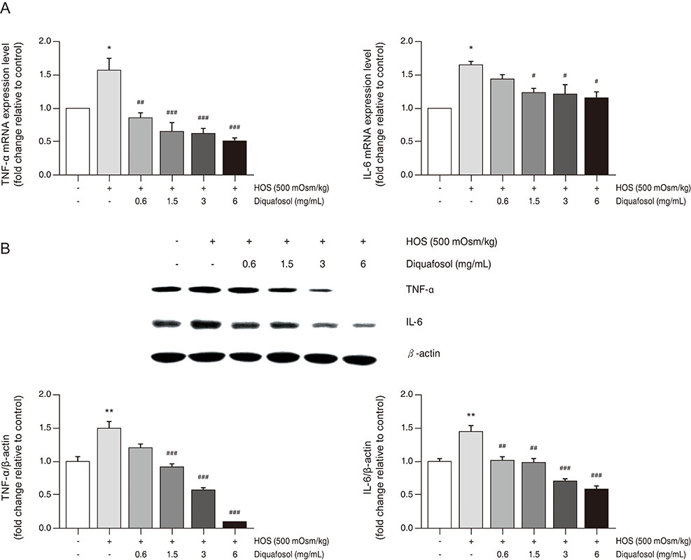
Fig. 2 Diquafosol downregulates mRNA and protein expression of tumor necrosis factor-α (TNF-α) and interleukin-6 (IL-6) in human corneal epithelial cells. (A) Total RNA was extracted from human corneal epithelial cells. RNA levels were measured by real-time polymerase chain reaction. The cells were exposed to hyperosmotic media (500 mOsm/kg DMEM/F12, serum-free) for 30 minutes, followed by diquafosol (0.6–6 mg/mL) for 4 hours. The graph of multiple analyses shows the relative mRNA levels of TNF-α and IL-6. Glyceraldehyde-3-phosphate dehydrogenase was used as the reference gene. (B) Representative western blots of TNF-α and IL-6. Cells were exposed to hyperosmotic media (500 mOsm/kg DMEM/F12, serum-free) for 30 minutes, followed by diquafosol (0.6–6 mg/mL) for 24 hours. Expression levels of TNF-α and IL-6 were determined using β-actin as a control. The densities of bands relative to those of β-actin were measured using ImageJ software. Data are expressed as the mean ± standard deviation from three separate experiments (*p < 0.01 and **p < 0.01, significantly different from the control; #p < 0.05, ##p < 0.01, and ###p < 0.001, significantly different from the hyperosmotic group). HOS = hyperosmotic stress.

Fig. 3 Fluorescence immunocytochemistry for tumor necrosis factor-α (TNF-α) and interleukin-6 (IL-6) in human corneal epithelial cells. Cells were exposed to hyperosmotic media (500 mOsm/kg DMEM/F12, serum-free) for 30 minutes, followed by diquafosol (DQF, 0.6–6 mg/mL) for 24 hours. Changes in the expression of cytoplasmic TNF-α and IL-6 were confirmed using fluorescence immunocytochemistry. Cells were counterstained with DAPI. The fluorescence intensity from green to blue was measured in ImageJ software using a color histogram. White scale bars: 100 µm (***p < 0.001, significantly different from the control; ###p < 0.001, significantly different from the hyperosmotic group). HOS = hyperosmotic stress.

Fig. 4 Effects of diquafosol on the nuclear factor-kappa B (NF-κB) signaling pathway in human corneal epithelial cells. (A) Expression levels of NF-κB, p-NF-κB, I-κB, and p-I-κB proteins in nuclear and cytosolic fractions were assessed using western blotting. The cells were exposed to hyperosmotic media (500 mOsm/kg DMEM/F12, serum-free) for 30 minutes, followed by diquafosol (1.5–6 mg/mL) for 30 minutes. Lamin B1 and ß-actin were used as standard proteins for quantitating the levels of the proteins of interest. (B) Expression of p-NF-κB in the nuclear fraction was confirmed using fluorescence immunocytochemistry. Cells were counterstained with DAPI. Fluorescence intensity and the density of bands were measured using ImageJ software. (C) Schematic diagram showing the inhibitory effects of diquafosol on hyperosmotic stress-induced inflammatory cytokine production via the NF-κB signaling pathway in human corneal epithelial cells. Data are expressed as the mean ± standard deviation from three separate experiments (***p < 0.001, significantly different from the control; #p < 0.05, ##p < 0.01 and ###p < 0.001, significantly different from the hyperosmotic group). HOS = hyperosmotic stress.
Reference
-
1. Gulati S, Jain S. Ocular pharmacology of tear film, dry eye, and allergic conjunctivitis. Handb Exp Pharmacol. 2017; 242:97–118.
Article2. McCarty CA, Bansal AK, Livingston PM, et al. The epidemiology of dry eye in Melbourne, Australia. Ophthalmology. 1998; 105:1114–1119.3. Kuang TM, Tsai SY, Hsu WM, et al. Correctable visual impairment in an elderly Chinese population in Taiwan: the Shihpai Eye Study. Invest Ophthalmol Vis Sci. 2007; 48:1032–1037.
Article4. Miljanovic B, Dana R, Sullivan DA, Schaumberg DA. Impact of dry eye syndrome on vision-related quality of life. Am J Ophthalmol. 2007; 143:409–415.5. Versura P, Profazio V, Schiavi C, Campos EC. Hyperosmolar stress upregulates HLA-DR expression in human conjunctival epithelium in dry eye patients and in vitro models. Invest Ophthalmol Vis Sci. 2011; 52:5488–5496.
Article6. Bellotti M, Bast W, Berra A, Bonetto FJ. Effects of osmolarity on human epithelial conjunctival cells using an electrical technique. Graefes Arch Clin Exp Ophthalmol. 2011; 249:1875–1882.
Article7. Julio G, Lluch S, Pujol P, Merindano MD. Effects of tear hyperosmolarity on conjunctival cells in mild to moderate dry eye. Ophthalmic Physiol Opt. 2012; 32:317–323.
Article8. Li DQ, Chen Z, Song XJ, et al. Stimulation of matrix metalloproteinases by hyperosmolarity via a JNK pathway in human corneal epithelial cells. Invest Ophthalmol Vis Sci. 2004; 45:4302–4311.
Article9. Li DQ, Luo L, Chen Z, et al. JNK and ERK MAP kinases mediate induction of IL-1beta, TNF-alpha and IL-8 following hyperosmolar stress in human limbal epithelial cells. Exp Eye Res. 2006; 82:588–596.10. Cavet ME, Harrington KL, Ward KW, Zhang JZ. Mapracorat, a novel selective glucocorticoid receptor agonist, inhibits hyperosmolar-induced cytokine release and MAPK pathways in human corneal epithelial cells. Mol Vis. 2010; 16:1791–1800.11. Al-Ayyoubi S, Gali-Muhtasib H. Differential apoptosis by gallotannin in human colon cancer cells with distinct p53 status. Mol Carcinog. 2007; 46:176–186.
Article12. Neuhofer W. Role of NFAT5 in inflammatory disorders associated with osmotic stress. Curr Genomics. 2010; 11:584–590.
Article13. Schwartz L, Guais A, Pooya M, Abolhassani M. Is inflammation a consequence of extracellular hyperosmolarity? J Inflamm (Lond). 2009; 6:21.
Article14. Nichols KK, Yerxa B, Kellerman DJ. Diquafosol tetrasodium: a novel dry eye therapy. Expert Opin Investig Drugs. 2004; 13:47–54.
Article15. Gum SI, Kim YH, Jung JC, et al. Cyclosporine A inhibits TGF-β2-induced myofibroblasts of primary cultured human pterygium fibroblasts. Biochem Biophys Res Commun. 2017; 482:1148–1153.
Article16. Kanellopoulos AJ, Asimellis G. In pursuit of objective dry eye screening clinical techniques. Eye Vis (Lond). 2016; 3:1.
Article17. Lemp MA, Bron AJ, Baudouin C, et al. Tear osmolarity in the d iagnosis and management of d ry eye d isease. Am J Ophthalmol. 2011; 151:792–798.18. Corrales RM, Villarreal A, Farley W, et al. Strain-related cytokine profiles on the murine ocular surface in response to desiccating stress. Cornea. 2007; 26:579–584.
Article19. Gilbard JP, Carter JB, Sang DN, et al. Morphologic effect of hyperosmolarity on rabbit corneal epithelium. Ophthalmology. 1984; 91:1205–1212.
Article20. Luo L, Li DQ, Corrales RM, Pflugfelder SC. Hyperosmolar saline is a proinflammatory stress on the mouse ocular surface. Eye Contact Lens. 2005; 31:186–193.
Article21. Pan Z, Wang Z, Yang H, et al. TRPV1 activation is required for hypertonicity-stimulated inflammatory cytokine release in human corneal epithelial cells. Invest Ophthalmol Vis Sci. 2011; 52:485–493.
Article22. Lam H, Bleiden L, de Paiva CS, et al. Tear cytokine profiles in dysfunctional tear syndrome. Am J Ophthalmol. 2009; 147:198–205.
Article23. Enriquez-de-Salamanca A, Castellanos E, Stern ME, et al. Tear cytokine and chemokine analysis and clinical correlations in evaporative-type dry eye disease. Mol Vis. 2010; 16:862–873.24. Koh S. Clinical utility of 3% diquafosol ophthalmic solution in the treatment of dry eyes. Clin Ophthalmol. 2015; 9:865–872.
Article25. Park JH, Moon SH, Kang DH, et al. Diquafosol sodium inhibits apoptosis and inf lammation of corneal epithelial cells via activation of Erk1/2 and RSK: in vitro and in vivo dry eye model. Invest Ophthalmol Vis Sci. 2018; 59:5108–5115.26. Lan W, Petznick A, Heryati S, et al. Nuclear Factor-κB: central regulator in ocular surface inflammation and diseases. Ocul Surf. 2012; 10:137–148.
Article27. Shi H, Berger EA. Characterization of site-specific phosphorylation of NF-κB p65 in retinal cells in response to high glucose and cytokine polarization. Mediators Inflamm. 2018; 2018:3020675.
Article28. Guzman M, Keitelman I, Sabbione F, et al. Desiccating stress-induced disruption of ocular surface immune tolerance drives dry eye disease. Clin Exp Immunol. 2016; 184:248–256.
Article29. Fujihara T, Murakami T, Fujita H, et al. Improvement of corneal barrier function by the P2Y(2) agonist INS365 in a rat dry eye model. Invest Ophthalmol Vis Sci. 2001; 42:96–100.30. Massingale ML, Li X, Vallabhajosyula M, et al. Analysis of inflammatory cytokines in the tears of dry eye patients. Cornea. 2009; 28:1023–1027.
Article
- Full Text Links
-
- Actions
-
Cited
- CITED
-
- Close
- Share
-
- Similar articles
-
- Role of Tumor Necrosis Factor Alpha, Interleukin 8 and Dexamethasone in the FAK Expression by Human Nucleus Pulposus Cells
- The Effect of Induced Heat Shock Protein 33 in Human Corneal Epithelial Cell
- The Effect of the Corneal Epithelium on the Keratocyte Apoptosis
- Effects of Platelet-rich Plasma on Ocular Surface in Patients with Dry Eye Syndrome: Clinico-experimental Analysis
- CXC and CC Chemokine Expression by Intestinal Epithelial Cells in Response to Clostridium difficile Toxin A
