Long Intergenic Non-Protein Coding RNA 665 Regulates Viability, Apoptosis, and Autophagy via the MiR-186-5p/MAP4K3 Axis in Hepatocellular Carcinoma
- Affiliations
-
- 1Department of General Surgery, Jinchang Central Hospital, Jinchang, Gansu, China. intersection88@126.com
- 2Department of Ultrasound, Shenzhen Hospital of Southern Medical University, Shenzhen, China.
- KMID: 2457480
- DOI: http://doi.org/10.3349/ymj.2019.60.9.842
Abstract
- PURPOSE
Long intergenic non-protein coding RNA 665 (LINC00665) plays a vital role in the development of cancer. Its function in hepatocellular carcinoma (HCC), however, remains largely unknown.
MATERIALS AND METHODS
The expressions of LINC00665, miR-186-5p, and MAP4K3 were determined by qRT-PCR. Cell viability and apoptosis were evaluated by MTT and flow cytometry, respectively. Autophagic puncta formation was observed by fluorescence microscopy. Bioinformatics analysis, luciferase reporter assay, RNA immunoprecipitation, and RNA pulldown were performed to identify associations among LINC00665, miR-186-5p, and MAP4K3. Western blot was utilized to examine the expressions of MAP4K3, Beclin-1, and LC3. Tumor growth was evaluated in a xenograft model.
RESULTS
Elevations in LINC00665 were observed in HCC tissues and cells. The overall survival of HCC patients with high levels of LINC00665 was shorter than those with low levels. In vitro, LINC00665 depletion inhibited viability and induced apoptosis and autophagy. miR-186-5p interacted with LINC00665 and was downregulated in HCC tissues and cells. Upregulation of miR-186-5p inhibited viability and induced apoptosis and autophagy, which were attenuated by upregulation of LINC00665. MAP4K3 was found to possess binding sites with miR-186-5p and was upregulated in HCC tissues and cells. MAP4K3 depletion inhibited viability and induced apoptosis and autophagy, which were attenuated by miR-186-5p inhibitor. In vivo, miR-186-5p expression was negatively correlated with LINC00665 or MAP4K3 in HCC tissues, while LINC00665 was positively correlated with MAP4K3. LINC00665 knockdown suppressed tumor growth.
CONCLUSION
LINC00665 was involved in cell viability, apoptosis, and autophagy in HCC via miR-186-5p/MAP4K3 axis, which may provide a new approach for HCC treatment.
Keyword
MeSH Terms
Figure
-
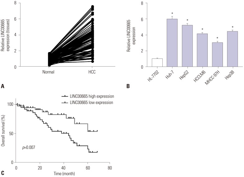
Fig. 1 LINC00665 expression upregulated in HCC. qRT-PCR assay was performed to measure the expression of LINC00665 in HCC tissues and cells. (A) The expression of LINC00665 in HCC tissues and matched normal adjacent tissues. (B) The expression of LINC00665 in HCC cell lines (Huh-7, HepG2, HCCLM6, MHCC-97H, and Hep3B) and human liver cell line HL-7702. (C) The correlation between the LINC00665 gene expression and overall survival in 24 patients with HCC. *p<0.05. LINC00665, long intergenic non-protein coding RNA 665; HCC, hepatocellular carcinoma.
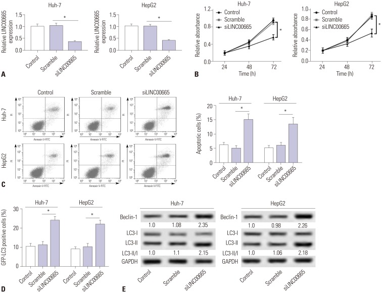
Fig. 2 Downregulation of LINC00665 inhibits hepatocellular carcinoma proliferation and induces apoptosis and autophagy in vitro. HepG2 and Huh-7 cells were transfected with siLINC00665 or siNC. (A) LINC00665 expression was detected in HepG2 and Huh-7 cells by qRT-PCR. (B) MTT assay was performed to evaluate cell proliferation. (C) Cell apoptotic rate was detected by flow cytometry. (D) Autophagy formation was analyzed by transfection of GFP-LC3 into HepG2 and Huh-7 cells. (E) Western blot was performed to detect the protein expression of Beclin-1 and LC3. *p<0.05. LINC00665, long intergenic non-protein coding RNA 665.
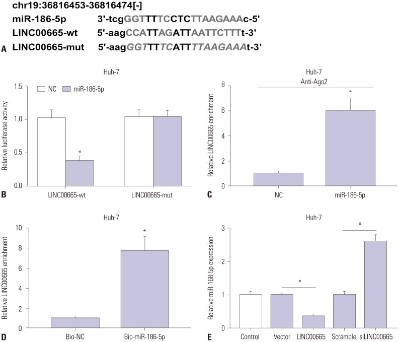
Fig. 3 LINC00665 interacts with miR-186-5p. (A) The binding sites between miR-186-5p and LINC00665 were predicted by starBase v2.0, and the luciferase reporter plasmids containing the wild-type (wt) or mutated (mut) LINC00665 binding sites of miR-186-5p were established. (B) The luciferase activity was measured in Huh-7 cells co-transfected with LINC00665-wt or LINC00665-mut luciferase reporter and miR-186-5p mimic or miR-NC. (C) The RIP assay was performed, and expression of LINC00665 was detected in samples bound to the Ago2 antibody or IgG in Huh-7 cells. (D) Detection of LINC00665 expression levels using qRT-PCR in samples pulled down by biotinylated miR-186-5p or negative control. (E) Expression levels of miR-186-5p in Huh-7 cells transfected with siLINC00665, siNC, pcDNA LINC00665, or pcDNA3.0 vector. *p<0.05. LINC00665, long intergenic non-protein coding RNA 665.
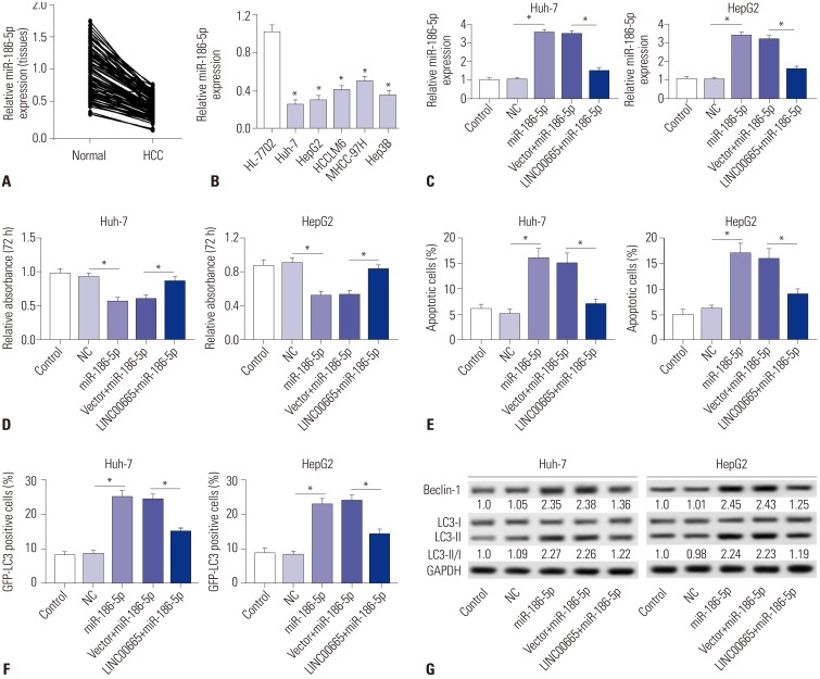
Fig. 4 LINC00665 attenuates the tumor-suppressive effect of miR-186-5p in HCC cells in vitro. (A and B) The expression levels of miR-186-5p were measured in HCC tissues and cell lines by qRT-PCR. (C–G) HepG2 and Huh-7 cells were transfected with miR-NC, miR-186-5p mimic, pcDNA3.0 vector+miR-186-5p mimic, and pcDNA LINC00665+miR-186-5p mimic. (C) miR-186-5p expression was detected in HepG2 and Huh-7 cells by qRT-PCR. (D) Cell proliferation was detected by MTT assay. (E) Cell apoptotic rate was analyzed by flow cytometry. (F) Autophagy formation was analyzed by transfection of GFP-LC3 into HepG2 and Huh-7 cells. (G) Western blot was performed to detect the protein expression of Beclin-1 and LC3. *p<0.05. LINC00665, long intergenic non-protein coding RNA 665; HCC, hepatocellular carcinoma.
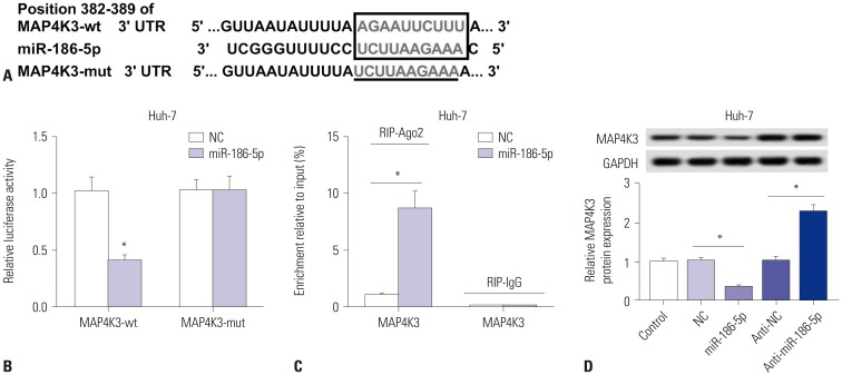
Fig. 5 MAP4K3 a target gene of miR-186-5p. (A) The binding sites between miR-186-5p and MAP4K3 were predicted by TargetScan, and the luciferase reporter plasmids containing the wild-type (wt) or mutated (mut) MAP4K3 binding sites of miR-186-5p were established. (B) The luciferase activity was measured in Huh-7 cells co-transfected with MAP4K3-wt or MAP4K3-mut luciferase reporter and miR-186-5p mimic or miR-NC. (C) The RIP assay was performed, and expression of MAP4K3 was detected in the samples bound to the Ago2 antibody or IgG in Huh-7 cells transfected with miR-186-5p mimic or miR-NC. (D) Detection of MAP4K3 expression level using qRT-PCR in Huh-7 cells transfected with NC mimic, miR-186-5p mimic, NC inhibitor, or miR-186-5p inhibitor. *p<0.05. LINC00665, long intergenic non-protein coding RNA 665.
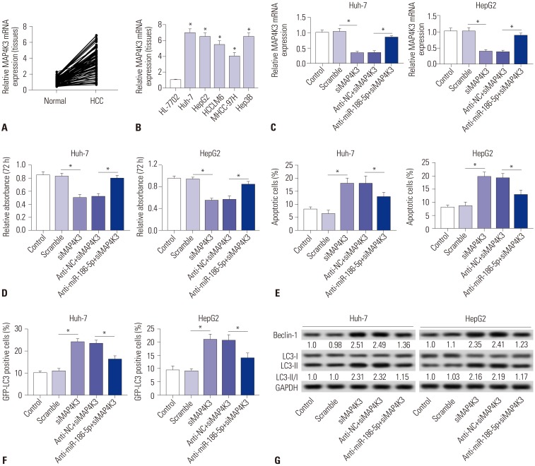
Fig. 6 Downregulation of miR-186-5p attenuates the tumor-suppressive effect of MAP4K3 knockout in HCC cells in vitro. (A and B) The expression levels of MAP4K3 were measured in HCC tissues and cell lines by qRT-PCR. (C–G) HepG2 and Huh-7 cells were transfected with si-NC, siMAP4K3, inhibitor negative control+siMAP4K3, and miR-186-5p inhibitor+siMAP4K3. (C) MAP4K3 expression was detected in HepG2 and Huh-7 cells by qRT-PCR. (D) Cell proliferation was detected by MTT assay. (E) Cell apoptotic rate was analyzed by flow cytometry. (F) Autophagy formation was analyzed by transfection of GFP-LC3 into HepG2 and Huh-7 cells. (G) Western blot was performed to detect the protein expression of Beclin-1 and LC3. *p<0.05. HCC, hepatocellular carcinoma.
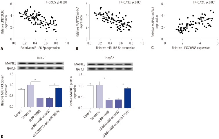
Fig. 7 LINC00665 regulates the expression of MAP4K3 by interacting with miR-186-5p. (A) The correlation between LINC00665 and miR-186-5p levels was analyzed in HCC tissues. (B) The correlation between MAP4K3 and miR-186-5p levels was analyzed in HCC tissues. (C) The correlation between LINC00665 and MAP4K3 levels was analyzed in HCC tissues. (D) Expression levels of MAP4K3 in HepG2 and Huh-7 cells transfected with siNC, siLINC00665, inhibitor negative control+ siLINC00665, and miR-186-5p inhibitor+siLINC00665. *p<0.05. LINC00665, long intergenic non-protein coding RNA 665; HCC, hepatocellular carcinoma.
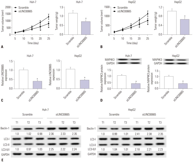
Fig. 8 Knockdown of LINC00665 inhibits tumor growth in vivo. HepG2 and Huh-7 cells transfected with siLINC00665 or siNC were subcutaneously injected into nude mice. (A and B) Tumor volume was measured at 5, 10, 15, 20, and 25 days after implantation of HepG2 or Huh-7 cells, and tumor weight was measured at 25 days after implantation of HepG2 or Huh-7 cells. (C and D) Mice were euthanized for the analysis of LINC00665 and MAP4K3 by qRT-PCR and Western blot at 25 days after implantation of HepG2 or Huh-7 cells. (E) Western blot was performed to examine the expression of Beclin-1 and LC3. *p<0.05. LINC00665, long intergenic non-protein coding RNA 665.
Reference
-
1. Mak LY, Cruz-Ramón V, Chinchilla-López P, Torres HA, LoConte NK, Rice JP, et al. Global epidemiology, prevention, and management of hepatocellular carcinoma. Am Soc Clin Oncol Educ Book. 2018; 38:262–279. PMID: 30231359.
Article2. Margini C, Dufour JF. The story of HCC in NAFLD: from epidemiology, across pathogenesis, to prevention and treatment. Liver Int. 2016; 36:317–324. PMID: 26601627.
Article3. Yang JD, Roberts LR. Hepatocellular carcinoma: a global view. Nat Rev Gastroenterol Hepatol. 2010; 7:448–458. PMID: 20628345.
Article4. Rastogi A. Changing role of histopathology in the diagnosis and management of hepatocellular carcinoma. World J Gastroenterol. 2018; 24:4000–4013. PMID: 30254404.
Article5. Llovet JM, Montal R, Sia D, Finn RS. Molecular therapies and precision medicine for hepatocellular carcinoma. Nat Rev Clin Oncol. 2018; 15:599–616. PMID: 30061739.
Article6. Peng JC, Shen J, Ran ZH. Transcribed ultraconserved region in human cancers. RNA Biol. 2013; 10:1771–1777. PMID: 24384562.
Article7. He Y, Meng XM, Huang C, Wu BM, Zhang L, Lv XW, et al. Long noncoding RNAs: novel insights into hepatocelluar carcinoma. Cancer Lett. 2014; 344:20–27. PMID: 24183851.
Article8. Huang JL, Zheng L, Hu YW, Wang Q. Characteristics of long noncoding RNA and its relation to hepatocellular carcinoma. Carcinogenesis. 2014; 35:507–514. PMID: 24296588.
Article9. Li G, Zhang H, Wan X, Yang X, Zhu C, Wang A, et al. Long noncoding RNA plays a key role in metastasis and prognosis of hepatocellular carcinoma. Biomed Res Int. 2014; 2014:780521. PMID: 24757675.
Article10. Sun J, Bie B, Zhang S, Yang J, Li Z. Long non-coding RNAs: critical players in hepatocellular carcinoma. Int J Mol Sci. 2014; 15:20434–20448. PMID: 25387074.11. Wang F, Ying HQ, He BS, Pan YQ, Deng QW, Sun HL, et al. Upregulated lncRNA-UCA1 contributes to progression of hepatocellular carcinoma through inhibition of miR-216b and activation of FGFR1/ERK signaling pathway. Oncotarget. 2015; 6:7899–7917. PMID: 25760077.
Article12. Li T, Xie J, Shen C, Cheng D, Shi Y, Wu Z, et al. Amplification of long noncoding RNA ZFAS1 promotes metastasis in hepatocellular carcinoma. Cancer Res. 2015; 75:3181–3191. PMID: 26069248.
Article13. Wen DY, Lin P, Pang YY, Chen G, He Y, Dang YW, et al. Expression of the long intergenic non-protein coding RNA 665 (LINC00665) gene and the cell cycle in hepatocellular carcinoma using The Cancer Genome Atlas, the Gene Expression Omnibus, and Quantitative Real-Time Polymerase Chain Reaction. Med Sci Monit. 2018; 24:2786–2808. PMID: 29728556.
Article14. Di Leva G, Garofalo M, Croce CM. MicroRNAs in cancer. Annu Rev Pathol. 2014; 9:287–314. PMID: 24079833.
Article15. Song Y, Wang F, Huang Q, Cao Y, Zhao Y, Yang C. MicroRNAs contribute to hepatocellular carcinoma. Mini Rev Med Chem. 2015; 15:459–466. PMID: 25807945.
Article16. Guo W, Tan W, Liu S, Huang X, Lin J, Liang R, et al. MiR-570 inhibited the cell proliferation and invasion through directly targeting B7-H1 in hepatocellular carcinoma. Tumour Biol. 2015; 36:9049–9057. PMID: 26084609.
Article17. Oksuz Z, Serin MS, Kaplan E, Dogen A, Tezcan S, Aslan G, et al. Serum microRNAs; miR-30c-5p, miR-223-3p, miR-302c-3p and miR-17-5p could be used as novel non-invasive biomarkers for HCV-positive cirrhosis and hepatocellular carcinoma. Mol Biol Rep. 2015; 42:713–720. PMID: 25391771.
Article18. Arbogast F, Gros F. Lymphocyte autophagy in homeostasis, activation, and inflammatory diseases. Front Immunol. 2018; 9:1801. PMID: 30127786.
Article19. Wang Z, Choi ME. Autophagy in kidney health and disease. Antioxid Redox Signal. 2014; 20:519–537. PMID: 23642034.
Article20. Singh SS, Vats S, Chia AY, Tan TZ, Deng S, Ong MS, et al. Dual role of autophagy in hallmarks of cancer. Oncogene. 2018; 37:1142–1158. PMID: 29255248.
Article21. Folkerts H, Hilgendorf S, Vellenga E, Bremer E, Wiersma VR. The multifaceted role of autophagy in cancer and the microenvironment. Med Res Rev. 2019; 39:517–560. PMID: 30302772.
Article22. Su Z, Yang Z, Xu Y, Chen Y, Yu Q. MicroRNAs in apoptosis, autophagy and necroptosis. Oncotarget. 2015; 6:8474–8490. PMID: 25893379.
Article23. Zhai H, Fesler A, Ju J. MicroRNA: a third dimension in autophagy. Cell Cycle. 2013; 12:246–250. PMID: 23255136.24. Du F, Feng Y, Fang J, Yang M. MicroRNA-143 enhances chemosensitivity of Quercetin through autophagy inhibition via target GABARAPL1 in gastric cancer cells. Biomed Pharmacother. 2015; 74:169–177. PMID: 26349981.
Article25. Lan T, Yan X, Li Z, Xu X, Mao Q, Ma W, et al. Long non-coding RNA PVT1 serves as a competing endogenous RNA for miR-186-5p to promote the tumorigenesis and metastasis of hepatocellular carcinoma. Tumour Biol. 2017; 39:1010428317705338. PMID: 28656879.
Article26. Li C, Chen J, Zhang K, Feng B, Wang R, Chen L. Progress and prospects of long noncoding RNAs (lncRNAs) in hepatocellular carcinoma. Cell Physiol Biochem. 2015; 36:423–434. PMID: 25968300.
Article27. Yuan JH, Yang F, Wang F, Ma JZ, Guo YJ, Tao QF, et al. A long noncoding RNA activated by TGF-β promotes the invasion-metastasis cascade in hepatocellular carcinoma. Cancer Cell. 2014; 25:666–681. PMID: 24768205.
Article28. Huang YH, Al-Aidaroos AQ, Yuen HF, Zhang SD, Shen HM, Rozycka E, et al. A role of autophagy in PTP4A3-driven cancer progression. Autophagy. 2014; 10:1787–1800. PMID: 25136802.
Article29. Kang R, Zeh HJ, Lotze MT, Tang D. The Beclin 1 network regulates autophagy and apoptosis. Cell Death Differ. 2011; 18:571–580. PMID: 21311563.
Article30. Qiu DM, Wang GL, Chen L, Xu YY, He S, Cao XL, et al. The expression of beclin-1, an autophagic gene, in hepatocellular carcinoma associated with clinical pathological and prognostic significance. BMC Cancer. 2014; 14:327. PMID: 24885292.
Article31. Hu D, Wu J, Xu L, Zhang R, Chen L. A method for the establishment of a cell line with stable expression of the GFP-LC3 reporter protein. Mol Med Rep. 2012; 6:783–786. PMID: 22825056.
Article32. Li J, Xia L, Zhou Z, Zuo Z, Xu C, Song H, et al. MiR-186-5p upregulation inhibits proliferation, metastasis and epithelial-to-mesenchymal transition of colorectal cancer cell by targeting ZEB1. Arch Biochem Biophys. 2018; 640:53–60. PMID: 29325758.
Article33. Islam F, Gopalan V, Vider J, Wahab R, Ebrahimi F, Lu CT, et al. MicroRNA-186-5p overexpression modulates colon cancer growth by repressing the expression of the FAM134B tumour inhibitor. Exp Cell Res. 2017; 357:260–270. PMID: 28549913.
Article34. Wang H, Shen Q, Zhang X, Yang C, Cui S, Sun Y, et al. The long noncoding RNA XIST controls non-small cell lung cancer proliferation and invasion by modulating miR-186-5p. Cell Physiol Biochem. 2017; 41:2221–2229. PMID: 28448993.
Article35. Jones DZ, Schmidt ML, Suman S, Hobbing KR, Barve SS, Gobejishvili L, et al. Micro-RNA-186-5p inhibition attenuates proliferation, anchorage independent growth and invasion in metastatic prostate cancer cells. BMC Cancer. 2018; 18:421. PMID: 29653561.
Article36. Cargnello M, Roux PP. Activation and function of the MAPKs and their substrates, the MAPK-activated protein kinases. Microbiol Mol Biol Rev. 2011; 75:50–83. PMID: 21372320.
Article
- Full Text Links
-
- Actions
-
Cited
- CITED
-
- Close
- Share
-
- Similar articles
-
- LncRNA STARD7-AS1 suppresses cervical cancer cell proliferation while promoting autophagy by regulating miR-31-5p/TXNIP axis to inactivate the mTOR signaling
- Long Noncoding RNA Cytoskeleton Regulator RNA Suppresses Apoptosis in Hepatoma Cells by Modulating the miR-125a-5p/ HS1-Associated Protein X-1 Axis to Induce Caspase-9 Inactivation
- Long Non-Coding RNA NORAD Inhibits Breast Cancer Cell Proliferation and Metastasis by Regulating miR-155-5p/ SOCS1 Axis
- Electroacupuncture-Modulated MiR-106b-5p Expression Enhances Autophagy by Targeting Beclin-1 to Promote Motor Function Recovery After Spinal Cord Injury in Rats
- Long Noncoding-RNA Component of Mitochondrial RNA Processing Endoribonuclease Promotes Carcinogenesis in Triple-Negative Breast Cancer Cells via the Competing Endogenous RNA Mechanism
