Dement Neurocogn Disord.
2015 Mar;14(1):48-51. 10.12779/dnd.2015.14.1.48.
Reversible Amygdala and Parahippocampal Lesions of Brain ¹â¸Fluorodeoxy Glucose-Positron Emission Tomography in Neuropsychiatric Systemic Lupus Erythematosus
- Affiliations
-
- 1Department of Neurology, Myongji Hospital, Seonam University College of Medicine, Goyang, Korea. neurohan5403@gmail.com
- KMID: 2443043
- DOI: http://doi.org/10.12779/dnd.2015.14.1.48
Abstract
- BACKGROUND
Systemic lupus erythematosus (SLE) is an autoimmune disease that is a significant source of morbidity and mortality when it manifests in the central nervous system. The early detection and treatment of neuropsychiatric SLE (NPSLE) is very important, but a confirmative diagnostic tool has yet to be developed.
CASE REPORT
We report here a case of neuropsychiatric manifestations in a patient that were associated with SLE, and evidence of reversal of bilateral amygdala and parahippocampal lesions in the brain revealed by 18fluorodeoxy glucose-positron emission tomography.
CONCLUSIONS
We are suggestive of 18fluorodeoxy glucose-positron emission tomography appear to be more sensitive in detecting subtle brain changes in NPSLE.
Keyword
MeSH Terms
Figure
-
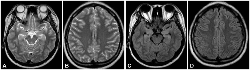
Fig. 1 Brain magnetic resonance imaging on admission. T2-weighted image (A and B), fluid-attenuated inversion recovery (C and D) image showed unremarkable findings.
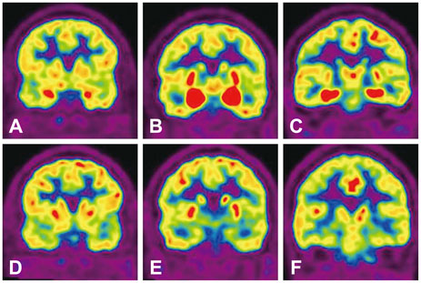
Fig. 2 Brain coronal 18fluorodeoxy glucose-positron emission tomography (PET) revealed increased glucose metabolism in bilateral amygdala and parahippocampal gyrus (A, B, and C). Follow-up PET showed no findings in the same areas (D, E, and F).
Reference
-
1. Borowoy AM, Pope JE, Silverman E, Fortin PR, Pineau C, Smith CD, et al. Neuropsychiatric lupus: the prevalence and autoantibody associations depend on the definition: results from the 1000 faces of lupus cohort. Semin Arthritis Rheum. 2012; 42:179–185.
Article2. Fanouriakis A, Boumpas DT, Bertsias GK. Pathogenesis and treatment of CNS lupus. Curr Opin Rheumatol. 2013; 25:577–583.
Article3. Govoni M, Castellino G, Padovan M, Borrelli M, Trotta F. Recent advances and future perspective in neuroimaging in neuropsychiatric systemic lupus erythematosus. Lupus. 2004; 13:149–158.
Article4. Peterson PL, Axford JS, Isenberg D. Imaging in CNS lupus. Best Pract Res Clin Rheumatol. 2005; 19:727–739.
Article5. Folstein MF, Folstein SE, McHugh PR. "Mini-mental state". A practical method for grading the cognitive state of patients for the clinician. J Psychiatr Res. 1975; 12:189–198.
Article6. Kim HG. Assessment of memory disorders using Rey-Kim Memory Test. Korean J Rehabil Psychol. 2001; 8:29–48.
Article7. Kim HG. Clinical evaluation of the frontal lobe syndrome using Kims frontal-executive neuropsychological test. Korean J Rehabil Psychol. 2001; 8:173–190.
Article8. Kim H, Na DL. Normative data on the Korean version of the Boston Naming Test. J Clin Exp Neuropsychol. 1999; 21:127–133.
Article9. Glanz BI, Schur PH, Lew RA, Khoshbin S. Lateralized cognitive dysfunction in patients with systemic lupus erythematosus. Lupus. 2005; 14:896–902.
Article10. Emori A, Matsushima E, Aihara O, Ohta K, Koike R, Miyasaka N, et al. Cognitive dysfunction in systemic lupus erythematosus. Psychiatry Clin Neurosci. 2005; 59:584–589.
Article11. Kozora E, Arciniegas DB, Filley CM, West SG, Brown M, Miller D, et al. Cognitive and neurologic status in patients with systemic lupus erythematosus without major neuropsychiatric syndromes. Arthritis Rheum. 2008; 59:1639–1646.
Article12. DeGiorgio LA, Konstantinov KN, Lee SC, Hardin JA, Volpe BT, Diamond B. A subset of lupus anti-DNA antibodies cross-reacts with the NR2 glutamate receptor in systemic lupus erythematosus. Nat Med. 2001; 7:1189–1193.
Article13. Watson P, Storbeck J, Mattis P, Mackay M. Cognitive and emotional abnormalities in systemic lupus erythematosus: evidence for amygdala dysfunction. Neuropsychol Rev. 2012; 22:252–270.14. Sato S, Nakajima J, Shimura M, Kawashima H, Yoshio T, Hara Y. Reversible basal ganglia lesions in neuropsychiatric lupus: a report of three pediatric cases. Int J Rheum Dis. 2014; 17:274–279.15. Poil AR, Yousef Khan F, Lutf A, Hammoudeh M. Chorea as the first and only manifestation of systemic lupus erythematosus. Case Rep Rheumatol. 2012; 2012:907402.
Article16. Sibbitt WL Jr, Sibbitt RR, Brooks WM. Neuroimaging in neuropsychiatric systemic lupus erythematosus. Arthritis Rheum. 1999; 42:2026–2038.
Article17. Abreu MR, Jakosky A, Folgerini M, Brenol JC, Xavier RM, Kapczinsky F. Neuropsychiatric systemic lupus erythematosus: correlation of brain MR imaging, CT, and SPECT. Clin Imaging. 2005; 29:215–221.
Article18. Luyendijk J, Steens SC, Ouwendijk WJ, Steup-Beekman GM, Bollen EL, van der Grond J, et al. Neuropsychiatric systemic lupus erythematosus: lessons learned from magnetic resonance imaging. Arthritis Rheum. 2011; 63:722–732.
Article19. Curiel R, Akin EA, Beaulieu G, DePalma L, Hashefi M. PET/CT imaging in systemic lupus erythematosus. Ann N Y Acad Sci. 2011; 1228:71–80.
Article20. Lee SW, Park MC, Lee SK, Park YB. The efficacy of brain (18)F-fluorodeoxyglucose positron emission tomography in neuropsychiatric lupus patients with normal brain magnetic resonance imaging findings. Lupus. 2012; 21:1531–1537.
Article
- Full Text Links
-
- Actions
-
Cited
- CITED
-
- Close
- Share
-
- Similar articles
-
- Neuropsychiatric Lupus Diagnosed by Brain PET: A Case Report
- Systemic Lupus Erythematosus Associated Pitfalls on ¹â¸F-FDG PET/CT: Reactive Follicular Hyperplasia, Kikuchi-Fujimoto Disease, Inflammation and Lymphoid Hyperplasia of the Spleen Mimicking Lymphoma
- The Normalization of Brain ¹â¸F-fluorodeoxy-D-glucose Positron Emission Tomography Hypometabolism following Electroconvulsive Therapy in a 55-year-old Woman with Treatment-resistant Late Onset Depression: A Case Report
- Changes in Cerebral Cortex and Limbic Brain Functions after Short-Term Paroxetine Treatment in Panic Disorder: An [18F]FDG-PET Pilot Study
- Successful treatment with rituximab in a patient with lupus cerebritis and posterior reversible encephalopathy syndrome: a case report
