A Case of Metronidazole-Induced Encephalopathy: Atypical Involvement of the Brain on MRI
- Affiliations
-
- 1Department of Radiology, Hallym University Dongtan Sacred Heart Hospital, Hwaseong, Gyeonggi-do, Korea. csk1270@hallym.or.kr
- KMID: 2421554
- DOI: http://doi.org/10.13104/imri.2018.22.3.200
Abstract
- Metronidazole is an antimicrobial agent widely used for the treatment of anaerobic infection or antibiotics-associated diarrhea. It is generally thought to be safe, but can induce reversible toxic encephalopathy in the case of excessive or cumulative over-dose. Metronidazole-induced encephalopathy generally demonstrates the characteristic features of typical lesion location and bilaterality on magnetic resonance imaging (MRI). We report a case of metronidazole-induced encephalopathy with the involvement of asymmetric white matter. To our knowledge, only a few cases have been reported with respect to white matter lesion characteristics on MRI with diffusion-weighted images.
MeSH Terms
Figure
-
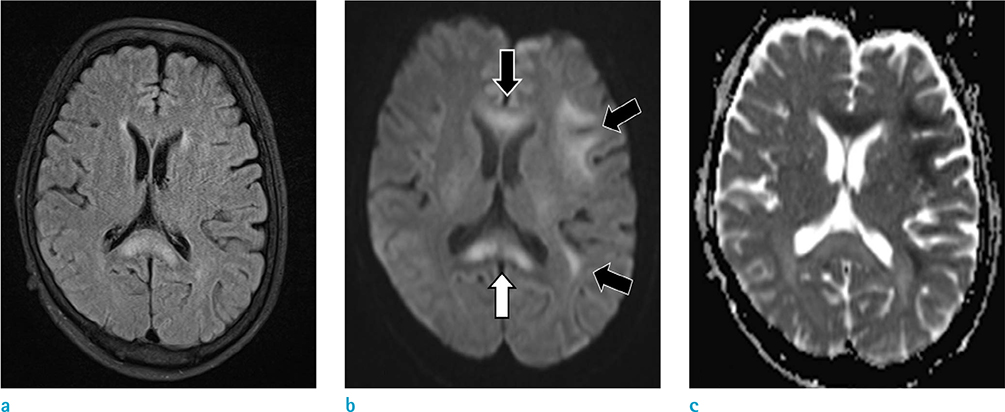
Fig. 1 Initial MRI of the brain of a 58-year-old woman (a–c). Diffusion-weighted imaging (b) showed multifocal high signal intensities at the genu and splenium of the bilateral corpus callosum and subcortical white matter of the left cerebral hemisphere (black and white arrows). The splenium of the corpus callosum (white arrow) showed no increased value on the apparent diffusion coefficient (ADC) map (c) and high signal intensity on a T2 fluid attenuation inversion recovery (FLAIR) image (a). The others showed a decreased value on the ADC map and isointensity on a T2 FLAIR image.
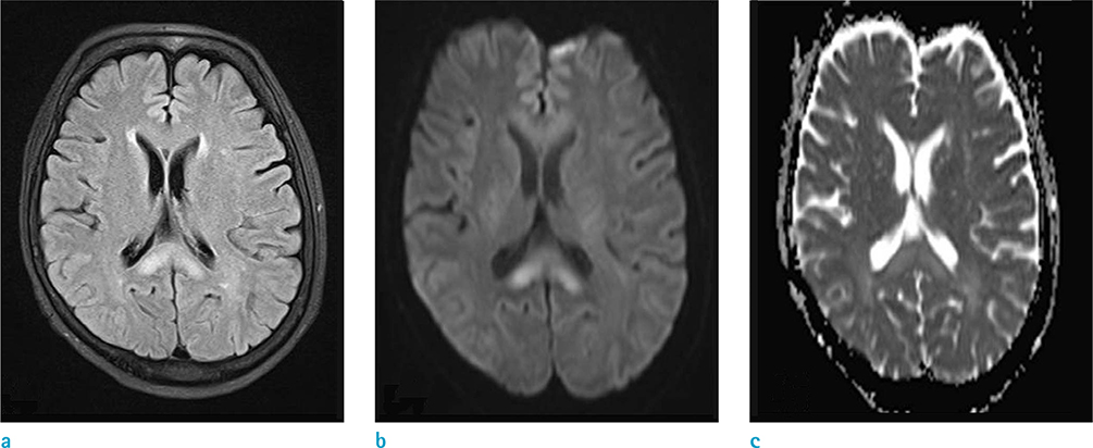
Fig. 2 One-week follow-up MRI of the brain after discontinuation of metronidazole (a–c). The previous lesions are markedly resolved on diffusion-weighted imaging (DWI) (b) and apparent diffusion coefficient (ADC) map (c), although the lesion in the splenium of the corpus callosum remained on a T2 fluid attenuation inversion recovery (FLAIR) image (a) and DWI.
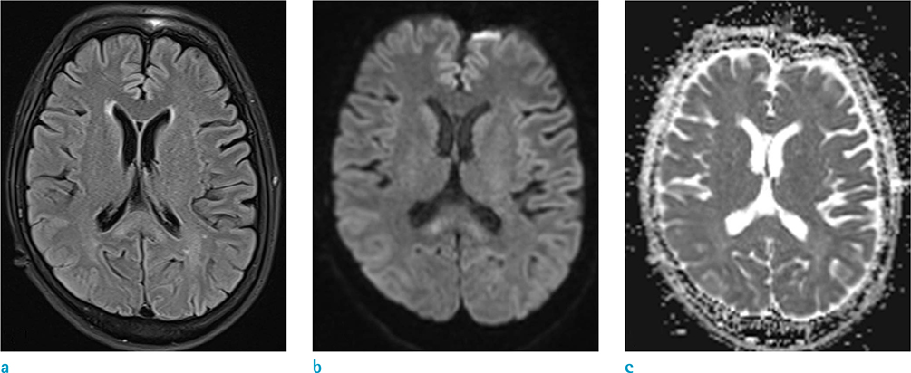
Fig. 3 One-month follow-up MRI of the brain (a–c). Previously-noted lesions are nearly completely resolved.
Reference
-
1. Bahn Y, Kim E, Park C, Park HC. Metronidazole induced encephalopathy in a patient with brain abscess. J Korean Neurosurg Soc. 2010; 48:301–304.
Article2. Kim E, Na DG, Kim EY, Kim JH, Son KR, Chang KH. MR imaging of metronidazole-induced encephalopathy: lesion distribution and diffusion-weighted imaging findings. AJNR Am J Neuroradiol. 2007; 28:1652–1658.
Article3. Park HS, Han DS. Management of antibiotics-associated diarrhea. Korean J Gastroenterol. 2009; 54:5–12.
Article4. Graves TD, Condon M, Loucaidou M, Perry RJ. Reversible metronidazole-induced cerebellar toxicity in a multiple transplant recipient. J Neurol Sci. 2009; 285:238–240.
Article5. Dow SW, LeCouteur RA, Poss ML, Beadleston D. Central nervous system toxicosis associated with metronidazole treatment of dogs: five cases (1984–1987). J Am Vet Med Assoc. 1989; 195:365–368.6. Olson EJ, Morales SC, McVey AS, Hayden DW. Putative metronidazole neurotoxicosis in a cat. Vet Pathol. 2005; 42:665–669.
Article7. Scharer K. Selective alterations of Purkinje cells in the dog after oral administration of high doses of nitroimidazole derivatives (author's transl). Verh Dtsch Ges Pathol. 1972; 56:407–410.8. von Rogulja P, Kovac W, Schmid H. Metronidazol encephalopathy in rats. Acta Neuropathol. 1973; 25:36–45.9. Ahmed A, Loes DJ, Bressler EL. Reversible magnetic resonance imaging findings in metronidazole-induced encephalopathy. Neurology. 1995; 45:588–589.
Article
- Full Text Links
-
- Actions
-
Cited
- CITED
-
- Close
- Share
-
- Similar articles
-
- Metronidazole Induced Encephalopathy in a Patient with Brain Abscess
- Reversible Encephalopathy Induced by Metronidazole
- Atypical Metronidazole-Induced Encephalopathy in Anaerobic Brain Abscess
- Two Cases of Metronidazole-induced Encephalopathy
- A case of metronidazole induced encephalopathy in a cirrhotic patient
