Nat Prod Sci.
2018 Jun;24(2):99-102. 10.20307/nps.2018.24.2.99.
Ameliorative Effects of Ombuoside on Dopamine Biosynthesis in PC12 Cells
- Affiliations
-
- 1College of Pharmacy, Chungbuk National University, Cheongju 28160, Korea. myklee@chungbuk.ac.kr
- 2Research Center for Bioresource and Health, Chungbuk National University, Cheongju 28160, Korea.
- 3Department of Food and Nutrition, Chungcheong University, Cheongju 28171, Republic of Korea.
- KMID: 2416069
- DOI: http://doi.org/10.20307/nps.2018.24.2.99
Abstract
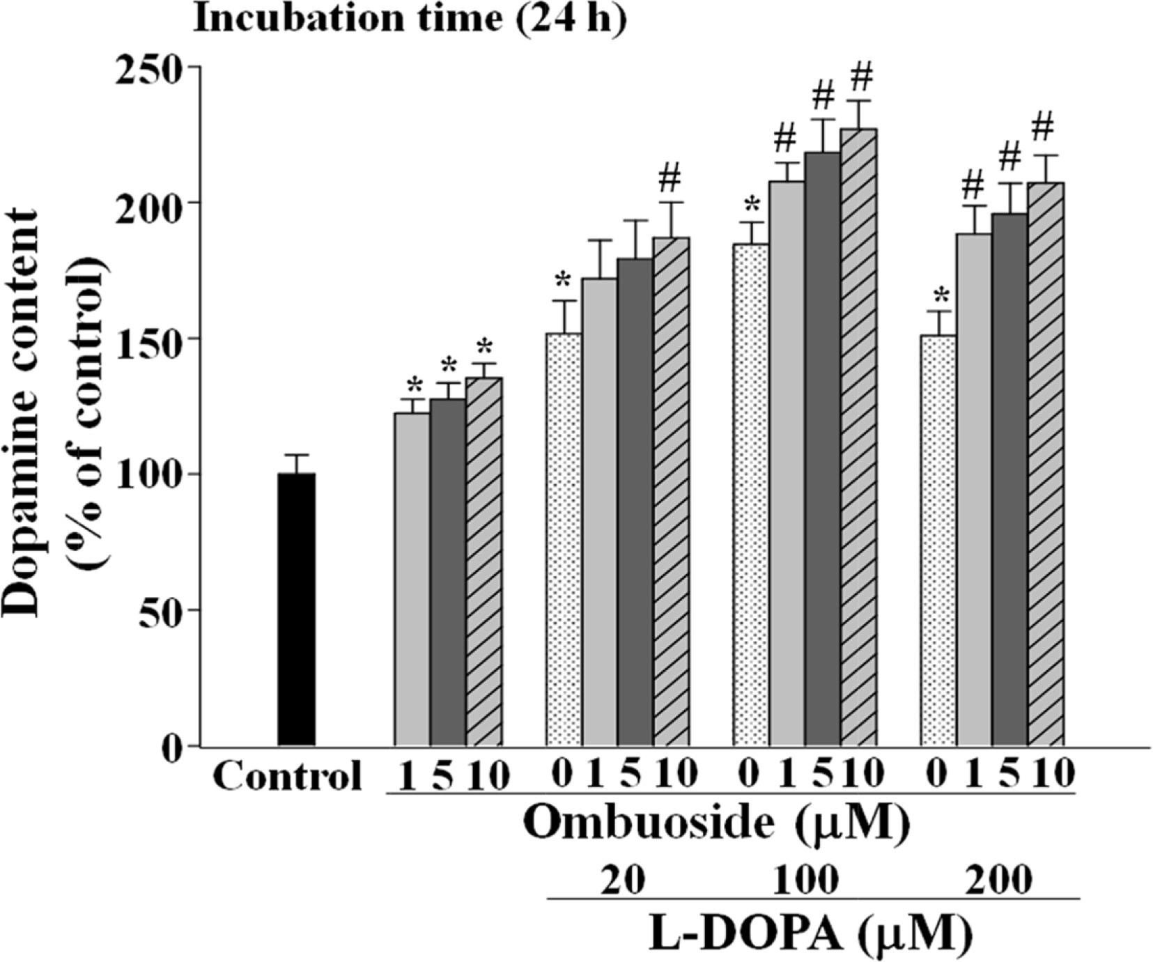
- This study investigated the effects of ombuoside, a flavonol glycoside, on dopamine biosynthesis in PC12 cells. Ombuoside at concentrations of 1, 5, and 10 µM increased intracellular dopamine levels at 1 - 24 h. Ombuoside (1, 5, and 10 µM) also significantly increased the phosphorylation of tyrosine hydroxylase (TH) (Ser40) and cyclic AMP-response element binding protein (CREB) (Ser133) at 0.5 - 6 h. In addition, ombuoside (1, 5, and 10 µM) combined with L-DOPA (20, 100, and 200 µM) further increased intracellular dopamine levels for 24 h compared to L-DOPA alone. These results suggest that ombuoside regulates dopamine biosynthesis by modulating TH and CREB activation in PC12 cells.
Keyword
MeSH Terms
Figure
Reference
-
References
(1). Fahn S.Ann. NY Acad. Sci. 2003. 991:1–14.(2). Nagatsu T.., Levitt M.., Udenfriend S. J.Biol. Chem. 1964. 239:2910–2917.(3). Kilbourne E. J.., Nankova B. B.., Lewis E. J.., McMahon A.., Osaka H.., Sabban D. B.., Sabban E. L. J.Biol. Chem. 1992. 267:7563–7569.(4). Kim K. S.., Lee M. K.., Carroll J.., Joh T. H. J.Biol. Chem. 1993. 268:15689–15695.(5). Campbell D. G.., Hardie D. G.., Vulliet P. R. J.Biol. Chem. 1986. 261:10489–10492.(6). Haycock J. W.Neurochem. Res. 1993. 18:15–26.(7). Kim K. S.., Park D. H.., Wessel T. C.., Song B.., Wagner J. A.., Joh T. H.Proc. Natl. Acad. Sci. USA. 1993. 90:3471–3475.(8). Marsden C. D.., Parkes J. D.Lancet. 1977. 1:345–349.(9). Migheli R.., Godani C.., Sciola L.., Delogu M. R.., Serra P. A.., Zangani D.., De Natale G.., Miele E.., Desole M. S. J.Neurochem. 1999. 73:1155–1163.(10). Jin C. M.., Yang Y. J.., Huang H. S.., Lim S. C.., Kai M.., Lee M. K.Eur. J. Pharmacol. 2008. 591:88–95.(11). Razmovski-Naumovski V.., Huang T. H. W.., Tran V. H.., Li G. Q.., Duke C. C.., Roufogalis B. D.Phytochem. Rev. 2005. 14:197–219.(12). Choi H. S.., Zhao T. T.., Shin K. S.., Kim S. H.., Hwang B. Y.., Lee C. K.., Lee M. K.Molecules. 2013. 18:4342–4356.(13). Choi H. S.., Park M. S.., Kim S. H.., Hwang B. Y.., Lee C. K.., Lee M. K.Molecules. 2010. 15:2814–2824.(14). Shin K. S.., Zhao T. T.., Park K. H.., Park H. J.., Hwang B. Y.., Lee C. K.., Lee M. K.BMC Neurosci. 2015. 16:23.(15). Shin K. S.., Zhao T. T.., Choi H. S.., Hwang B. Y.., Lee C. K.., Lee M. K.Brain Res. 2014. 1567:57–65.(16). Rice-Evans C. A.., Miller N. J.., Paganga G.Free Radic. Biol. Med. 1996. 20:933–956.(17). Amaro-Luis J. M.., Adrián M.., Díaz C.Ann. Pharm. Fr. 1997. 55:262–268.(18). Pollard S. E.., Kuhnle G. G.., Vauzour D.., Vafeiadou K.., Tzounis X.., Whiteman M.., Rice-Evans C.., Spencer J. P.Biochem. Biophys. Res. Commun. 2006. 350:960–968.(19). Kumar S.., Pandey A. K.Scientific World Journal. 2013. 2013:162750.(20). Tischler A. S.., Perlman R. L.., Morse G. M.., Sheard B. E. J.Neurochem. 1983. 40:364–370.(21). Mosmann T. J.Immunol. Methods. 1983. 65:55–63.(22). Lowry O. H.., Rosebrough N. J.., Farr A. L.., Randall R. L. J.Biol. Chem. 1951. 193:265–275.(23). Gonzalez G. A.., Montminy M. R.Cell. 1989. 59:675–680.(24). Basma A. N.., Morris E. J.., Nicklas W. J.., Geller H. M. J.Neurochem. 1995. 64:825–832.
- Full Text Links
- Actions
-
Cited
- CITED
-
- Close
- Share
- Similar articles
-
- Amphetamine-induced ERM Proteins Phosphorylation Is through PKCbeta Activation in PC12 Cells
- Influence of Phentolamine on the centrally induced Renal effects of Norepinephrine and Dopamine in the Rabbit
- Anti-stress effects of ginseng via down-regulation of tyrosine hydroxylase (TH) and dopamine beta-hydroxylase (DBH) gene expression in immobilization-stressed rats and PC12 cells
- Nerve growth factor(NGF) induces mRNA expression of the new transcription factor protein p48ZnF
- Roles of Dopamine in Proliferation of Gastric-Cancer Cells


