Effect of BIS depletion on HSF1-dependent transcriptional activation in A549 non-small cell lung cancer cells
- Affiliations
-
- 1Department of Biochemistry, College of Medicine, The Catholic University of Korea, Seoul 06591, Korea. leejh@catholic.ac.kr
- 2The Institute for Aging and Metabolic Diseases, College of Medicine, The Catholic University of Korea, Seoul 06591, Korea.
- 3Laboratory of Genomic Instability and Cancer Therapeutics, Cancer Mutation Research Center, Chosun University School of medicine, Gwangju 61452, Korea.
- 4Genome Editing Research Center, KRIBB, Daejeon 34141, Korea.
- 5Department of Biomolecular Science, Korea University of Science and Technology, Daejeon 34113, Korea.
- KMID: 2414270
- DOI: http://doi.org/10.4196/kjpp.2018.22.4.457
Abstract
- The expression of BCL-2 interacting cell death suppressor (BIS), an anti-stress or anti-apoptotic protein, has been shown to be regulated at the transcriptional level by heat shock factor 1 (HSF1) upon various stresses. Recently, HSF1 was also shown to bind to BIS, but the significance of these protein-protein interactions on HSF1 activity has not been fully defined. In the present study, we observed that complete depletion of BIS using a CRISPR/Cas9 system in A549 non-small cell lung cancer did not affect the induction of heat shock protein (HSP) 70 and HSP27 mRNAs under various stress conditions such as heat shock, proteotoxic stress, and oxidative stress. The lack of a functional association of BIS with HSF1 activity was also demonstrated by transient downregulation of BIS by siRNA in A549 and U87 glioblastoma cells. Endogenous BIS mRNA levels were significantly suppressed in BIS knockout (KO) A549 cells compared to BIS wild type (WT) A549 cells at the constitutive and inducible levels. The promoter activities of BIS and HSP70 as well as the degradation rate of BIS mRNA were not influenced by depletion of BIS. In addition, the expression levels of the mutant BIS construct, in which 14 bp were deleted as in BIS-KO A549 cells, were not different from those of the WT BIS construct, indicating that mRNA stability was not the mechanism for autoregulation of BIS. Our results suggested that BIS was not required for HSF1 activity, but was required for its own expression, which involved an HSF1-independent pathway.
MeSH Terms
Figure
-
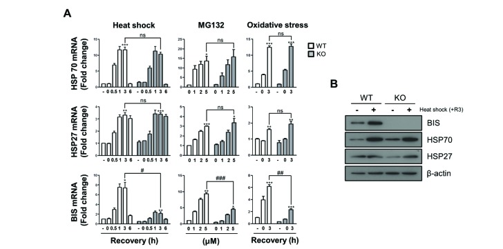
Fig. 1 The induction of HSP70 and HSP27 mRNA was not affected by BIS depletion under stress conditions.(A) Wild type (WT) A549 cells and BIS-knockout (KO) A549 cells were exposed to heat shock (43℃) for 30 min and subsequent recovery for up to 6 h, to MG132 for 6 h, or to glucose free and serum free conditions for 3 h followed by 3 h of recovery. The mRNA expression levels at the indicated times or indicated treatment concentrations were measured by qRT-PCR analyses. The fold induction was determined as the relative value of each mRNA level compared to the untreated WT A549 cells, which was designated arbitrarily as 1.0. Data are represented as the mean±SEM from three independent experiments. *p≤0.05, **p≤0.01, ***p≤0.001 vs. control in each group; #p≤0.05, ##p≤0.01, ###p≤0.001 vs. WT at the indicated times or concentrations. ns, statistically non-significant. (B) The protein levels of BIS, HSP70, and HSP27 in the WT and BIS-KO A549 were determined by western blotting of samples after 3 h of recovery (+R3) following heat shock and control cell samples. Actin expression was used as a loading control.
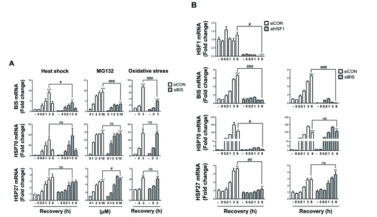
Fig. 2 BIS knockdown did not affect HSP70 and HSP27 induction by various stress conditions.(A) BIS expression was transiently suppressed by 200 nM of control siRNA (siCON) and BIS specific siRNA (siBIS) for 48 h in A549 cells, and the fold induction was compared for BIS, HSP70, and HSP27 mRNA expression in response to heat shock, MG132, or oxidative stress as described in Fig. 1. (B) U87 cells were transfected with HSF1 siRNA (siHSF1) or BIS siRNA (siBIS) for 48 h and exposed to heat shock. The expression levels of HSF1, BIS, HSP70, and HSP27 mRNA were determined as in Fig. 1. *p≤0.05, **p≤0.01, ***p≤0.001 vs. untreated control cells in each group; #p≤0.05, ##p≤0.01, ###p≤0.001 vs. siCON at the indicated times or treatment concentrations. ns, statistically non-significant.
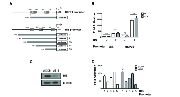
Fig. 3 The promoter activities of BIS and HSP70 were not affected by BIS depletion.(A) Schematic diagram for the relative position of heat shock element (HSE) to ATG in the promoter region of the HSP70 and BIS genes. (B) WT A549 and BIS-KO A549 cells were transfected with BIS or HSP70 promoter constructs for 24 h and then exposed to heat shock (HS) at 43℃ for 30 min, and then further at 37℃ for 1 h. Transcriptional activation of the BIS (P3) and HSP70 promoters are presented as fold changes compared to the value of the pGL3 basic construct in untreated conditions in WT and BIS-KO A549 cells. (C) Western blotting shows that BIS protein expression was sufficiently repressed by siRNA. (D) Following suppression of BIS in A549 cells with BIS siRNA (siBIS), the reporter constructs were expressed and compared to the control (siCON). After normalization with renilla activity, the results are presented as fold change compared to the activity from the pGL3 basic vector (mean±SE, n=3). #p≤0.05 vs. siCON for P1. ns, statistically non-significant.

Fig. 4 Autoregulation of BIS was not mediated by the alteration of mRNA stability.(A) The sequencing results of genotyping in BIS-KO A549 cells show the deletion in exon 1 of the BIS gene and consequent generation of a premature stop codon. (B) WT and BIS-KO A549 cells were treated with actinomycin D for up to 24 h. At the indicated times, mRNA was extracted, and the remaining BIS mRNA was measured and compared to the initial value with qRT-PCR analysis (mean±SE, n=3). (C) The coding region of the WT BIS and the deletion mutant of the BIS gene, in which 14 bp were deleted (Δ14), were cloned into the pEGFP-C1 construct (left). After transfection for 48 h, the GFP mRNA levels representing WT and Δ14-BIS were determined with PCR amplification and agarose electrophoresis by loading different quantities of PCR products (right). (D) The BIS pre-mRNA levels were determined in WT A549 and BIS-KO A549 cells using qRT-PCR analysis with specific primers for intron 2 (solid arrows). The BIS mature mRNA was levels were also determined by the primers covering exon 2 and 3 (dotted arrows). Reverse transcription was performed as described in Method section. *p≤0.05 vs. WT.
Reference
-
1. Lee JH, Takahashi T, Yasuhara N, Inazawa J, Kamada S, Tsujimoto Y. Bis, a Bcl-2-binding protein that synergizes with Bcl-2 in preventing cell death. Oncogene. 1999; 18:6183–6190. PMID: 10597216.
Article2. Takayama S, Xie Z, Reed JC. An evolutionarily conserved family of Hsp70/Hsc70 molecular chaperone regulators. J Biol Chem. 1999; 274:781–786. PMID: 9873016.
Article3. Rosati A, Graziano V, De Laurenzi V, Pascale M, Turco MC. BAG3: a multifaceted protein that regulates major cell pathways. Cell Death Dis. 2011; 2:e141. PMID: 21472004.
Article4. Rosati A, Ammirante M, Gentilella A, Basile A, Festa M, Pascale M, Marzullo L, Belisario MA, Tosco A, Franceschelli S, Moltedo O, Pagliuca G, Lerose R, Turco MC. Apoptosis inhibition in cancer cells: a novel molecular pathway that involves BAG3 protein. Int J Biochem Cell Biol. 2007; 39:1337–1342. PMID: 17493862.
Article5. Zhu H, Liu P, Li J. BAG3: a new therapeutic target of human cancers? Histol Histopathol. 2012; 27:257–261. PMID: 22237703.6. Selcen D, Muntoni F, Burton BK, Pegoraro E, Sewry C, Bite AV, Engel AG. Mutation in BAG3 causes severe dominant childhood muscular dystrophy. Ann Neurol. 2009; 65:83–89. PMID: 19085932.
Article7. Odgerel Z, Sarkozy A, Lee HS, McKenna C, Rankin J, Straub V, Lochmüller H, Paola F, D'Amico A, Bertini E, Bushby K, Goldfarb LG. Inheritance patterns and phenotypic features of myofibrillar myopathy associated with a BAG3 mutation. Neuromuscul Disord. 2010; 20:438–442. PMID: 20605452.
Article8. Lei Z, Brizzee C, Johnson GV. BAG3 facilitates the clearance of endogenous tau in primary neurons. Neurobiol Aging. 2015; 36:241–248. PMID: 25212465.
Article9. Seidel K, Vinet J, Dunnen WF, Brunt ER, Meister M, Boncoraglio A, Zijlstra MP, Boddeke HW, Rüb U, Kampinga HH, Carra S. The HSPB8-BAG3 chaperone complex is upregulated in astrocytes in the human brain affected by protein aggregation diseases. Neuropathol Appl Neurobiol. 2012; 38:39–53. PMID: 21696420.
Article10. Merabova N, Sariyer IK, Saribas AS, Knezevic T, Gordon J, Turco MC, Rosati A, Weaver M, Landry J, Khalili K. WW domain of BAG3 is required for the induction of autophagy in glioma cells. J Cell Physiol. 2015; 230:831–841. PMID: 25204229.
Article11. Behl C. Breaking BAG: the Co-Chaperone BAG3 in health and disease. Trends Pharmacol Sci. 2016; 37:672–688. PMID: 27162137.
Article12. Rosati A, Basile A, Falco A, d'Avenia M, Festa M, Graziano V, De Laurenzi V, Arra C, Pascale M, Turco MC. Role of BAG3 protein in leukemia cell survival and response to therapy. Biochim Biophys Acta. 2012; 1826:365–369. PMID: 22710027.
Article13. Wang HQ, Liu HM, Zhang HY, Guan Y, Du ZX. Transcriptional upregulation of BAG3 upon proteasome inhibition. Biochem Biophys Res Commun. 2008; 365:381–385. PMID: 17996194.
Article14. Rosati A, Leone A, Del Valle L, Amini S, Khalili K, Turco MC. Evidence for BAG3 modulation of HIV-1 gene transcription. J Cell Physiol. 2007; 210:676–683. PMID: 17187345.
Article15. Gentilella A, Khalili K. BAG3 expression is sustained by FGF2 in neural progenitor cells and impacts cell proliferation. Cell Cycle. 2010; 9:4245–4247. PMID: 20962586.
Article16. Liu J, Qu CB, Xue YX, Li Z, Wang P, Liu YH. MiR-143 enhances the antitumor activity of shikonin by targeting BAG3 expression in human glioblastoma stem cells. Biochem Biophys Res Commun. 2015; 468:105–112. PMID: 26541455.
Article17. Flum M, Kleemann M, Schneider H, Weis B, Fischer S, Handrick R, Otte K. miR-217-5p induces apoptosis by directly targeting PRKCI, BAG3, ITGAV and MAPK1 in colorectal cancer cells. J Cell Commun Signal. 2018; 12:451–466. PMID: 28905214.
Article18. d'Avenia M, Citro R, De Marco M, Veronese A, Rosati A, Visone R, Leptidis S, Philippen L, Vitale G, Cavallo A, Silverio A, Prota C, Gravina P, De Cola A, Carletti E, Coppola G, Gallo S, Provenza G, Bossone E, Piscione F, Hahne M, De Windt LJ, Turco MC, De Laurenzi V. A novel miR-371a-5p-mediated pathway, leading to BAG3 upregulation in cardiomyocytes in response to epinephrine, is lost in Takotsubo cardiomyopathy. Cell Death Dis. 2015; 6:e1948. PMID: 26512958.19. Ben Aicha S, Lessard J, Pelletier M, Fournier A, Calvo E, Labrie C. Transcriptional profiling of genes that are regulated by the endoplasmic reticulum-bound transcription factor AIbZIP/CREB3L4 in prostate cells. Physiol Genomics. 2007; 31:295–305. PMID: 17712038.20. Gentilella A, Passiatore G, Deshmane S, Turco MC, Khalili K. Activation of BAG3 by Egr-1 in response to FGF-2 in neuroblastoma cells. Oncogene. 2008; 27:5011–5018. PMID: 18469860.
Article21. Cesaro E, Montano G, Rosati A, Crescitelli R, Izzo P, Turco MC, Costanzo P. WT1 protein is a transcriptional activator of the antiapoptotic bag3 gene. Leukemia. 2010; 24:1204–1206. PMID: 20410921.
Article22. Song S, Kole S, Precht P, Pazin MJ, Bernier M. Activation of heat shock factor 1 plays a role in pyrrolidine dithiocarbamate-mediated expression of the co-chaperone BAG3. Int J Biochem Cell Biol. 2010; 42:1856–1863. PMID: 20692357.
Article23. Yoo HJ, Im CN, Youn DY, Yun HH, Lee JH. Bis is induced by oxidative stress via activation of HSF1. Korean J Physiol Pharmacol. 2014; 18:403–409. PMID: 25352760.24. Jacobs AT, Marnett LJ. HSF1-mediated BAG3 expression attenuates apoptosis in 4-hydroxynonenal-treated colon cancer cells via stabilization of anti-apoptotic Bcl-2 proteins. J Biol Chem. 2009; 284:9176–9183. PMID: 19179333.
Article25. Franceschelli S, Rosati A, Lerose R, De Nicola S, Turco MC, Pascale M. Bag3 gene expression is regulated by heat shock factor 1. J Cell Physiol. 2008; 215:575–577. PMID: 18286539.
Article26. Gentilella A, Khalili K. Autoregulation of co-chaperone BAG3 gene transcription. J Cell Biochem. 2009; 108:1117–1124. PMID: 19777443.
Article27. Gentilella A, Khalili K. BAG3 expression in glioblastoma cells promotes accumulation of ubiquitinated clients in an Hsp70-dependent manner. J Biol Chem. 2011; 286:9205–9215. PMID: 21233200.
Article28. Chen Y, Yang LN, Cheng L, Tu S, Guo SJ, Le HY, Xiong Q, Mo R, Li CY, Jeong JS, Jiang L, Blackshaw S, Bi LJ, Zhu H, Tao SC, Ge F. Bcl2-associated athanogene 3 interactome analysis reveals a new role in modulating proteasome activity. Mol Cell Proteomics. 2013; 12:2804–2819. PMID: 23824909.
Article29. Dai C, Sampson SB. HSF1: Guardian of proteostasis in cancer. Trends Cell Biol. 2016; 26:17–28. PMID: 26597576.
Article30. Dai C, Santagata S, Tang Z, Shi J, Cao J, Kwon H, Bronson RT, Whitesell L, Lindquist S. Loss of tumor suppressor NF1 activates HSF1 to promote carcinogenesis. J Clin Invest. 2012; 122:3742–3754. PMID: 22945628.
Article31. Cui MN, Yun HH, Lee NE, Kim HY, Im CN, Kim YS, Lee JH. Depletion of BIS sensitizes A549 cells to treatment with cisplatin. Mol Cell Toxicol. 2016; 12:63–71.
Article32. Baek JY, Yun HH, Im CN, Ko JH, Jeong SM, Lee JH. BIS overexpression does not affect the sensitivity of HEK 293T cells against apoptosis. Mol Cell Toxicol. 2017; 13:95–103.
Article33. Jelluma N, Yang X, Stokoe D, Evan GI, Dansen TB, Haas-Kogan DA. Glucose withdrawal induces oxidative stress followed by apoptosis in glioblastoma cells but not in normal human astrocytes. Mol Cancer Res. 2006; 4:319–330. PMID: 16687487.
Article34. Liu Y, Song XD, Liu W, Zhang TY, Zuo J. Glucose deprivation induces mitochondrial dysfunction and oxidative stress in PC12 cell line. J Cell Mol Med. 2003; 7:49–56. PMID: 12767261.
Article35. Schweingruber C, Rufener SC, Zünd D, Yamashita A, Mühlemann O. Nonsense-mediated mRNA decay - mechanisms of substrate mRNA recognition and degradation in mammalian cells. Biochim Biophys Acta. 2013; 1829:612–623. PMID: 23435113.
Article36. Karousis ED, Nasif S, Mühlemann O. Nonsense-mediated mRNA decay: novel mechanistic insights and biological impact. Wiley Interdiscip Rev RNA. 2016; 7:661–682. PMID: 27173476.
Article37. Kim HY, Kim YS, Yun HH, Im CN, Ko JH, Lee JH. ERK-mediated phosphorylation of BIS regulates nuclear translocation of HSF1 under oxidative stress. Exp Mol Med. 2016; 48:e260. PMID: 27659916.
Article38. Jin YH, Ahn SG, Kim SA. BAG3 affects the nucleocytoplasmic shuttling of HSF1 upon heat stress. Biochem Biophys Res Commun. 2015; 464:561–567. PMID: 26159920.
Article39. Im CN, Yun HH, Lee JH. Heat shock factor 1 depletion sensitizes A172 glioblastoma cells to temozolomide via suppression of cancer stem cell-like properties. Int J Mol Sci. 2017; 18:E468. PMID: 28241425.
Article40. Fang X, Bogomolovas J, Wu T, Zhang W, Liu C, Veevers J, Stroud MJ, Zhang Z, Ma X, Mu Y, Lao DH, Dalton ND, Gu Y, Wang C, Wang M, Liang Y, Lange S, Ouyang K, Peterson KL, Evans SM, Chen J. Loss-of-function mutations in co-chaperone BAG3 destabilize small HSPs and cause cardiomyopathy. J Clin Invest. 2017; 127:3189–3200. PMID: 28737513.
Article41. Li J, Labbadia J, Morimoto RI. Rethinking HSF1 in stress, development, and organismal health. Trends Cell Biol. 2017; 27:895–905. PMID: 28890254.
Article42. Su KH, Dai C. Metabolic control of the proteotoxic stress response: implications in diabetes mellitus and neurodegenerative disorders. Cell Mol Life Sci. 2016; 73:4231–4248. PMID: 27289378.
Article43. Jiang S, Tu K, Fu Q, Schmitt DC, Zhou L, Lu N, Zhao Y. Multifaceted roles of HSF1 in cancer. Tumour Biol. 2015; 36:4923–4931. PMID: 26108999.
Article
- Full Text Links
-
- Actions
-
Cited
- CITED
-
- Close
- Share
-
- Similar articles
-
- Bis is Induced by Oxidative Stress via Activation of HSF1
- Heme Oxygenase-1 via Transcriptional Activation of Nrf2 Attenuates Apoptosis in Lung Cancer Cells
- The Effect of Inhibition of Heme Oxygenase-1 on Chemosensitivity of Cisplatin in Lung Cancer Cells
- Antiproliferative effect of Citrus junos extracts on A549 human non-smallcell lung cancer cells
- Antimetastatic effect of fucoidan against non-small cell lung cancer by suppressing non-receptor tyrosine kinase and extracellular signalrelated kinase pathway
