Various Wavelengths of Light-Emitting Diode Light Regulate the Proliferation of Human Dermal Papilla Cells and Hair Follicles via Wnt/β-Catenin and the Extracellular Signal-Regulated Kinase Pathways
- Affiliations
-
- 1Department of Dermatology, St. Paul's Hospital, College of Medicine, The Catholic University of Korea, Seoul, Korea. johnkang@catholic.ac.kr
- KMID: 2395183
- DOI: http://doi.org/10.5021/ad.2017.29.6.747
Abstract
- BACKGROUND
The human dermal papilla cells (hDPCs) play an important role in regulation of hair cycling and growth.
OBJECTIVE
The aim of this study was to investigate the effect of different wavelengths of light-emitting diode (LED) irradiation on the proliferation of cultured hDPCs and on the growth of human hair follicles (HFs) in vitro.
METHODS
We examined the effect of LED irradiation on Wnt/β-catenin signaling and mitogen-activated protein kinase (MAPK) pathways in hDPCs. Anagen HFs were cultured with LED irradiation and elongation of each hair shaft was measured.
RESULTS
The most potent wavelength in promoting the hDPC proliferation is 660 nm and 830 nm promoted hDPC proliferation to a lesser extent than 660 nm. Various wavelengths significantly increased β-catenin, Axin2, Wnt3a, Wnt5a and Wnt10b mRNA expression. LED irradiation significantly increased β-catenin and cyclin D expression, and the phosphorylation of MAPK and extracellular signal-regulated kinase (ERK). HFs irradiated with 415 nm and 660 nm grew longer than control.
CONCLUSION
Our result suggests that LED has a potential to stimulate hDPC proliferation via the activation of Wnt/β-catenin signaling and ERK pathway. To our best knowledge, this is the first report which investigated that the effect of various wavelengths of LED on hDPC proliferation and the underlying mechanisms.
Keyword
MeSH Terms
Figure
-
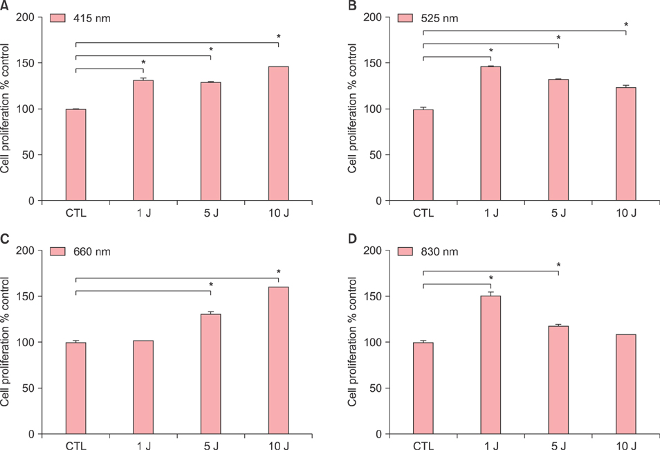
Fig. 1 Effects of various wavelengths and doses of light-emitting diode light (A: 415 nm, B: 525 nm, C: 660 nm, D: 830 nm) on human dermal papilla cell (hDPC) proliferation. The hDPCs (2×10⁴ cells/well) were cultured in serum-free Dulbecco's modified Eagle medium for 24 hours. Then the cultured cells were irradiated immediately with indicated doses and wavelengths, and then incubated for 48 hours. MTT assay was assessed at 48 hours and the relative level is shown as mean±standard deviation from triplicate samples. Statistically significant differences were determined by one-way ANOVA (*p<0.05) compared to control (CTL).

Fig. 2 Effects of light-emitting diode (LED) light on the mRNA expression of β-catenin (A), Axin2 (B), Wnt3a (C), Wnt5a (D) and Wnt10b (E) in human dermal papilla cells (hDPCs). Effects of LED irradiation on the protein expression of β-catenin (F) and cyclin D1 (G) in hDPCs. The hDPCs (2×10⁵ cells/well) were cultured in serum-free Dulbecco's modified Eagle medium for 24 hours and then irradiated with indicated doses and wavelengths, and incubated for 24 hours. The mRNA expression was examined by using real time-polymerase chain reaction and normalized against GAPDH (A~E). The protein level of β-catenin and cyclin D1 was examined by Western blot and normalized against β-actin expression (F, G). The relative level is shown as mean±standard deviation from triplicate samples. Statistically significant differences were determined by one-way ANOVA (*p<0.05) compared to control (CTL).

Fig. 3 Effects of light-emitting diode (LED) light on the phosphorylation of MEK (A), extracellular signal-regulated kinase (ERK) (B), and c-Jun N-terminal kinase (JNK) (C) in human dermal papilla cells (hDPCs). Effects of treatment with PD98059 on the proliferation of hDPCs and the counteract effect of LED irradiation on the inhibitory effect of PD98059 on hDPC proliferation (D~H). The hDPCs (2×10⁵ cells/well) were cultured in serum-free Dulbecco's modified Eagle medium for 24 hours and then irradiated with indicated doses and wavelengths, and incubated for 24 hours (A~C). The protein level of phosphorylated form of MEK (MEK1/2) (A), ERK (ERK1/2) (B) and JNK (C) was examined by Western blot and the relative level is shown as mean±standard deviation (SD) from triplicate samples. Next, the hDPCs were pretreated with 20 µM of PD98059 for 1 hour, and then irradiated with indicated doses and wavelengths, and incubated for 72 hours (D~H). Positive control group in which LED irradiation without PD98059 was shown in (I~L). MTT assay was assessed at 72 hours and the relative level is shown as mean±SD from triplicate samples. Statistically significant differences were determined by one-way ANOVA (*p<0.05) compared to CTL (A~C, I~K). Statistically significant differences were determined by t-test compared to CTL (†p<0.05); one-way ANOVA (#p<0.05) compared to PD98059 treatment group (D~H). CTL: control, pMEK: phosphorylated MEK, pERK: phosphorylated ERK, pJNK: phosphorylated JNK.

Fig. 4 Effects of various wavelengths and doses of light-emitting diode light (A: 415 nm, B: 525 nm, C: 660 nm, D: 830 nm) on the elongation of hair follicles (HFs) in ex vivo culture of whole human scalp HFs. Thirty nine HFs from one individual were cultured with light of various wavelengths and doses for 3 days. Elongation of each hair shaft was measured under a microscope on 3 day. The relative length of each hair shaft is shown as mean±standard deviation from three HFs in percent change compared to control. Statistically significant differences were determined by t-test (*p<0.05) compared to 0 day (0 day; non-treatment). CTL: control.
Cited by 1 articles
-
Synthesized Ceramide Induces Growth of Dermal Papilla Cells with Potential Contribution to Hair Growth
Jee Hye Oh, Kwan Ho Jeong, Jung Eun Kim, Hoon Kang
Ann Dermatol. 2019;31(2):164-174. doi: 10.5021/ad.2019.31.2.164.
Reference
-
1. Mester E, Szende B, Gärtner P. The effect of laser beams on the growth of hair in mice. Radiobiol Radiother (Berl). 1968; 9:621–626.2. Avci P, Gupta GK, Clark J, Wikonkal N, Hamblin MR. Low-level laser (light) therapy (LLLT) for treatment of hair loss. Lasers Surg Med. 2014; 46:144–151.
Article3. Lee WJ, Lee KC, Kim MJ, Jang YH, Lee SJ, Kim DW. Efficacy of red or infrared light-emitting diodes in a mouse model of propionibacterium acnes-induced inflammation. Ann Dermatol. 2016; 28:186–191.
Article4. Wikramanayake TC, Rodriguez R, Choudhary S, Mauro LM, Nouri K, Schachner LA, et al. Effects of the Lexington LaserComb on hair regrowth in the C3H/HeJ mouse model of alopecia areata. Lasers Med Sci. 2012; 27:431–436.
Article5. King LE Jr, Silva KA, Kennedy VE, Sundberg JP. Lack of response to laser comb in spontaneous and graft-induced alopecia areata in C3H/HeJ mice. J Invest Dermatol. 2014; 134:264–266.
Article6. Messenger AG, Senior HJ, Bleehen SS. The in vitro properties of dermal papilla cell lines established from human hair follicles. Br J Dermatol. 1986; 114:425–430.
Article7. Twentyman PR, Luscombe M. A study of some variables in a tetrazolium dye (MTT) based assay for cell growth and chemosensitivity. Br J Cancer. 1987; 56:279–285.
Article8. Philpott MP, Green MR, Kealey T. Human hair growth in vitro. J Cell Sci. 1990; 97:463–471.
Article9. Kishimoto J, Burgeson RE, Morgan BA. Wnt signaling maintains the hair-inducing activity of the dermal papilla. Genes Dev. 2000; 14:1181–1185.
Article10. Sheen YS, Fan SM, Chan CC, Wu YF, Jee SH, Lin SJ. Visible red light enhances physiological anagen entry in vivo and has direct and indirect stimulative effects in vitro. Lasers Surg Med. 2015; 47:50–59.
Article11. Simpson CR, Kohl M, Essenpreis M, Cope M. Near-infrared optical properties of ex vivo human skin and subcutaneous tissues measured using the Monte Carlo inversion technique. Phys Med Biol. 1998; 43:2465–2478.
Article12. Weiss RA, McDaniel DH, Geronemus RG, Weiss MA. Clinical trial of a novel non-thermal LED array for reversal of photoaging: clinical, histologic, and surface profilometric results. Lasers Surg Med. 2005; 36:85–91.
Article13. Shimizu H, Morgan BA. Wnt signaling through the betacatenin pathway is sufficient to maintain, but not restore, anagen-phase characteristics of dermal papilla cells. J Invest Dermatol. 2004; 122:239–245.
Article14. Ouji Y, Nakamura-Uchiyama F, Yoshikawa M. Canonical Wnts, specifically Wnt-10b, show ability to maintain dermal papilla cells. Biochem Biophys Res Commun. 2013; 438:493–499.
Article15. Shefer G, Oron U, Irintchev A, Wernig A, Halevy O. Skeletal muscle cell activation by low-energy laser irradiation: a role for the MAPK/ERK pathway. J Cell Physiol. 2001; 187:73–80.
Article16. Dent P, Yacoub A, Fisher PB, Hagan MP, Grant S. MAPK pathways in radiation responses. Oncogene. 2003; 22:5885–5896.
Article17. Botchkarev VA, Kishimoto J. Molecular control of epithelial-mesenchymal interactions during hair follicle cycling. J Investig Dermatol Symp Proc. 2003; 8:46–55.
Article
- Full Text Links
-
- Actions
-
Cited
- CITED
-
- Close
- Share
-
- Similar articles
-
- Synergistic Proliferating Effects of Electrical Stimulation and Minoxidil on Human Dermal Papilla Cells
- Myristoleic Acid Promotes Anagen Signaling by Autophagy through Activating Wnt/β-Catenin and ERK Pathways in Dermal Papilla Cells
- 3-Deoxysappanchalcone Promotes Proliferation of Human Hair Follicle Dermal Papilla Cells and Hair Growth in C57BL/6 Mice by Modulating WNT/β-Catenin and STAT Signaling
- Vanillic Acid Stimulates Anagen Signaling via the PI3K/Akt/ β-Catenin Pathway in Dermal Papilla Cells
- Synthesized Ceramide Induces Growth of Dermal Papilla Cells with Potential Contribution to Hair Growth
