Korean J Neurotrauma.
2016 Oct;12(2):112-117. 10.13004/kjnt.2016.12.2.112.
Effectiveness of Cortical Atrophy Scale and Indirect Indices of Brain Atrophy to Predict Chronic Subdural Hematoma in Older Patients
- Affiliations
-
- 1Department of Neurosurgery, School of Medicine, Chungnam National University Hospital, Daejeon, Korea. swchoi@cnu.ac.kr
- KMID: 2356783
- DOI: http://doi.org/10.13004/kjnt.2016.12.2.112
Abstract
OBJECTIVE
To determine whether baseline cerebral atrophy can predict the rate of future chronic subdural hematoma (CSDH) after head trauma and compare indirect markers of brain atrophy with volumetric analysis of computed tomography (CT).
METHODS
Single institution case-control study involving 1,476 patients who visited our hospital after head trauma from January 2009 to December 2014. Forty-one patients with delayed CSDH were identified and age, gender matched 41 patients were selected as control group. Both volumetric analyze on CT and Evans index, frontal horn index, bicaudate ratio, sylvian fissure ratio and cortical atrophy scale of 82 patients were estimated by different raters and relationship of those factors with CSDH was analyzed.
RESULTS
Every indirect indices except cortical atrophy scale were not enough to give a good estimate of CSDH. Brain atrophy and cortical atrophy scale were predisposing factors of CSDH on multivariate analysis with statistical significance.
CONCLUSION
Brain atrophy was a potential prognostic factor of CSDH after trauma. In practice, patients with a value of cortical atrophy scale over moderate grade needed more attention for CSDH.
MeSH Terms
Figure
-
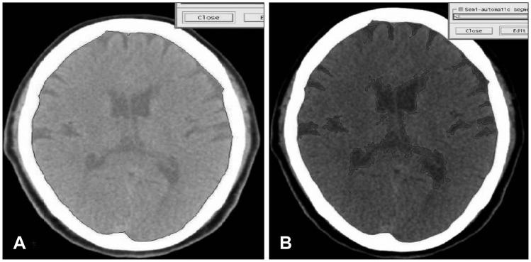
FIGURE 1 Volume measurement of Gamma-Plan program (Elekta instrument AB, Stockholm, Sweden) by sequential volume mapping. (A) Black line contain Intracranial volume. (B) Black lilne contain cerebrospinal fluid volume.
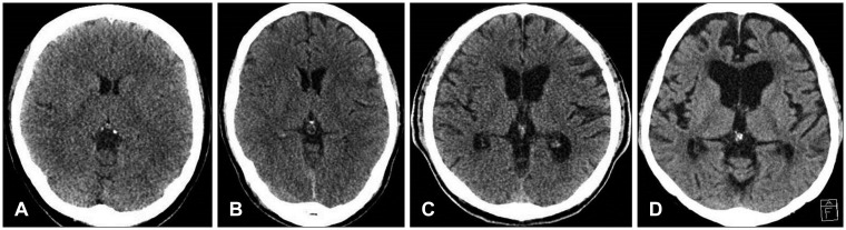
FIGURE 2 Brain computed tomography examples of cortical atrophy scale. (A) Absent atrophy. (B) Mild atrophy. (C) Moderate atrophy. (D) Severe atrophy.
Reference
-
1. Ahmed E, Aurangzeb A, Khan SA, Maqbool S, Ali A, Zadran KK, et al. Frequency of conservatively managed traumatic acute subdural haematoma changing into chronic subdural haematoma. J Ayub Med Coll Abbottabad. 2012; 24:71–74.2. Bermel RA, Bakshi R, Tjoa C, Puli SR, Jacobs L. Bicaudate ratio as a magnetic resonance imaging marker of brain atrophy in multiple sclerosis. Arch Neurol. 2002; 59:275–280. PMID: 11843699.
Article3. Evans W. An encephalographic ratio for estimating ventricular enlargement and cerebral atrophy. Arch Neurol Psychiatry. 1942; 47:931–937.
Article4. Goldszal AF, Davatzikos C, Pham DL, Yan MX, Bryan RN, Resnick SM. An image-processing system for qualitative and quantitative volumetric analysis of brain images. J Comput Assist Tomogr. 1998; 22:827–837. PMID: 9754125.
Article5. Jack A, O'Kelly C, McDougall C, Findlay JM. Predicting recurrence after chronic subdural haematoma drainage. Can J Neurol Sci. 2015; 42:34–39. PMID: 25557536.
Article6. Koedam EL, Lehmann M, van der Flier WM, Scheltens P, Pijnenburg YA, Fox N, et al. Visual assessment of posterior atrophy development of a MRI rating scale. Eur Radiol. 2011; 21:2618–2625. PMID: 21805370.
Article7. Möller C, van der Flier WM, Versteeg A, Benedictus MR, Wattjes MP, Koedam EL, et al. Quantitative regional validation of the visual rating scale for posterior cortical atrophy. Eur Radiol. 2014; 24:397–404. PMID: 24092044.
Article8. Markwalder TM. Chronic subdural hematomas: a review. J Neurosurg. 1981; 54:637–645. PMID: 7014792.
Article9. Marmarou A, Bergsneider M, Klinge P, Relkin N, Black PM. The value of supplemental prognostic tests for the preoperative assessment of idiopathic normal-pressure hydrocephalus. Neurosurgery. 2005; 57:S17–S28. PMID: 16160426.
Article10. Sajanti J, Majamaa K. High concentrations of procollagen propeptides in chronic subdural haematoma and effusion. J Neurol Neurosurg Psychiatry. 2003; 74:522–524. PMID: 12640081.
Article11. Scheltens P, Pasquier F, Weerts JG, Barkhof F, Leys D. Qualitative assessment of cerebral atrophy on MRI: inter- and intra-observer reproducibility in dementia and normal aging. Eur Neurol. 1997; 37:95–99. PMID: 9058064.
Article12. Son S, Yoo CJ, Lee SG, Kim EY, Park CW, Kim WK. Natural course of initially non-operated cases of acute subdural hematoma: the risk factors of hematoma progression. J Korean Neurosurg Soc. 2013; 54:211–219. PMID: 24278650.13. Tugcu B, Tanriverdi O, Baydin S, Hergunsel B, Günaldı Ö, Ofluoglu E, et al. Can recurrence of chronic subdural hematoma be predicted? A retrospective analysis of 292 cases. J Neurol Surg A Cent Eur Neurosurg. 2014; 75:37–41. PMID: 23307307.
Article14. van Zagten M, Kessels F, Boiten J, Lodder J. Interobserver agreement in the assessment of cerebral atrophy on CT using bicaudate and sylvian-fissure ratios. Neuroradiology. 1999; 41:261–264. PMID: 10344510.
Article15. Walchenbach R, Geiger E, Thomeer RT, Vanneste JA. The value of temporary external lumbar CSF drainage in predicting the outcome of shunting on normal pressure hydrocephalus. J Neurol Neurosurg Psychiatry. 2002; 72:503–506. PMID: 11909911.16. Winn HR. Youmans neurological surgery. ed 5. Philadelphia, PA: W. B. Saunders;2004.17. Yamamoto H, Hirashima Y, Hamada H, Hayashi N, Origasa H, Endo S. Independent predictors of recurrence of chronic subdural hematoma: results of multivariate analysis performed using a logistic regression model. J Neurosurg. 2003; 98:1217–1221. PMID: 12816267.
Article18. Yamazaki Y, Tachibana S, Kitahara Y, Ohwada T. Promotive factors of chronic subdural hematoma in relation to age. No Shinkei Geka. 1996; 24:47–51. PMID: 8559264.
- Full Text Links
-
- Actions
-
Cited
- CITED
-
- Close
- Share
-
- Similar articles
-
- CT findings of brain atrophy after chemotheraphy in acute leukemia
- Chronic Subdural Hematoma in the Aged, Trauma or Degeneration?
- Comparision between Brain Atrophy and Subdural Volume to Predict Chronic Subdural Hematoma: Volumetric CT Imaging Analysis
- Chronic Subdural Hematoma Superimposed on Posttraumatic Subdural Hygroma: A Report of Three Cases
- Rapid Spontaneous Resolution of Contralateral Lentiform Acute Subdural Hematoma After Burr Hole Trephination for Chronic Subdural Hematoma
