Comparision between Brain Atrophy and Subdural Volume to Predict Chronic Subdural Hematoma: Volumetric CT Imaging Analysis
- Affiliations
-
- 1Department of Neurosurgery, School of Medicine, Chungnam National University, Daejeon, Korea. neons@cnu.ac.kr
- KMID: 2378264
- DOI: http://doi.org/10.13004/kjnt.2015.11.2.87
Abstract
OBJECTIVE
Brain atrophy and subdural hygroma were well known factors that enlarge the subdural space, which induced formation of chronic subdural hematoma (CSDH). Thus, we identified the subdural volume that could be used to predict the rate of future CSDH after head trauma using a computed tomography (CT) volumetric analysis.
METHODS
A single institution case-control study was conducted involving 1,186 patients who visited our hospital after head trauma from January 1, 2010 to December 31, 2014. Fifty-one patients with delayed CSDH were identified, and 50 patients with age and sex matched for control. Intracranial volume (ICV), the brain parenchyme, and the subdural space were segmented using CT image-based software. To adjust for variations in head size, volume ratios were assessed as a percentage of ICV [brain volume index (BVI), subdural volume index (SVI)]. The maximum depth of the subdural space on both sides was used to estimate the SVI.
RESULTS
Before adjusting for cranium size, brain volume tended to be smaller, and subdural space volume was significantly larger in the CSDH group (p=0.138, p=0.021, respectively). The BVI and SVI were significantly different (p=0.003, p=0.001, respectively). SVI [area under the curve (AUC), 77.3%; p=0.008] was a more reliable technique for predicting CSDH than BVI (AUC, 68.1%; p=0.001). Bilateral subdural depth (sum of subdural depth on both sides) increased linearly with SVI (p<0.0001).
CONCLUSION
Subdural space volume was significantly larger in CSDH groups. SVI was a more reliable technique for predicting CSDH. Bilateral subdural depth was useful to measure SVI.
Keyword
MeSH Terms
Figure
-

FIGURE 1 Relationship between the subdural volume index (SVI) and brain volume index (BVI) in the control and chronic subdural hematoma (CSDH) groups. Cumulative distribution of BVI and SVI between the control and CSDH groups. The cut-off value of SVI was 6.6% (sensitivity, 37.2% and specificity, 86%) with area under the curve (AUC) of 77.3%. The cut-off value of BVI was 94.8% (sensitivity, 82.9% and specificity, 47.8%) with AUC of 68.1%. Note that CSDH group was markedly higher than SVI and BVI cut-off values.
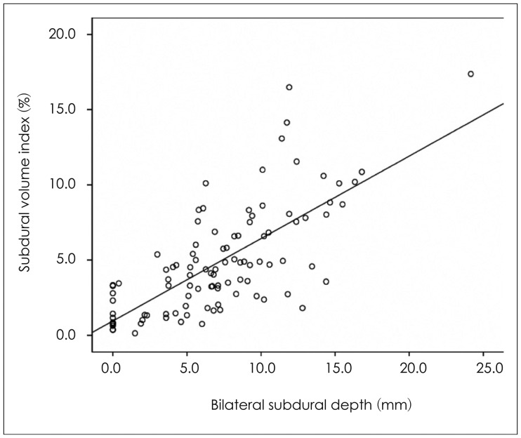
FIGURE 2 Relation between the subdural volume index (SVI) and bilateral subdural depth. The relationship between the SVI and bilateral subdural depth. The correlation coefficient was 0.72 (p<0.0001), indicating that the SVI increases as bilateral subdural depth increases.
Reference
-
1. Ambarki K, Israelsson H, Wåhlin A, Birgander R, Eklund A, Malm J. Brain ventricular size in healthy elderly: comparison between Evans index and volume measurement. Neurosurgery. 2010; 67:94–99. discussion 99PMID: 20559096.2. Aspegren OP, Åstrand R, Lundgren MI, Romner B. Anticoagulation therapy a risk factor for the development of chronic subdural hematoma. Clin Neurol Neurosurg. 2013; 115:981–984. PMID: 23128014.
Article3. Baechli H, Nordmann A, Bucher HC, Gratzl O. Demographics and prevalent risk factors of chronic subdural haematoma: results of a large single-center cohort study. Neurosurg Rev. 2004; 27:263–266. PMID: 15148652.
Article4. Camel M, Grubb RL Jr. Treatment of chronic subdural hematoma by twist-drill craniotomy with continuous catheter drainage. J Neurosurg. 1986; 65:183–187. PMID: 3723175.5. Ge Y, Grossman RI, Babb JS, Rabin ML, Mannon LJ, Kolson DL. Age-related total gray matter and white matter changes in normal adult brain. Part I: volumetric MR imaging analysis. AJNR Am J Neuroradiol. 2002; 23:1327–1333. PMID: 12223373.6. Gebel JM, Sila CA, Sloan MA, Granger CB, Weisenberger JP, Green CL, et al. Comparison of the ABC/2 estimation technique to computer-assisted volumetric analysis of intraparenchymal and subdural hematomas complicating the GUSTO-1 trial. Stroke. 1998; 29:1799–1801. PMID: 9731597.
Article7. Goldszal AF, Davatzikos C, Pham DL, Yan MX, Bryan RN, Resnick SM. An image-processing system for qualitative and quantitative volumetric analysis of brain images. J Comput Assist Tomogr. 1998; 22:827–837. PMID: 9754125.
Article8. Mori K, Mitsuoka H, Cho K, Tajima A, Maeda M. Rate constant of gadolinium (Gd)-DTPA transfer into chronic subdural hematomas. Neurol Res. 1996; 18:126–134. PMID: 9162866.
Article9. Sato S, Suzuki J. Ultrastructural observations of the capsule of chronic subdural hematoma in various clinical stages. J Neurosurg. 1975; 43:569–578. PMID: 1181389.
Article10. Winn HR. Youmans neurological surgery. ed 5. Philadelphia: WB Saunders;2004.
- Full Text Links
-
- Actions
-
Cited
- CITED
-
- Close
- Share
-
- Similar articles
-
- Chronic Subdural Hematoma Superimposed on Posttraumatic Subdural Hygroma: A Report of Three Cases
- Effectiveness of Cortical Atrophy Scale and Indirect Indices of Brain Atrophy to Predict Chronic Subdural Hematoma in Older Patients
- Evolution of Chronic Subdural Hematoma based on Brain CT findings and Appropriate Treatment Methods
- Bilateral Acute Subdural Hematoma Following Evacuation of Chronic Subdural Hematoma
- Intraoperative Development of Contralateral Subdural Hematoma during Evacuation of Acute Subdural Hematoma: Case Report
