The Anti-inflammatory Effect of Cobalt-Protoporphyrin for Rats with Epididymitis Induced by Escherichia coli Infection
- Affiliations
-
- 1Department of Urology, Wonkwang University School of Medicine, Iksan, Korea. seraph@wonkwang.ac.kr
- KMID: 2294243
- DOI: http://doi.org/10.4111/kju.2006.47.6.656
Abstract
-
PURPOSE: Heme oxygenase-1 (HO-1), an inducible heat shock protein, catalyzes the heme to iron, biliverdin and carbon monoxide. It also has an inhibitory effect on necrosis and inflammation. Cobalt (III)-protoporphyrin IX (CoPP) is known to be a HO-1 inducer. Our intension was to find whether CoPP has an anti-inflammatory effect through the induction of HO-1 in rats with epididymitis.
MATERIALS AND METHODS
Thirty two Sprague-Dawley male rats (age: 8-12 weeks, weight: 200-250gm) were selected for the experiments. Anesthesia was performed with an intraperitoneal injection of ketamine hydrochloride (140mg/kg). Four rats were taken and used as a control group. Epididymitis was induced in 28 rats by an injection of E. coli (1 x 10(5)/ml) to the epididymis. In the first step, groups of 4 rats were sacrificed serially after 4, 12, 48, and 72 hours for Hematoxylin & Eosin (H&E) staining and Western blot for inducible nitric oxide synthase (iNOS) and cyclooxygenase (COX)-2. In the second step, groups of 4 rats were injected with either dimethyl sulphoxide (DMSO) 7 microliter, DMSO 7 microliter with 50mg/ml CoPP or DMSO 7 microliter with 100mg/ml CoPP. They were then sacrificed 72 hours later for H&E staining and Western blot for iNOS and COX-2.
RESULTS
In the first step, increased inflammation was evident H&E staining over time. Western blots, iNOS expression was detected after 48 hours and COX-2 was after 12 hours. In the second step, decreased inflammation was evident H&E staining, and the expressions of iNOS and COX-2 were suppressed in the CoPP treated group.
CONCLUSIONS
CoPP can reduce the inflammation of epididymis in rats, and the mechanism may be related with HO-1.
Keyword
MeSH Terms
-
Anesthesia
Animals
Biliverdine
Blotting, Western
Carbon Monoxide
Cobalt
Dimethyl Sulfoxide
Eosine Yellowish-(YS)
Epididymis
Epididymitis*
Escherichia coli Infections*
Escherichia coli*
Escherichia*
Heat-Shock Proteins
Hematoxylin
Heme
Heme Oxygenase (Decyclizing)
Heme Oxygenase-1
Humans
Inflammation
Injections, Intraperitoneal
Iron
Ketamine
Male
Necrosis
Nitric Oxide Synthase Type II
Prostaglandin-Endoperoxide Synthases
Rats*
Rats, Sprague-Dawley
Biliverdine
Carbon Monoxide
Cobalt
Dimethyl Sulfoxide
Eosine Yellowish-(YS)
Heat-Shock Proteins
Hematoxylin
Heme
Heme Oxygenase (Decyclizing)
Heme Oxygenase-1
Iron
Ketamine
Nitric Oxide Synthase Type II
Prostaglandin-Endoperoxide Synthases
Figure
-
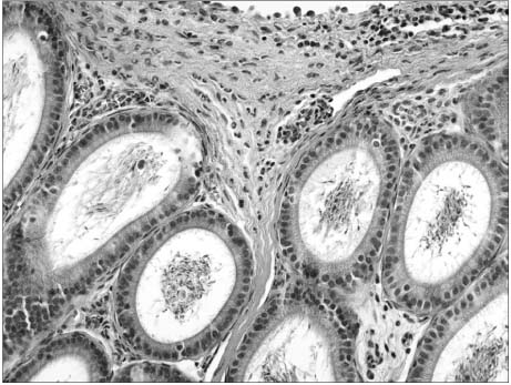
Fig. 1 Microscopic finding of epididymis. The epididymal duct is surrounded by infiltrated neutrophils and congested vessels 72 hours after an injection of E. coli to the epididymis (H&E stain, ×100).
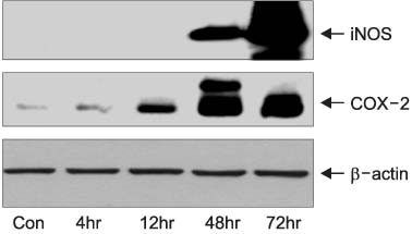
Fig. 2 Western blot analysis of epididymal tissue. The expressions of inducible nitric oxide synthase (iNOS) and cyclooxygenase (COX)-2 increases after an injection of E. coli to the epididymis.
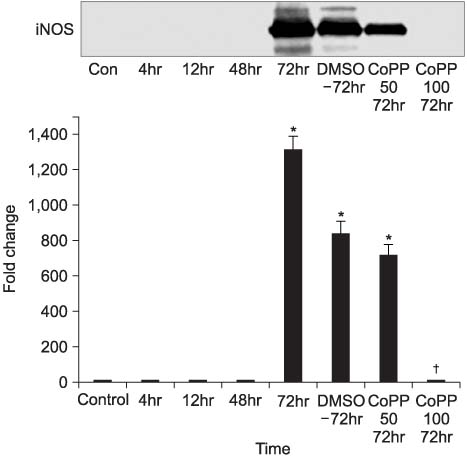
Fig. 3 Western blot analysis of epididymal tissue after the induction of inflammation and application of cobalt (III)-protoporphyrin IX (CoPP). The expression of inducible nitric oxide synthase (iNOS) increase for up to 72 hours after an injection of E. coli, but decreases after the application of CoPP (CoPP 50: CoPP 50mg/ml+dimethyl sulphoxide (DMSO) 7µl, CoPP 100: CoPP 100mg/ml+DMSO 7µl). *: significantly different from the control at p<0.05, †: significantly different from corresponding values at 72 hours and 72 hours after an injection of DMSO at p<0.05.
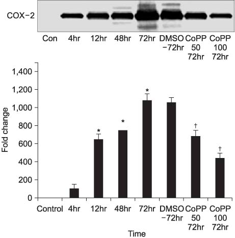
Fig. 4 Western blot analysis of epididymal tissue after the induction of inflammation and application of cobalt (III)-protoporphyrin IX (CoPP). The expression of cyclooxygenase (COX)-2 increase after an injection of E. coli, and decreases after the application of CoPP (CoPP 50: CoPP 50mg/ml+dimethyl sulphoxide (DMSO) 7 µl, CoPP 100: CoPP 100mg/ml+DMSO 7µl). *: significantly different from the control at p<0.05, †: significantly different from corresponding values at 72 hours and 72 hours after an injection of DMSO at p<0.05.
Reference
-
1. Torti PM, Torti SV. Regulation of ferritin genes and protein. Blood. 2002. 99:3505–3516.2. Montellano PR. The mechanism of heme oxygenase. Curr Opin Chem Biol. 2000. 4:221–227.3. Applegate LA, Luscher P, Tyrrell RM. Induction of heme oxygenase: a general response to oxidant stress in cultured mammalian cells. Cancer Res. 1991. 51:974–978.4. Maines MD. The heme oxygenase system: a regulator of second messenger gases. Annu Rev Pharmacol Toxicol. 1997. 37:517–554.5. Lee TS, Chau LY. Heme oxygenase-1 mediates the anti-inflammatory effect of interleukin-10 in mice. Nat Med. 2002. 8:240–246.6. Brasile L, Buelow R, Stubenitsky BM, Kootstra G. Induction of heme oxygenase-1 in kidneys during ex vivo warm perfusion. Transplantation. 2003. 76:1145–1149.7. Wagener FA, Volk HD, Willis D, Abraham NG, Soares MP, Adema GJ, et al. Different faces of the heme-heme oxygenase system in inflammation. Pharm Rev. 2003. 55:551–571.8. Choi BM, Pae HO, Jeong YR, Oh GS, Jun CD, Kim BR, et al. Overexpression of heme oxygenase (HO-1) renders Jurkat T cells resistant to fas-mediated apoptosis: involvement of iron released by HO-1. Free Radical Biol Med. 2004. 36:858–871.9. Weber CM, Eke BC, Maines MD. Corticosterone regulates heme oxygenase-2 and NO synthase transcription and protein expression in rat brain. J Neurochem. 1994. 63:953–962.10. McCoubrey WK Jr, Huang TJ, Maines MD. Isolation and characterization of a cDNA from the rat brain that encodes hemoprotein heme oxygenase-3. Eur J Biochem. 1997. 247:725–732.11. Siow RC, Sato H, Mann GE. Heme oxygenase carbon monoxide signalling pathway in atherosclerosis: anti-atherogenic actions of bilirubin and carbon monoxide? Cardiovasc Res. 1999. 41:385–394.12. Tenhunen R, Marver HS, Schmid R. The enzymatic conversion of heme to bilirubin by microsomal heme oxygenase. Proc Natl Acad USA. 1968. 61:748–755.13. McCarter SD, Akyea TG, Lu X, Bihari A, Scott JR, Badhwar A, et al. Endogenous heme oxygenase induction is a critical mechanism attenuating apoptosis and restoring microvascular perfusion following limb ischemia/reperfusion. Surgery. 2004. 136:67–75.14. Gopinathan V, Miller NJ, Milner AD, Rice-Evans CA. Bilirubin and ascorbate antioxidant activity in neonatal plasma. FEBS Lett. 1994. 349:197–200.15. Choi BM, Pae HO, Kim YM, Chung HT. Nitric oxide-mediated cytoprotection of hepatocytes from glucose deprivation-induced cytotoxicity: involvement of heme oxygenase-1. Hepatology. 2003. 37:810–823.16. Wagener FA, Eggert A, Boerman OC, Oyen WJ, Verhofstad A, Abraham NG, et al. Heme is a potent inducer of inflammation in mice and is counteracted by heme oxygenase. Blood. 2001. 98:1802–1811.17. Sass G, Soares MC, Yamashita K, Seyfried S, Zimmermann WH, Eschenhagen T, et al. Heme oxygenase-1 and its reaction product, carbon monoxide, prevent inflammation-related apoptotic liver damage in mice. Hepatology. 2003. 38:909–918.18. Hayashi S, Takamiya R, Yamaguchi T, Matsumoto K, Tojo SJ, Tamatani T, et al. Induction of heme oxygenase-1 suppresses venular leukocyte adhesion elicited by oxidative stress: role of bilirubin generated by the enzyme. Circ Res. 1999. 85:663–671.19. Suttner DM, Dennery PA. Reversal of HO-1 related cytoprotection with increased expression is due to reactive iron. FASEB J. 1999. 13:1800–1809.
- Full Text Links
-
- Actions
-
Cited
- CITED
-
- Close
- Share
-
- Similar articles
-
- Xanthogranulomatous Epididymitis Unresponsive to Antibiotic Treatment
- Escherichia coli O157: H7 Infection
- The Effect of Intravenous E. coli Injection on the Kidney after Ureteral Ligation in the Rat
- Escherichia coli O157:H7 Infection
- An outbreak of inapparent non-O157 enterohemorrhagic escherichia coli infection
