Kruppel-Like Factor 2 Suppression by High Glucose as a Possible Mechanism of Diabetic Vasculopathy
- Affiliations
-
- 1Cardiovascular Laboratory, Clinical Research Institute, Seoul National University Hospital, Seoul, Korea. hylee612@snu.ac.kr
- 2Department of Internal Medicine, Seoul National University College of Medicine, Seoul, Korea.
- KMID: 2225016
- DOI: http://doi.org/10.4070/kcj.2012.42.4.239
Abstract
- BACKGROUND AND OBJECTIVES
Endothelial dysfunction is widely observed in diabetes mellitus, resulting in diabetic vascular complications. Kruppel-like factor 2 (KLF2) is implicated as being a key molecule that maintains endothelial function. We evaluated the expression of KLF2 in endothelial cells cultured in high glucose and investigated its functional implication in a diabetic animal model.
SUBJECTS AND METHODS
Human umbilical vein endothelial cells (HUVECs) were cultured in physiologically high glucose (35 mM) condition. The Otsuka Long Evans Tokushima Fatty (OLETF) strain of rat was used as an excellent model of obese type II diabetes, and their lean littermates are Long Evans Tokushima Otsuka (LETO) rats.
RESULTS
In HUVECs cultured in physiologically high glucose condition, FOXO1 was activated whereas KLF2 and endothelial nitric oxide synthase (eNOS) expression was near completely abolished, which was completely reversed by FOXO1 small interfering ribonucleic acid. In the vessels harvested from the OLETF rats, the animal model of type II diabetes, KLF2 and eNOS expression were found depleted. When vascular remodeling was induced in the left common carotid artery by reduction of blood flow with partial ligation of the distal branches, greater neointimal hypertrophy was observed in OLETF rats compared with the control LETO rats.
CONCLUSION
KLF2 suppression in endothelial cells by high glucose is a possible mechanism of diabetic endothelial dysfunction. The strategy of replenishing KLF2 may be effective for preventing diabetic vascular dysfunction.
MeSH Terms
Figure
-
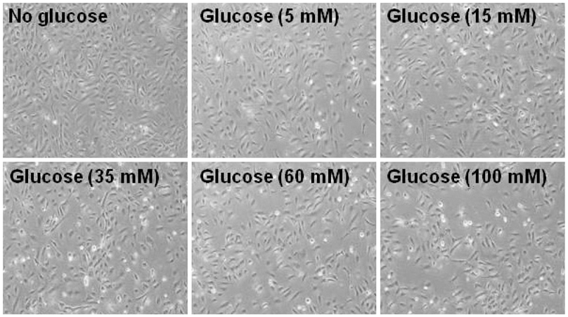
Fig. 1 Morphologic changes of HUVECs 72 hours after culture under high glucose conditions. HUVECs were cultured in high glucose with the indicated concentration. HUVECs: human umbilical vein endothelial cells.
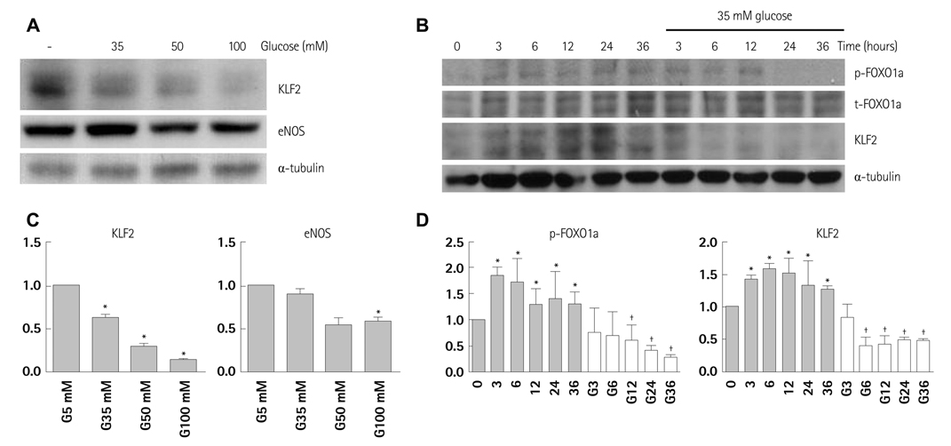
Fig. 2 Activation of FOXO1 and inhibition of KLF2 under high glucose conditions. A: KLF2 and eNOS protein expression at the indicated concentration of glucose. B: serial immunoblot analysis of phosphorylated FOXO1 on serine 256 (p-FOXO1), total FOXO1 (t-FOXO1), and KLF2 expression in endothelial cells incubated under physiologically high glucose conditions (35 mM). C: quantification of triplicate experiments of A by densitometry. D: quantification of triplicate experiments of B by densitometry. *p<0.05 compared to the control, †p<0.05 compared to the value of 36 hours. KLF2: Krüppel-like factor 2, eNOS: endothelial nitric oxide synthase.
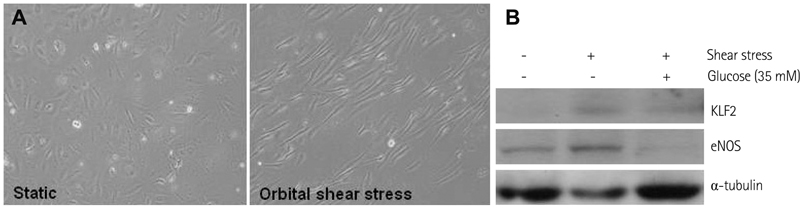
Fig. 3 Inhibition of shear flow-induced KLF2 expression under high glucose conditions. A: morphologic changes in HUVECs after shear stress application (24 hours, 12 dynes/cm2). B: immunoblot analysis evaluating the impact of shear stress and high glucose on the expression of KLF2 and eNOS. KLF2: Krüppel-like factor 2, HUVECs: human umbilical vein endothelial cells, eNOS: endothelial nitric oxide synthase.
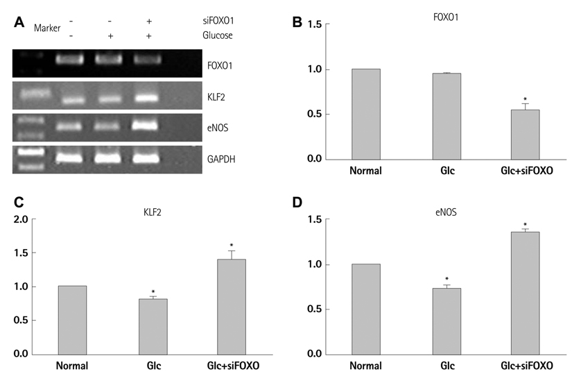
Fig. 4 Suppression of KLF2 and eNOS by FOXO1 in endothelial cells. A: representative RT-PCR analysis figure evaluating blocking effect of FOXO1 by FOXO1 siRNA on KLF2 and eNOS mRNA expression in high glucose condition. B-D: quantification of triplicate experiments of A. *p<0.05 compared to the control. KLF2: Krüppel-like factor 2, eNOS: endothelial nitric oxide synthase, RT-PCR: reverse transcriptase-polymer chain reaction, siRNA: small interfering ribonucleic acid, mRNA: messenger ribonucleic acid.
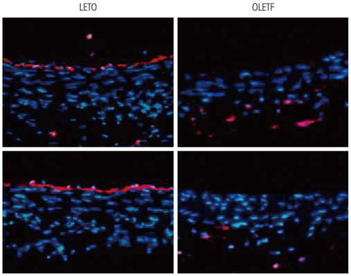
Fig. 5 Suppression of KLF2 in the blood vessels from diabetic, OLEFT rats. Representative immunofluorescent microscopic images of carotid arteries stained with KLF2 (colored-red) in the OLETF rats (left) and the control LETO rats (right). KLF2: Krüppel-like factor 2, OLEFT: Otsuka Long Evans Tokushima Fatty, LETO: Long Evans Tokushima Otsuka.

Fig. 6 Comparison of KLF2 expression in endothelial cells at abdominal-celiac branch point between OLETF and control LETO rats. A: schematic showing the branch point of the celiac artery off the abdominal aorta. The line shows the section through which the samples were taken for immunohistochemical examination of KLF2 expression. B: representative photographs of immunofluorescent staining of blood vessels with KLF2 in the OLETF rats and the LETO rats. KLF2 expression is stained red at the branching point between the abdominal aorta and the celiac artery of the indicated rats. Arrow indicates 'atherosclerosis-resistant flow-dividing' side and 'atherosclerosis-prone lateral' side. C: semi-quantitative graph of KLF2-stained luminal circumferential length (4 rats in each group). KLF2: Krüppel-like factor 2, OLEFT: Otsuka Long Evans Tokushima Fatty, LETO: Long Evans Tokushima Otsuka.

Fig. 7 Evaluation of neointimal formation after induction of vascular remodeling. A: representative photomicrographs of common carotid arteries sections from the indicated rats 1 week after ligation. At the left are photographs from the sham-operated artery. At the right are photographs from the ligation-operated artery. Yellow arrow indicates intima. B: comparison of intimal thickness. n=8 (two slide sections in one carotid artery sample) in each group (4 rats in each group). Statistical comparisons between ligation-operated and sham-operated arteries were performed using a paired t-test for comparison for intra-group comparisons and a Student's t-test for inter-group comparisons. OLEFT: Otsuka Long Evans Tokushima Fatty, LETO: Long Evans Tokushima Otsuka.
Reference
-
1. Haffner SM, Lehto S, Rönnemaa T, Pyörälä K, Laakso M. Mortality from coronary heart disease in subjects with type 2 diabetes and in nondiabetic subjects with and without prior myocardial infarction. N Engl J Med. 1998. 339:229–234.2. Hink U, Li H, Mollnau H, et al. Mechanisms underlying endothelial dysfunction in diabetes mellitus. Circ Res. 2001. 88:E14–E22.3. De Vriese AS, Verbeuren TJ, Van de Voorde J, Lameire NH, Vanhoutte PM. Endothelial dysfunction in diabetes. Br J Pharmacol. 2000. 130:963–974.4. Johnstone MT, Creager SJ, Scales KM, Cusco JA, Lee BK, Creager MA. Impaired endothelium-dependent vasodilation in patients with insulin-dependent diabetes mellitus. Circulation. 1993. 88:2510–2516.5. Balletshofer BM, Rittig K, Enderle MD, et al. Endothelial dysfunction is detectable in young normotensive first-degree relatives of subjects with type 2 diabetes in association with insulin resistance. Circulation. 2000. 101:1780–1784.6. Hattori Y, Kawasaki H, Abe K, Kanno M. Superoxide dismutase recovers altered endothelium-dependent relaxation in diabetic rat aorta. Am J Physiol. 1991. 261(4 Pt 2):H1086–H1094.7. Beckman JA, Goldfine AB, Gordon MB, Garrett LA, Creager MA. Inhibition of protein kinase Cbeta prevents impaired endothelium-dependent vasodilation caused by hyperglycemia in humans. Circ Res. 2002. 90:107–111.8. Steinberg HO, Chaker H, Leaming R, Johnson A, Brechtel G, Baron AD. Obesity/insulin resistance is associated with endothelial dysfunction: implications for the syndrome of insulin resistance. J Clin Invest. 1996. 97:2601–2610.9. Atkins GB, Jain MK. Role of Krüppel-like transcription factors in endothelial biology. Circ Res. 2007. 100:1686–1695.10. Samatar AA, Wang L, Mirza A, Koseoglu S, Liu S, Kumar CC. Transforming growth factor-beta 2 is a transcriptional target for Akt/protein kinase B via forkhead transcription factor. J Biol Chem. 2002. 277:28118–28126.11. Buteau J, Accili D. Regulation of pancreatic beta-cell function by the forkhead protein FoxO1. Diabetes Obes Metab. 2007. 9:Suppl 2. 140–146.12. Kamagate A, Qu S, Perdomo G, et al. FoxO1 mediates insulin-dependent regulation of hepatic VLDL production in mice. J Clin Invest. 2008. 118:2347–2364.13. Kim HS, Skurk C, Thomas SR, et al. Regulation of angiogenesis by glycogen synthase kinase-3beta. J Biol Chem. 2002. 277:41888–41896.14. Kanemoto N, Hishigaki H, Miyakita A, et al. Genetic dissection of "OLETF", a rat model for non-insulin-dependent diabetes mellitus. Mamm Genome. 1998. 9:419–425.15. Kumar A, Lindner V. Remodeling with neointima formation in the mouse carotid artery after cessation of blood flow. Arterioscler Thromb Vasc Biol. 1997. 17:2238–2244.16. Korshunov VA, Berk BC. Flow-induced vascular remodeling in the mouse: a model for carotid intima-media thickening. Arterioscler Thromb Vasc Biol. 2003. 23:2185–2191.17. Wang N, Miao H, Li YS, et al. Shear stress regulation of Krüppel-like factor 2 expression is flow pattern-specific. Biochem Biophys Res Commun. 2006. 341:1244–1251.18. Parmar KM, Larman HB, Dai G, et al. Integration of flow-dependent endothelial phenotypes by Kruppel-like factor 2. J Clin Invest. 2006. 116:49–58.19. Hurks R, Eisinger MJ, Goovaerts I, et al. Early endothelial dysfunction in young type 1 diabetics. Eur J Vasc Endovasc Surg. 2009. 37:611–615.20. Baris N, Akdeniz B, Uyar S, et al. Are complex coronary lesions more frequent in patients with diabetes mellitus? Can J Cardiol. 2006. 22:935–937.21. Iakovou I, Schmidt T, Bonizzoni E, et al. Incidence, predictors, and outcome of thrombosis after successful implantation of drug-eluting stents. JAMA. 2005. 293:2126–2130.22. Parmar KM, Nambudiri V, Dai G, Larman HB, Gimbrone MA Jr, García-Cardeña G. Statins exert endothelial atheroprotective effects via the KLF2 transcription factor. J Biol Chem. 2005. 280:26714–26719.23. Rossi J, Rouleau L, Tardif JC, Leask RL. Effect of simvastatin on Kruppel-like factor2, endothelial nitric oxide synthase and thrombomodulin expression in endothelial cells under shear stress. Life Sci. 2010. 87:92–99.24. Arslan F, Pasterkamp G, de Kleijn DP. Unraveling pleiotropic effects of statins: bit by bit, a slow case with perspective. Circ Res. 2008. 103:334–336.25. Thum T, Bauersachs J. Sports or statins for atheroprotection? New insight from Kruppel-like factor 2. Cardiovasc Res. 2006. 72:193–195.
- Full Text Links
-
- Actions
-
Cited
- CITED
-
- Close
- Share
-
- Similar articles
-
- Inhibition of Sarcoplasmic Reticulum Ca2+ Uptake by Pyruvate and Fatty Acid in H9c2 Cardiomyocytes: Implications for Diabetic Cardiomyopathy
- Effect of Antiplatelets in Diabetic Peripheral Vasculopathy: Comparison by Ankle-Brachial Index and Peak Wave Velocity
- Regulatory Effects of O-GlcNAcylation in Vascular Smooth Muscle Cells on Diabetic Vasculopathy
- The Role of the Kidney in Glucose Metabolism
- Effects of Troglitazone on the Expression of VEGF and TGF-beta in Cultured Rat Mesangial Cells
