J Korean Acad Conserv Dent.
2011 Sep;36(5):397-408. 10.5395/JKACD.2011.36.5.397.
Comparison of gene expression profiles of human dental pulp cells treated with mineral trioxide aggregate and calcium hydroxide
- Affiliations
-
- 1Program in Conservative Dentistry, Seoul National University Graduate School, Seoul, Korea.
- 2Department of Conservative Dentistry, Seoul National University School of Dentistry and Dental Research Institute, Seoul, Korea. baeks@snu.ac.kr
- KMID: 2176577
- DOI: http://doi.org/10.5395/JKACD.2011.36.5.397
Abstract
OBJECTIVES
This study investigated changes in gene expressions concerning of differentiation, proliferation, mineralization and inflammation using Human-8 expression bead arrays when white Mineral Trioxide Aggregate and calcium hydroxide-containing cement were applied in vitro to human dental pulp cells (HDPCs).
MATERIALS AND METHODS
wMTA (white ProRoot MTA, Dentsply) and Dycal (Dentsply Caulk) in a Teflon tube (inner diameter 10 mm, height 1 mm) were applied to HDPCs. Empty tube-applied HDPCs were used as negative control. Total RNA was extracted at 3, 6, 9 and 24 hr after wMTA and Dycal application. The results of microarray were confirmed by reverse transcriptase polymerase chain reaction.
RESULTS
Out of the 24,546 genes, 43 genes (e.g., BMP2, FOSB, THBS1, EDN1, IL11, COL10A1, TUFT1, HMOX1) were up-regulated greater than two-fold and 25 genes (e.g., SMAD6, TIMP2, DCN, SOCS2, CEBPD, KIAA1199) were down-regulated below 50% by wMTA. Two hundred thirty nine genes (e.g., BMP2, BMP6, SMAD6, IL11, FOS, VEGFA, PlGF, HMOX1, SOCS2, CEBPD, KIAA1199) were up-regulated greater than two-fold and 358 genes (e.g., EDN1, FGF) were down-regulated below 50% by Dycal.
CONCLUSIONS
Both wMTA and Dycal induced changes in gene expressions related with differentiation and proliferation of pulp cells. wMTA induced changes in gene expressions related with mineralization, and Dycal induced those related with angiogenesis. The genes related with inflammation were more expressed by Dycal than by wMTA. It was confirmed that both wMTA and Dycal were able to induce gene expression changes concerned with the pulp repair in different ways.
Keyword
MeSH Terms
-
Aluminum Compounds
Calcium
Calcium Compounds
Calcium Hydroxide
Dental Pulp
Dental Pulp Capping
Drug Combinations
Gene Expression
Glutamates
Guanine
Humans
Hydroxides
Inflammation
Interleukin-11
Minerals
Oxides
Polytetrafluoroethylene
Reverse Transcriptase Polymerase Chain Reaction
RNA
RNA-Directed DNA Polymerase
Silicates
Transcriptome
Pemetrexed
Aluminum Compounds
Calcium
Calcium Compounds
Calcium Hydroxide
Drug Combinations
Glutamates
Guanine
Hydroxides
Interleukin-11
Minerals
Oxides
Polytetrafluoroethylene
RNA
RNA-Directed DNA Polymerase
Silicates
Figure
-
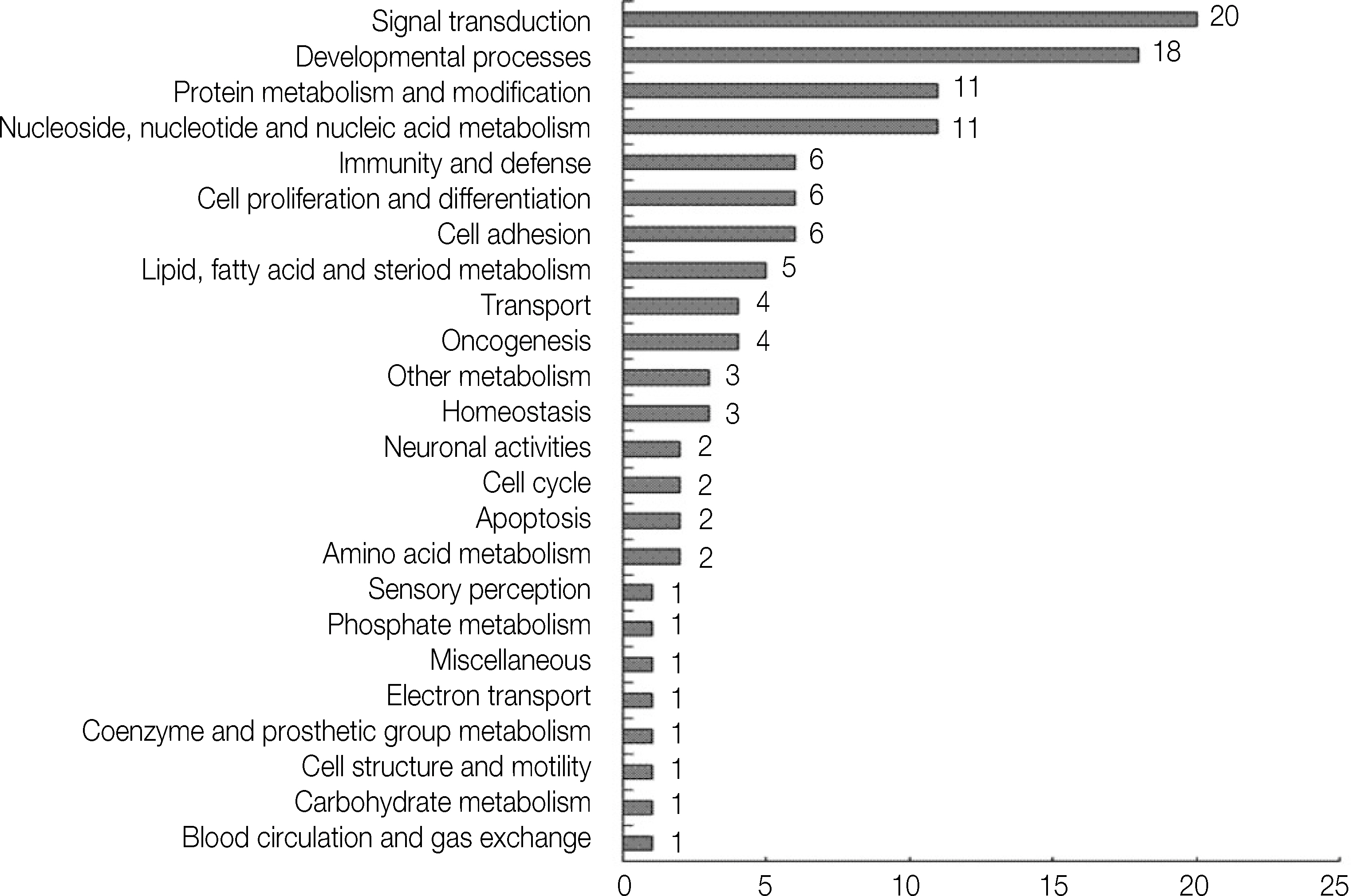
Figure 1. Classification of expressed genes by wMTA treatment. The genes which were up-or down-regulated by at least twofold following a cellular treatment with wMTA at any time point were classified using the panther classification system (http://www.pantherdb.org).
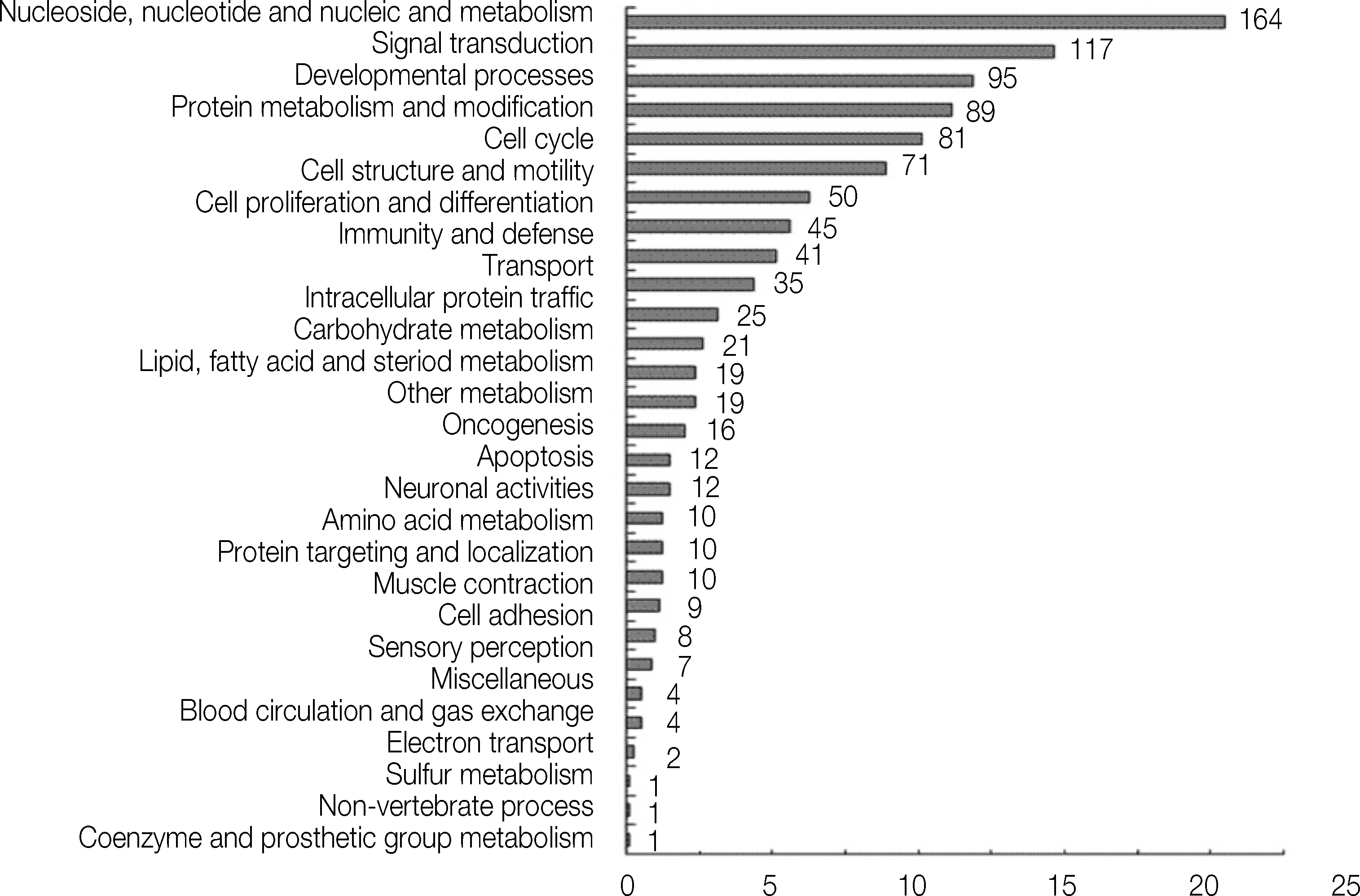
Figure 2. Classification of expressed genes by Dycal treatment. The genes which were up-or down-regulated by at least twofold following a cellular treatment with Dycal at any time point were classified using the panther classification system.
Cited by 1 articles
-
Analysis of gene expression during odontogenic differentiation of cultured human dental pulp cells
Min-Seock Seo, Kyung-Gyun Hwang, Hyongbum Kim, Seung-Ho Baek
Restor Dent Endod. 2012;37(3):142-148. doi: 10.5395/rde.2012.37.3.142.
Reference
-
References
1. American Association of Endodontists. Glossary of endodontic terms. 7th ed.p. pp40. 2003.2. Cox CF, Su¨bay RK, Ostro E, Suzuki S, Suzuki SH. Tunnel defects in dentin bridges: their formation following direct pulp capping. Oper Dent. 1996; 21:4–11.3. Cox CF, Hafez AA, Akimoto N, Otsuki M, Suzuki S, Tarim B. Biocompatibility of primer, adhesive and resin composite systems on non-exposed and exposed pulps of non-human primate teeth. Am J Dent. 1998; 11(Supplement):55–63.4. Cox CF, Tarim B, Kopel H, Gu¨rel G, Hafez A. Technique sensitivity: biological factors contributing to clinical success with various restorative materials. Adv Dent Res. 2001; 15:85–90.
Article5. Yun YR, Yang IS, Hwang YC, Hwang IN, Choi HR, Yoon SJ, Kim SH, Oh WM. Pulp response of mineral trioxide aggregate, calcium sulfate or calcium hydroxide. J Kor Acad Cons Dent. 2007; 32:95–101.
Article6. Bae JH, Kim YG, Yoon PY, Cho BH, Choi YH. Pulp response of beagle dog to direct pulp capping materials: histological study. J Kor Acad Cons Dent. 2010; 35:5–12.
Article7. Dominguez MS, Witherspoon DE, Gutmann JL, Opperman LA. Histological and scanning electron microscopy assessment of various vital pulp-therapy materials. J Endod. 2003; 29:324–333.
Article8. Ford TR, Torabinejad M, Abedi HR, Bakland LK, Kariyawasam SP. Using mineral trioxide aggregate as a pulp-capping material. J Am Dent Assoc. 1996; 127:1491–1494.
Article9. Faraco IM Jr, Holland R. Response of the pulp of dogs to capping with mineral trioxide aggregate or a calcium hydroxide cement. Dent Traumatol. 2001; 17:163–166.
Article10. Hauman CH, Love RM. Biocompatibility of dental materials used in contemporary endodontic therapy: a review. Part 2. Root canal-filling materials. Int Endod J. 2003; 36:147–160.11. Aeinehchi M, Eslami B, Ghanbariha M, Saffar AS. Mineral trioxide aggregate (MTA) and calcium hydroxide as pulp-capping agents in human teeth: a preliminary report. Int Endod J. 2003; 36:225–231.
Article12. Accorinte Mde L, Holland R, Reis A, Bortoluzzi MC, Murata SS, Dezan E Jr, Souza V, Alessandro LD. Evaluation of mineral trioxide aggregate and calcium hydroxide cement as pulp-capping agents in human teeth. J Endod. 2008; 34:1–6.13. Iwamoto CE, Adachi E, Pameijer CH, Barnes D, Romberg EE, Jeffries S. Clinical and histological evaluation of white ProRoot MTA in direct pulp capping. Am J Dent. 2006; 19:85–90.14. Yasuda Y, Ogawa M, Arakawa T, Kadowaki T, Saito T. The effect of mineral trioxide aggregate on the mineralization ability of rat dental pulp cells: an in vitro study. J Endod. 2008; 34:1057–1060.
Article15. Andelin WE, Shabahang S, Wright K, Torabinejad M. Identification of hard tissue after experimental pulp capping using dentin sialoprotein (DSP) as a marker. J Endod. 2003; 29:646–650.
Article16. Kuratate M, Yoshiba K, Shigetani Y, Yoshiba N, Ohshima H, Okiji T. Immunohistochemical analysis of nestin, osteopontin, and proliferating cells in the reparative process of exposed dental pulp capped with mineral trioxide aggregate. J Endod. 2008; 34:970–974.
Article17. Min KS, Yang SH, Kim EC. The combined effect of mineral trioxide aggregate and enamel matrix derivative on odontoblastic differentiation in human dental pulp cells. J Endod. 2009; 35:847–851.
Article18. Shi S, Robey PG, Gronthos S. Comparison of human dental pulp and bone marrow stromal stem cells by cDNA microarray analysis. Bone. 2001; 29:532–539.
Article19. McLachlan JL, Smith AJ, Bujalska IJ, Cooper PR. Gene expression profiling of pulpal tissue reveals the molecular complexity of dental caries. Biochim Biophys Acta. 2005; 1741:271–281.
Article20. Syudo M, Yamada S, Yanagiguchi K, Matsunaga T, Hayashi Y. Early gene expression analyzed by a genome microarray and real-time PCR in osteoblasts cultured with a 4-META/MMA-TBB adhesive resin sealer. Oral Surg Oral Med Oral Pathol Oral Radiol Endod. 2009; 107:e77–81.
Article21. Martinez ZR, Naruishi K, Yamashiro K, Myokai F, Yamada T, Matsuura K, Namba N, Arai H, Sasaki J, Abiko Y, Takashiba S. Gene profiles during root canal treatment in experimental rat periapical lesions. J Endod. 2007; 33:936–943.
Article22. Yokose S, Kadokura H, Tajima Y, Fujieda K, Katayama I, Matsuoka T, Katayama T. Establishment and characterization of a culture system for enzymatically released rat dental pulp cells. Calcif Tissue Int. 2000; 66:139–144.
Article23. Kim YB, Shon WJ, Lee WC, Kum KY, Baek SH, Bae KS. Gene Expression Profiling in Human Dental Pulp Cells Treated with Mineral Trioxide Aggregate. J Kor Acad Cons Dent. 35:152–163. 2010.
Article24. Goldberg M, Six N, Decup F, Lasfargues JJ, Salih E, Tompkins K, Veis A. Bioactive molecules and the future of pulp therapy. Am J Dent. 2003; 16:66–76.25. Almushayt A, Narayanan K, Zaki AE, George A. Dentin matrix protein 1 induces cytodifferentiation of dental pulp stem cells into odontoblasts. Gene Ther. 2006; 13:611–620.
Article26. Goldberg M, Farges JC, Lacerda-Pinheiro S, Six N, Jegat N, Decup F, Septier D, Carrouel F, Durand S, Chaussain-Miller C, Denbesten P, Veis A, Poliard A. Inflammatory and immunological aspects of dental pulp repair. Pharmacol Res. 2008; 58:137–147.
Article27. Reddi AH. Bone morphogenetic proteins: an unconventional approach to isolation of first mammalian mor-phogens. Cytokine Growth Factor Rev. 1997; 8:11–20.
Article28. Gu K, Smoke RH, Rutherford RB. Expression of genes for bone morphogenetic proteins and receptors in human dental pulp. Arch Oral Biol. 1996; 41:919–923.
Article29. Nakashima M, Nagasawa H, Yamada Y, Reddi AH. Regulatory role of transforming growth factor-β, bone morphogenetic protein-2, and protein-4 on gene expression of extracellular matrix proteins and differentiation of dental pulp cells. Dev Biol. 1994; 162:18–28.
Article30. Saito T, Ogawa M, Hata Y, Bessho K. Acceleration effect of human recombinant bone morphogenetic protein-2 on differentiation of human pulp cells into odon-toblasts. J Endod. 2004; 30:205–208.
Article31. Yasuda Y, Ogawa M, Arakawa T, Kadowaki T, Saito T. The effect of mineral trioxide aggregate on the mineralization ability of rat dental pulp cells: an in vitro study. J Endod. 2008; 34:1057–1060.
Article32. Wozney JM, Rosen V. Bone morphogenetic protein and bone morphogenetic protein gene family in bone formation and repair. Clin Orthop Relat Res. 1998; 346:26–37.
Article33. Takeda K, Oida S, Goseki M, Iimura T, Maruoka Y, Amagasa T, Sasaki S. Expression of bone morphogenetic protein genes in the human dental pulp cells. Bone. 1994; 15:467–470.
Article34. Zhang Q, Wang X, Chen Z, Liu G, Chen Z. Semi-quantitative RT-PCR analysis of LIM mineralization protein 1 and its associated molecules in cultured human dental pulp cells. Arch Oral Biol. 2007; 52:720–726.
Article35. Matsui S, Takeuchi H, Tsujimoto Y, Matsushima K. Effects of Smads and BMPs induced by Ga-Al-As laser irradiation on calcification ability of human dental pulp cells. J Oral Sci. 2008; 50:75–81.
Article36. Imamura T, Takase M, Nishihara A, Oeda E, Hanai J, Kawabata M, Miyazono K. Smad6 inhibits signaling by the TGF-βsuperfamily. Nature. 1997; 389:622–626.37. Lawler J. The functions of thrombospondin-1 and-2. Curr Opin Cell Biol. 2000; 12:634–640.
Article38. Murphy-Ullrich JE, Schultz-Cherry S, Ho¨o¨k M. Transforming growth factor-beta complexes with thrombospondin. Mol Biol Cell. 1992; 3:181–188.
Article39. Ueno A, Yamashita K, Nagata T, Tsurumi C, Miwa Y, Kitamura S, Inoue H. cDNA cloning of bovine thrombospondin 1 and its expression in odontoblasts and predentin. Biochim Biophys Acta. 1998; 1382:17–22.
Article40. Neuhaus SJ, Byers MR. Endothelin receptors and endothelin-1 in developing rat teeth. Arch Oral Biol. 2007; 52:655–662.
Article41. Yan Y, Liu Z, Zhang WG. In vitro study of the effects of endothelin-1 on human dental pulp cells. Chin J Dent Res. 1999; 2:5–13.42. Suga K, Saitoh M, Fukushima S, Takahashi K, Nara H, Yasuda S, Miyata K. Interleukin-11 induces osteoblast differentiation and acts synergistically with bone morphogenetic protein-2 in C3H10T1/2 cells. J Interferon Cytokine Res. 2001; 21:695–707.
Article43. Takeuchi Y, Watanabe S, Ishii G, Takeda S, Nakayama K, Fukumoto S, Kaneta Y, Inoue D, Matsumoto T, Harigaya K, Fujita T. Interleukin-11 as a stimulatory factor for bone formation prevents bone loss with advancing age in mice. J Biol Chem. 2002; 277:49011–49018.
Article44. Takayanaqi H, Kim S, Koqa T, Nishina H, Isshiki M, Yoshida H, Saiura A, Isobe M, Yokochi T, Inoue J, Wagner EF, Mak TW, Kodama T, Taniguchi T. Induction and activation of the transcription factor NFATc1 (NFAT2) integrate RANKL signaling in terminal differentiation of osteoclast. Dev Cell. 2002; 3:889–901.45. Grigoriadis AE, Wang ZQ, Cecchini MG, Hofstetter W, Felix R, Fleisch HA, Wagner EF. C-Fos: a key regulator of osteoclast-macrophage lineage determination and bone remodeling. Science. 1994; 266:443–448.
Article46. Grigoriadis AE, Schellander K, Wang ZQ, Wagner EF. Osteoblasts are target cells for transformation in c-fos transgenic mice. J Cell Biol. 1993; 122:685–701.
Article47. Gruda MC, van Amsterdam J, Rizzo CA, Durham SK, Lira S, Bravo R. Expression of FosB during mouse development: normal development of FosB knockout mice. Oncogene. 1996; 12:2177–2185.48. Smith AJ, Murray PE, Lumley PJ. Preserving the vital pulp in operative dentistry: 1. a biological approach. Dent Update. 2002; 29:64–69.
Article49. Keyt BA, Nguyen HV, Berleau LT, Duarte CM, Park J, Chen H, Ferrara N. Identification of vascular endothelial growth factor determinants for binding KDR and Flt-1 receptors: generation of receptorselective VEGF variants by site-directed mutagenesis. J Biol Chem. 1996; 271:5638–5646.50. Matsushita K, Motani R, Sakuta T, Yamaguchi N, Koga T, Matsuo K, Nagaoka S, Abeyama K, Maruyama I, Torii M. The role of vascular endothelial growth factor in human dental pulp cells: induction of chemotaxis, proliferation, and differentiation and activation of the AP-1-dependent signaling pathway. J Dent Res. 2000; 79:1596–1603.
Article51. Roberts-Clark DJ, Smith AJ. Angiogenic growth factors in human dentine matrix. Arch Oral Biol. 2000; 45:1013–1016.
Article52. Ribatti D. The crucial role of vascular permeability fac-tor/vascular endothelial growth factor in angiogenesis: a historical review. Br J Haematol. 2005; 128:303–309.
Article53. Heissig B, Hattori K, Friedrich M, Rafii S, Werb Z. Angiogenesis: vascular remodeling of the extracellular matrix involves metalloproteinases. Curr Opin Hematol. 2003; 10:136–141.
Article54. Seo DW, Li H, Guedez L, Wingfield PT, Diaz T, Salloum R, Wei BY, Stetler-Stevenson WG. TIMP-2 mediated inhibition of angiogenesis: an MMP-independent mechanism. Cell. 2003; 114:171–180.55. Murphy AN, Unsworth EJ, Stetler-Stevenson WG. Tissue inhibitor of metalloproteinases-2 inhibits bFGF-induced human microvascular endothelial cell proliferation. J Cell Physiol. 1993; 157:351–358.
Article56. Mochida Y, Duarte WR, Tanzawa H, Paschalis EP, Yamauchi M. Decorin modulates matrix mineralization in vitro. Biochem Biophys Res Commun. 2003; 305:6–9.
Article57. Alini M, Marriott A, Chen T, Abe S, Poole AR. A novel angiogenic molecule produced at the time of chondrocyte hypertrophy during endochondral bone formation. Dev Biol. 1996; 176:124–132.
Article58. Felszeghy S, Hollo′ K, Mo′dis L, Lammi MJ. Type X collagen in human enamel development: a possible role in mineralization. Acta Odontol Scand. 2000; 58:171–176.
Article59. Deutsch D, Palmon A, Fisher LW, Kolodny N, Termine JD, Young MF. Sequencing of bovine enamelin (“tuftelin”) a novel acidic enamel protein. J Biol Chem. 1991; 266:16021–10628.
Article60. Paine CT, Paine ML, Luo W, Okamoto CT, Lyngstadaas SP, Snead ML. A tuftelin-interacting protein (TIP39) localizes to the apical secretory pole of mouse ameloblasts. J Biol Chem. 2000; 275:22284–22292.
Article61. Otterbein LE, Choi AE. Heme oxygenase: colors of defense against cellular stress. Am J Physiol Lung Cell Mol Physiol. 2000; 279:L1029–1037.
Article62. Foresti R, Motterlini R. The heme oxygenase pathway and its interaction with nitric oxide in the control of cellular homeostasis. Free Radic Res. 1999; 31:459–475.
Article63. Min KS, Lee HJ, Kim SH, Lee SK, Kim HR, Pae HO, Chung HT, Shin HI, Lee SK, Kim EC. Hydrogen peroxide induces heme oxygenase-1 and dentin sialophos-phoprotein mRNA in human pulp cells. J Endod. 2008; 34:983–989.
Article64. Min KS, Kwon YY, Lee HJ, Lee SK, Kang KH, Lee SK, Kim EC. Effects of proinflammatory cytokines on the expression of mineralization markers and heme oxygenase-1 in human pulp cells. J Endod. 2006; 32:39–43.
Article65. Krebs DL, Hilton DJ. SOCS proteins: negative regulators of cytokine signaling. Stem Cells. 2001; 19:378–387.
Article66. Machado FS, Johndrow JE, Esper L, Dias A, Bafica A, Serhan CN, Aliberti J. Anti-inflammatory actions of lipoxin A(4) and aspirin-triggered lipoxin are SOCS-2 dependent. Nat Med. 2006; 12:330–334.
Article67. Alexander WS, Hilton DJ. The role of suppressors of cytokine signaling (SOCS) proteins in regulation of the immune response. Annu Rev Immunol. 2004; 22:503–529.
Article68. Menezes R, Garlet TP, Trombone AP, Repeke CE, Letra A, Granjeiro JM, Campanelli AP, Garlet GP. The potential role of suppressors of cytokine signaling in the attenuation of inflammatory reaction and alveolar bone loss associated with apical periodontitis. J Endod. 2008; 34:1480–1484.
Article69. Schrem H, Klempnauer J, Borlak J. Liver-enriched transcription factors in liver fuction and development. Part II: The C/EBPs and D site binding protein in cell cycle control, carcinogenesis, circadian gene regulation, liver regulation, apoptosis, and liver-specific gene regulation. Pharmacol Rev. 2004; 56:291–330.70. Liu YW, Chen CC, Wang JM, Chang WC, Huang YC, Chung SY, Chen BK, Hung JJ. Role of transcriptional factors Sp1, c-Rel, and c-Jun in LPS-induced C/EBPdelta gene expression of mouse macrophages. Cell Mol Life Sci. 2007; 64:3282–3294.71. Caivano M, Gorgoni B, Cohen P, Poli V. The induction of cyclooxygenage-2 mRNA in macrophages is biphasic and requires both CCAAT/enhancer-binding protein beta (C/EBP beta) and C/EBP delta transcription factors. J Biol Chem. 2001; 276:48693–48701.
- Full Text Links
-
- Actions
-
Cited
- CITED
-
- Close
- Share
-
- Similar articles
-
- Gene expression profiling in human dental pulp cells treated with mineral trioxide aggregate
- Chemical characteristics of mineral trioxide aggregate and its hydration reaction
- Use of mineral trioxide aggregate in the treatment of horizontal root fracture with a 4-year follow-up: case report
- Effect of Mineral Trioxide Aggregate and Calcium Hydroxide on Reparative Dentin Formation in Rats
- A preliminary report on histological outcome of pulpotomy with endodontic biomaterials vs calcium hydroxide
