Int J Stem Cells.
2016 May;9(1):44-52. 10.15283/ijsc.2016.9.1.44.
Comparative Evaluation for Potential Differentiation of Endothelial Progenitor Cells and Mesenchymal Stem Cells into Endothelial-Like Cells
- Affiliations
-
- 1Medical Biochemistry and Molecular Biology, Faculty of Medicine, Cairo University, Cairo, Egypt. dinasabry@kasralainy.edu.eg
- 2Obstetrics and Gynecology Department, Faculty of Medicine, Cairo University, Cairo, Egypt.
- KMID: 2164160
- DOI: http://doi.org/10.15283/ijsc.2016.9.1.44
Abstract
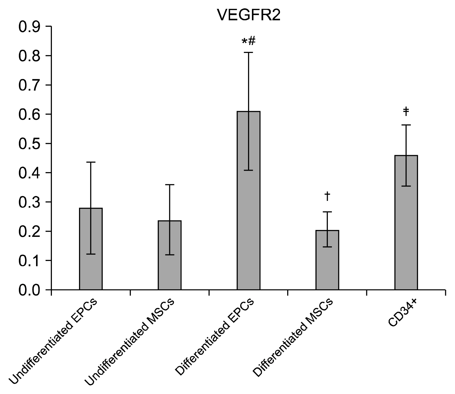
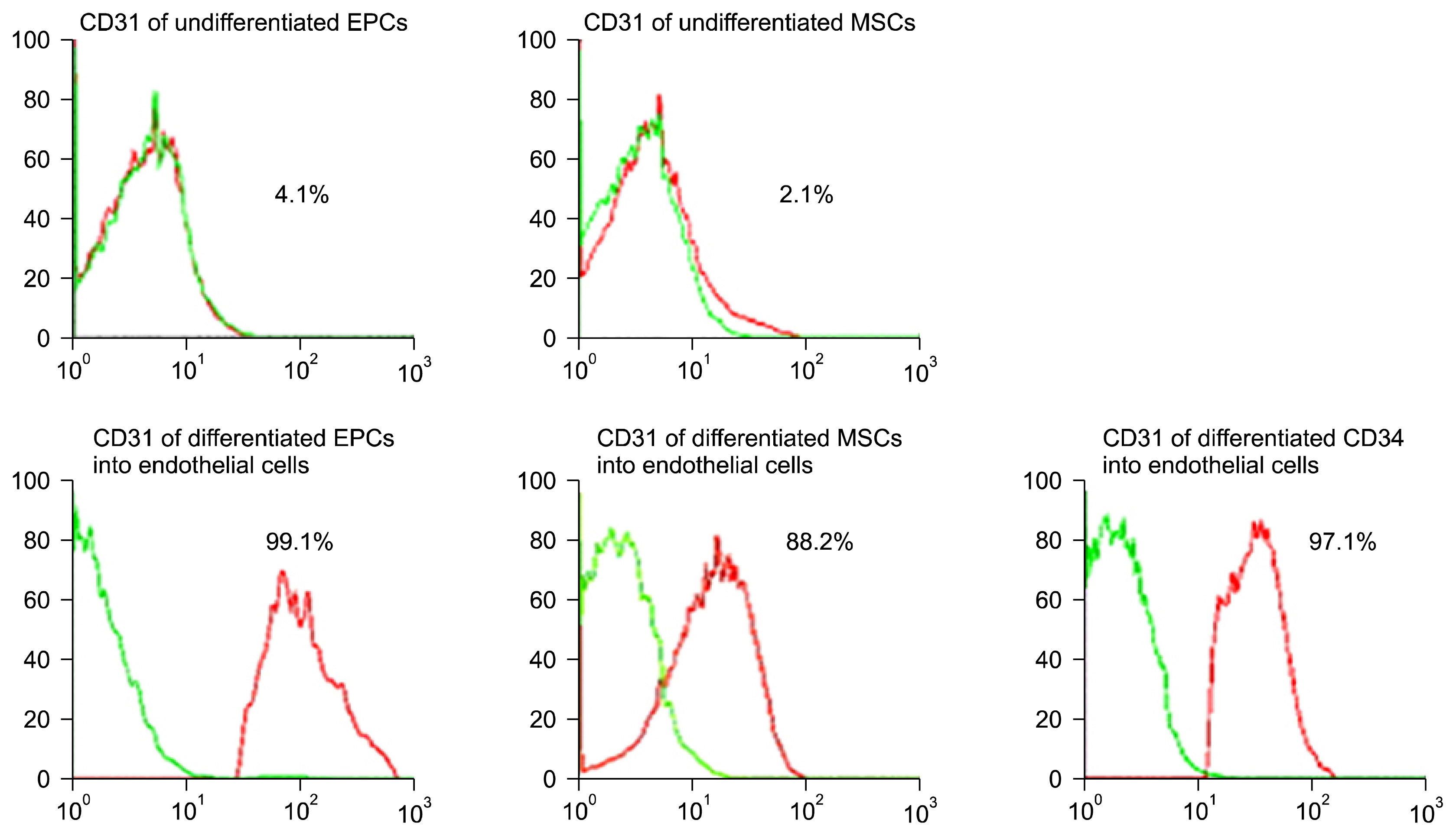
- Understanding the mechanisms of vascular remodeling could lead to more effective treatments for ischemic conditions. We aimed to compare between the abilities of both human Wharton jelly derived mesenchymal stem cells (hMSCs) and human cord blood endothelial progenitor cells (hEPCs) and CD34+ to induce angiogenesis in vitro. hMSCs, hEPCs, and CD34+ were isolated from human umbilical cord blood using microbead (MiniMacs). The cells characterization was assessed by flow cytometry following culture and real-time PCR for vascular endothelial growth factor receptor 2 (VEGFR2) and von Willebrand factor (vWF) to prove stem cells differentiation. The study revealed successful isolation of hEPCs, CD34+, and hMSCs. The hMSCs were identified by gaining CD29+ and CD44+ using FACS analysis. The hEPCs were identified by having CD133+, CD34+, and KDR. The potential ability of hEPCs and CD34+ to differentiate into endothelial-like cells was more than hMSCs. This finding was assessed morphologically in culture and by higher significant VEGFR2 and vWF genes expression (p<0.05) in differentiated hEPCs and CD34+ compared to differentiated hMSCs. hEPCs and CD34+ differentiation into endothelial-like cells were much better than that of hMSCs.
Keyword
MeSH Terms
Figure
Reference
-
References
1. Marti HJ, Bernaudin M, Bellail A, Schoch H, Euler M, Petit E, Risau W. Hypoxia-induced vascular endothelial growth factor expression precedes neovascularization after cerebral ischemia. Am J Pathol. 2000; 156:965–976. DOI: 10.1016/S0002-9440(10)64964-4. PMID: 10702412. PMCID: 1876841.
Article2. Li Y, Zhang D, Zhang Y, He G, Zhang F. Augmentation of neovascularization in murine hindlimb ischemia by combined therapy with simvastatin and bone marrow-derived mesenchymal stem cells transplantation. J Biomed Sci. 2010; 17:75. DOI: 10.1186/1423-0127-17-75. PMID: 20846454. PMCID: 2946286.
Article3. Ho TK, Shiwen X, Abraham D, Tsui J, Baker D. Stromal-cell-derived factor-1 (SDF-1)/CXCL12 as potential target of therapeutic angiogenesis in critical leg ischaemia. Cardiol Res Pract. 2012; 2012:143209. PMID: 22462026. PMCID: 3296148.
Article4. Liman TG, Endres M. New vessels after stroke: postischemic neovascularization and regeneration. Cerebrovasc Dis. 2012; 33:492–499. DOI: 10.1159/000337155. PMID: 22517438.
Article5. Aviles RJ, Annex BH, Lederman RJ. Testing clinical therapeutic angiogenesis using basic fibroblast growth factor (FGF-2). Br J Pharmacol. 2003; 140:637–646. DOI: 10.1038/sj.bjp.0705493. PMID: 14534147. PMCID: 1350957.
Article6. Awata T, Inoue K, Kurihara S, Ohkubo T, Watanabe M, Inukai K, Inoue I, Katayama S. A common polymorphism in the 5′-untranslated region of the VEGF gene is associated with diabetic retinopathy in type 2 diabetes. Diabetes. 2002; 51:1635–1639. DOI: 10.2337/diabetes.51.5.1635. PMID: 11978667.
Article7. Harris VK, Coticchia CM, Kagan BL, Ahmad S, Wellstein A, Riegel AT. Induction of the angiogenic modulator fibroblast growth factor-binding protein by epidermal growth factor is mediated through both MEK/ERK and p38 signal transduction pathways. J Biol Chem. 2000; 275:10802–10811. DOI: 10.1074/jbc.275.15.10802.
Article8. Schultz GS, Grant MB. Neovascular growth factors. Eye (Lond). 1991; 5:170–180. DOI: 10.1038/eye.1991.31.
Article9. Strasly M, Doronzo G, Cappello P, Valdembri D, Arese M, Mitola S, Moore P, Alessandri G, Giovarelli M, Bussolino F. CCL16 activates an angiogenic program in vascular endothelial cells. Blood. 2004; 103:40–49. DOI: 10.1182/blood-2003-05-1387.
Article10. Sameermahmood Z, Balasubramanyam M, Saravanan T, Rema M. Curcumin modulates SDF-1alpha/CXCR4-induced migration of human retinal endothelial cells (HRECs). Invest Ophthalmol Vis Sci. 2008; 49:3305–3311. DOI: 10.1167/iovs.07-0456. PMID: 18660423.
Article11. Weiss ML, Medicetty S, Bledsoe AR, Rachakatla RS, Choi M, Merchav S, Luo Y, Rao MS, Velagaleti G, Troyer D. Human umbilical cord matrix stem cells: preliminary characterization and effect of transplantation in a rodent model of Parkinson’s disease. Stem Cells. 2006; 24:781–792. DOI: 10.1634/stemcells.2005-0330.
Article12. Volpe G, Santodirocco M, Di Mauro L, Miscio G, Boscia FM, Muto B, Volpe N. Four phases of checks for exclusion of umbilical cord blood donors. Blood Transfus. 2011; 9:286–291. PMID: 21627927. PMCID: 3136596.13. Matsuo Y, Imanishi T, Hayashi Y, Tomobuchi Y, Kubo T, Hano T, Akasaka T. The effect of endothelial progenitor cells on the development of collateral formation in patients with coronary artery disease. Intern Med. 2008; 47:127–134. DOI: 10.2169/internalmedicine.47.0284. PMID: 18239320.
Article14. Anzalone R, Corrao S, Lo Iacono M, Loria T, Corsello T, Cappello F, Di Stefano A, Giannuzzi P, Zummo G, Farina F, La Rocca G. Isolation and characterization of CD276+/HLA-E+ human subendocardial mesenchymal stem cells from chronic heart failure patients: analysis of differentiative potential and immunomodulatory markers expression. Stem Cells Dev. 2013; 22:1–17. DOI: 10.1089/scd.2012.0402.
Article15. Goerke SM, Kiefer LS, Stark GB, Simunovic F, Finkenzeller G. miR-126 modulates angiogenic growth parameters of peripheral blood endothelial progenitor cells. Biol Chem. 2015; 396:245–252. DOI: 10.1515/hsz-2014-0259.
Article16. Abd El Aziz MT, Abd El Nabi EA, Abd El Hamid M, Sabry D, Atta HM, Rahed LA, Shamaa A, Mahfouz S, Taha FM, Elrefaay S, Gharib DM, Elsetohy KA. Endothelial progenitor cells regenerate infracted myocardium with neovascularisation development. J Adv Res. 2015; 6:133–144. DOI: 10.1016/j.jare.2013.12.006. PMID: 25750747. PMCID: 4348451.
Article17. Fons P, Gueguen-Dorbes G, Herault JP, Geronimi F, Tuyaret J, Frédérique D, Schaeffer P, Volle-Challier C, Herbert JM, Bono F. Tumor vasculature is regulated by FGF/FGFR signaling-mediated angiogenesis and bone marrow-derived cell recruitment: this mechanism is inhibited by SSR128129E, the first allosteric antagonist of FGFRs. J Cell Physiol. 2015; 230:43–51. DOI: 10.1002/jcp.24656.
Article18. Zhang C, Li Y, Wang J, Li K. The Supernatant Obtained from Cultured Anip973 Cells Enhances the Biological Activities of HUVEC. Zhongguo Fei Ai Za Zhi. 2015; 18:668–673. PMID: 26582221.19. Obi S, Masuda H, Akimaru H, Shizuno T, Yamamoto K, Ando J, Asahara T. Dextran induces differentiation of circulating endothelial progenitor cells. Physiol Rep. 2014; 2:e00261. DOI: 10.1002/phy2.261. PMID: 24760515. PMCID: 4002241.
Article
- Full Text Links
- Actions
-
Cited
- CITED
-
- Close
- Share
- Similar articles
-
- Endothelial progenitor cells and mesenchymal stem cells from human cord blood
- Adult Stem Cells: Beyond Regenerative Tool, More as a Bio-Marker in Obesity and Diabetes
- The Differentiation of Pluripotent Stem Cells towards Endothelial Progenitor Cells – Potential Application in Pulmonary Arterial Hypertension
- Differentiation of Osteoblast Progenitor Cells from Human Umbilical Cord Blood
- Therapeutic Angiogenesis with Somatic Stem Cell Transplantation


