Biocompatibility of two experimental scaffolds for regenerative endodontics
- Affiliations
-
- 1Faculty of Dentistry, National University of Singapore, and University Dental Cluster (Endodontics), National University Hospital Singapore, Singapore, Singapore. dephdep@gmail.com
- 2Department of Endodontics, School of Dental Medicine, University of Pennsylvania, Philadelphia, PA, USA.
- KMID: 2163291
- DOI: http://doi.org/10.5395/rde.2016.41.2.98
Abstract
OBJECTIVES
The biocompatibility of two experimental scaffolds for potential use in revascularization or pulp regeneration was evaluated.
MATERIALS AND METHODS
One resilient lyophilized collagen scaffold (COLL), releasing metronidazole and clindamycin, was compared to an experimental injectable poly(lactic-co-glycolic) acid scaffold (PLGA), releasing clindamycin. Human dental pulp stem cells (hDPSCs) were seeded at densities of 1.0 × 10(4), 2.5 × 10(4) and 5.0 × 10(4). The cells were investigated by light microscopy (cell morphology), MTT assay (cell proliferation) and a cytokine (IL-8) ELISA test (biocompatibility).
RESULTS
Under microscope, the morphology of cells coincubated for 7 days with the scaffolds appeared healthy with COLL. Cells in contact with PLGA showed signs of degeneration and apoptosis. MTT assay showed that at 5.0 × 10(4) hDPSCs, COLL demonstrated significantly higher cell proliferation rates than cells in media only (control, p < 0.01) or cells co-incubated with PLGA (p < 0.01). In ELISA test, no significant differences were observed between cells with media only and COLL at 1, 3, and 6 days. Cells incubated with PLGA expressed significantly higher IL-8 than the control at all time points (p < 0.01) and compared to COLL after 1 and 3 days (p < 0.01).
CONCLUSIONS
The COLL showed superior biocompatibility and thus may be suitable for endodontic regeneration purposes.
MeSH Terms
Figure
-

Figure 1 Comparison of representative cell appearances under the light microscope at ×100 and ×200. 'S' denotes scaffolds. (a - d) Cells grown in proximity to COLL scaffolds demonstrated a generally healthy appearance similar to other areas of the culture wells and controls (arrows 1); (e - f) Co-incubation with the PLGA scaffold often displayed signs of apoptosis and cell degeneration, particularly close to the actual material. Cell debris (arrow 2) and rounded cell morphologies (arrow 3) suggested non-attachment. COLL, lyophilized collagen; PLGA, poly-lactic-co-glycolic acid.

Figure 2 MTT assay. Absorbance measured using a multiplate reader. hDPSCs were seeded at 3 different densities (1.0 × 10⁴, 2.5 × 10⁴, and 5.0 × 10⁴) with media only (red), PLGA scaffold (blue) or COLL scaffold (green). Mean ± SEM for N = 6 per condition. *p < 0.05, by 2-way ANOVA and Bonferroni post hoc test. hDPSC, human dental pulp stem cells; PLGA, poly-lactic-co-glycolic acid; COLL, lyophilized collagen.
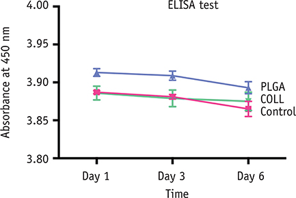
Figure 3 IL-8 ELISA test. Absorbance measured using a multiplate reader at 3 different time points (Days 1, 3, and 6). hDPSCs were seeded at 5.0 × 10⁴ cell density with media only (red), PLGA scaffold (blue) or COLL scaffold (green). Mean ± SEM for N = 6 per condition. *p < 0.05, by 2-way ANOVA and Bonferroni post hoc test. hDPSC, human dental pulp stem cells; PLGA, poly-lactic-coglycolic acid; COLL, lyophilized collagen.
Reference
-
1. de Chevigny C, Dao TT, Basrani BR, Marquis V, Farzaneh M, Abitbol S, Friedman S. Treatment outcome in endodontics: the Toronto study-phase 4: initial treatment. J Endod. 2008; 34:258–263.
Article2. Ricucci D, Russo J, Rutberg M, Burleson JA, Spångberg LS. A prospective cohort study of endodontic treatments of 1,369 root canals: results after 5 years. Oral Surg Oral Med Oral Pathol Oral Radiol Endod. 2011; 112:825–842.
Article3. Nirmalanandhan VS, Levy MS, Huth AJ, Butler DL. Effects of cell seeding density and collagen concentration on contraction kinetics of mesenchymal stem cell-seeded collagen constructs. Tissue Eng. 2006; 12:1865–1872.
Article4. Nirmalanandhan VS, Rao M, Sacks MS, Haridas B, Butler DL. Effect of length of the engineered tendon construct on its structure-function relationships in culture. J Biomech. 2007; 40:2523–2529.
Article5. Chandrahasa S, Murray PE, Namerow KN. Proliferation of mature ex vivo human dental pulp using tissue engineering scaffolds. J Endod. 2011; 37:1236–1239.
Article6. Huang GT, Sonoyama W, Liu Y, Liu H, Wang S, Shi S. The hidden treasure in apical papilla: the potential role in pulp/dentin regeneration and bioroot engineering. J Endod. 2008; 34:645–651.
Article7. Zhang W, Walboomers XF, Jansen JA. The formation of tertiary dentin after pulp capping with a calcium phosphate cement, loaded with PLGA microparticles containing TGF-beta1. J Biomed Mater Res A. 2008; 85:439–444.8. Murray PE, Garcia-Godoy F, Hargreaves KM. Regenerative endodontics: a review of current status and a call for action. J Endod. 2007; 33:377–390.
Article9. Sachlos E, Czernuszka JT. Making tissue engineering scaffolds work. Review: the application of solid freeform fabrication technology to the production of tissue engineering scaffolds. Eur Cell Mater. 2003; 5:29–39.
Article10. Inuyama Y, Kitamura C, Nishihara T, Morotomi T, Nagayoshi M, Tabata Y, Matsuo K, Chen KK, Terashita M. Effects of hyaluronic acid sponge as a scaffold on odontoblastic cell line and amputated dental pulp. J Biomed Mater Res B Appl Biomater. 2010; 92:120–128.
Article11. Zhang W, Walboomers XF, van Kuppevelt TH, Daamen WF, Bian Z, Jansen JA. The performance of human dental pulp stem cells on different three-dimensional scaffold materials. Biomaterials. 2006; 27:5658–5668.
Article12. Iohara K, Zheng L, Ito M, Ishizaka R, Nakamura H, Into T, Matsushita K, Nakashima M. Regeneration of dental pulp after pulpotomy by transplantation of CD31(-)/ CD146(-) side population cells from a canine tooth. Regen Med. 2009; 4:377–385.
Article13. Iohara K, Murakami M, Takeuchi N, Osako Y, Ito M, Ishizaka R, Utunomiya S, Nakamura H, Matsushita K, Nakashima M. A novel combinatorial therapy with pulp stem cells and granulocyte colony-stimulating factor for total pulp regeneration. Stem Cells Transl Med. 2013; 2:521–533.
Article14. Kodonas K, Gogos C, Papadimitriou S, Kouzi-Koliakou K, Tziafas D. Experimental formation of dentinlike structure in the root canal implant model using cryopreserved swine dental pulp progenitor cells. J Endod. 2012; 38:913–919.
Article15. El-Backly RM, Massoud AG, El-Badry AM, Sherif RA, Marei MK. Regeneration of dentine/pulp-like tissue using a dental pulp stem cell/poly(lactic-co-glycolic) acid scaffold construct in New Zealand white rabbits. Aust Endod J. 2008; 34:52–67.
Article16. Pagonis TC, Chen J, Fontana CR, Devalapally H, Ruggiero K, Song X, Foschi F, Dunham J, Skobe Z, Yamazaki H, Kent R, Tanner AC, Amiji MM, Soukos NS. Nanoparticlebased endodontic antimicrobial photodynamic therapy. J Endod. 2010; 36:322–328.
Article17. Cheng CF, Lee YY, Chi LY, Chen YT, Hung SL, Ling LJ. Bacterial penetration through antibiotic-loaded guided tissue regeneration membranes. J Periodontol. 2009; 80:1471–1478.
Article18. Bottino MC, Kamocki K, Yassen GH, Platt JA, Vail MM, Ehrlich Y, Spolnik KJ, Gregory RL. Bioactive nanofibrous scaffolds for regenerative endodontics. J Dent Res. 2013; 92:963–969.
Article19. Albuquerque MT, Valera MC, Moreira CS, Bresciani E, de Melo RM, Bottino MC. Effects of ciprofloxacincontaining scaffolds on enterococcus faecalis biofilms. J Endod. 2015; 41:710–714.
Article20. Kamocki K, Nör JE, Bottino MC. Effects of ciprofloxacincontaining antimicrobial scaffolds on dental pulp stem cell viability-in vitro studies. Arch Oral Biol. 2015; 60:1131–1137.
Article21. Huang GT, Al-Habib M, Gauthier P. Challenges of stem cell-based pulp and dentin regeneration: a clinical perspective. Endod Topics. 2013; 28:51–60.
Article22. Grossman LI. Polyantibiotic treatment of pulpless teeth. J Am Dent Assoc. 1951; 43:265–278.
Article23. Sato T, Hoshino E, Uematsu H, Noda T. In vitro antimicrobial susceptibility to combinations of drugs on bacteria from carious and endodontic lesions of human deciduous teeth. Oral Microbiol Immunol. 1993; 8:172–176.
Article24. Hoshino E, Kurihara-Ando N, Sato I, Uematsu H, Sato M, Kota K, Iwaku M. In-vitro antibacterial susceptibility of bacteria taken from infected root dentine to a mixture of ciprofloxacin, metronidazole and minocycline. Int Endod J. 1996; 29:125–130.
Article25. Iwaya SI, Ikawa M, Kubota M. Revascularization of an immature permanent tooth with apical periodontitis and sinus tract. Dent Traumatol. 2001; 17:185–187.
Article26. Banchs F, Trope M. Revascularization of immature permanent teeth with apical periodontitis: new treatment protocol. J Endod. 2004; 30:196–200.
Article27. Windley W 3rd, Teixeira F, Levin L, Sigurdsson A, Trope M. Disinfection of immature teeth with a triple antibiotic paste. J Endod. 2005; 31:439–443.
Article28. Dabbagh B, Alvaro E, Vu DD, Rizkallah J, Schwartz S. Clinical complications in the revascularization of immature necrotic permanent teeth. Pediatr Dent. 2012; 34:414–417.29. Bezgin T, Yılmaz AD, Celik BN, Sönmez H. Concentrated platelet-rich plasma used in root canal revascularization: 2 case reports. Int Endod J. 2014; 47:41–49.
Article30. Moioli EK, Clark PA, Xin X, Lal S, Mao JJ. Matrices and scaffolds for drug delivery in dental, oral and craniofacial tissue engineering. Adv Drug Deliv Rev. 2007; 59:308–324.
Article31. Enkel B, Dupas C, Armengol V, Akpe Adou J, Bosco J, Daculsi G, Jean A, Laboux O, LeGeros RZ, Weiss P. Bioactive materials in endodontics. Expert Rev Med Devices. 2008; 5:475–494.
Article32. Kitamura C, Nishihara T, Terashita M, Tabata Y, Washio A. Local regeneration of dentin-pulp complex using controlled release of fgf-2 and naturally derived spongelike scaffolds. Int J Dent. 2012; 2012:190561.
Article33. Yang X, Han G, Pang X, Fan M. Chitosan/collagen scaffold containing bone morphogenetic protein-7 DNA supports dental pulp stem cell differentiation in vitro and in vivo. J Biomed Mater Res A. 2012; 02. 18. [Epub ahead of print]. DOI: 10.1002/jbm.a.34064.34. Gilad JZ, Teles R, Goodson M, White RR, Stashenko P. Development of a clindamycin-impregnated fiber as an intracanal medication in endodontic therapy. J Endod. 1999; 25:722–727.
Article35. Molander A, Dahlén G. Evaluation of the antibacterial potential of tetracycline or erythromycin mixed with calcium hydroxide as intracanal dressing against Enterococcus faecalis in vivo. Oral Surg Oral Med Oral Pathol Oral Radiol Endod. 2003; 96:744–750.
Article36. Lin S, Levin L, Peled M, Weiss EI, Fuss Z. Reduction of viable bacteria in dentinal tubules treated with clindamycin or tetracycline. Oral Surg Oral Med Oral Pathol Oral Radiol Endod. 2003; 96:751–756.
Article37. Ruparel NB, Teixeira FB, Ferraz CC, Diogenes A. Direct effect of intracanal medicaments on survival of stem cells of the apical papilla. J Endod. 2012; 38:1372–1375.
Article38. Chuensombat S, Khemaleelakul S, Chattipakorn S, Srisuwan T. Cytotoxic effects and antibacterial efficacy of a 3-antibiotic combination: an in vitro study. J Endod. 2013; 39:813–819.
Article39. Soares Ade J, Lins FF, Nagata JY, Gomes BP, Zaia AA, Ferraz CC, de Almeida JF, de Souza-Filho FJ. Pulp revascularization after root canal decontamination with calcium hydroxide and 2% chlorhexidine gel. J Endod. 2013; 39:417–420.
Article40. Andreasen JO, Farik B, Munksgaard EC. Long-term calcium hydroxide as a root canal dressing may increase risk of root fracture. Dent Traumatol. 2002; 18:134–137.
Article
- Full Text Links
-
- Actions
-
Cited
- CITED
-
- Close
- Share
-
- Similar articles
-
- A novel antimicrobial-containing nanocellulose scaffold for regenerative endodontics
- Immunomodulatory Effects of Adipose Tissue-Derived Stem Cells on Elastin Scaffold Remodeling in Diabetes
- Chitosan-Poly(Vinyl Alcohol) Nanofibers by Free Surface Electrospinning for Tissue Engineering Applications
- Biocompatibility of root-end filling materials: recent update
- Microfluidic Spinning of the Fibrous Alginate Scaffolds for Modulation of the Degradation Profile
