J Korean Ophthalmol Soc.
2019 Dec;60(12):1205-1215. 10.3341/jkos.2019.60.12.1205.
Electroretinography Changes in Feline Model of Iodoacetic Acid-induced Retinal Degeneration
- Affiliations
-
- 1Department of Ophthalmology, Kosin University College of Medicine, Busan, Korea. hhiatus@gmail.com
- 2Department of Ophthalmology, Inje University College of Medicine, Busan, Korea.
- KMID: 2466172
- DOI: http://doi.org/10.3341/jkos.2019.60.12.1205
Abstract
- PURPOSE
We explored changes in photoreceptor function and histology in an iodoacetic acid (IAA)-induced model of feline retinal degeneration.
METHODS
From January to October 2014, we studied 11 adult felines (22 eyes) over 2 years of age divided into two groups (two in a control and nine in an IAA group). The mean body weights of these two groups were 1.75 ± 0.35 and 1.61 ± 0.19 kg, and the male:female sex ratios 1:1 and 2:7, respectively. Electroretinograms (ERGs) were obtained before injection and at 1-4 week post-injection (20 mg/kg IAA). Standard paraffin retinal sections were stained with hematoxylin/eosin and other sections subjected to immunohistochemistry. We histologically evaluated the outer nuclear layer, and photoreceptor cone and rod cells.
RESULTS
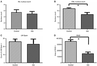
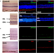
In ERGs of the IAA group, both the rod and cone mean b wave amplitudes decreased significantly from week 1 to week 4 after injection (27.43, 29.41, 64.17, and 56.03; and 61.04, 51.25, 131.36, and 136.68 µV, respectively) compared to baseline (322.48 and 610.00 µV respectively) (p < 0.01). Optical microscopy revealed a significant decrease in the cell count of the outer nuclear retinal layer (16.83 ± 0.89 in the control and 11.98 ± 3.55 in the IAA groups, p < 0.01). Fluorescence microscopy revealed a significant reduction in the mean area per unit length of the rod cell layer (35,225.67 ± 2,477.02 and 14,903.62 ± 2,319.65 in the control and IAA groups, p < 0.01), but not in the cone cell count (26.16 ± 1.34 and 23.98 ± 6.16 in the control and IAA groups, p = 0.075).
CONCLUSIONS
ERGs revealed that functional b wave amplitudes fell after IAA-induce retinal degeneration in felines; histology showed that this was accompanied by reductions in the numbers of outer nuclear layers and rod cells. IAA induces photoreceptor degeneration in felines; further study is necessary.
MeSH Terms
Figure
Reference
-
1. Narayan DS, Chidlow G, Wood JP, Casson RJ. Glucose metabolism in mammalian photoreceptor inner and outer segments. Clin Exp Ophthalmol. 2017; 45:730–741.2. Borsch O, Santos-Ferreira T, Ader M. Photoreceptor transplantation into the degenerative retina. Klin Monbl Augenheilkd. 2017; 234:343–353.3. Berson EL. Retinitis pigmentosa. The Friedenwald Lecture. Invest Ophthalmol Vis Sci. 1993; 34:1659–1676.4. Hartong DT, Berson EL, Dryja TP. Retinitis pigmentosa. Lancet. 2006; 368:1795–1809.5. Birch DG, Anderson JL, Fish GE. Yearly rates of rod and cone functional loss in retinitis pigmentosa and cone-rod dystrophy. Ophthalmology. 1999; 106:258–268.6. Clarke G, Collins RA, Leavitt BR, et al. A one-hit model of cell death in inherited neuronal degenerations. Nature. 2000; 406:195–199.7. Cheng YP, Ke CY, Kuo CC, Lee YJ. Effect of a complex lutein formula in an animal model for light-induced retinal degeneration. Chin J Physiol. 2016; 59:202–209.8. Taylor L, Arnér K, Ghosh F. N-methyl-N-nitrosourea-induced neuronal cell death in a large animal model of retinal degeneration in vitro. Exp Eye Res. 2016; 148:55–64.9. Nagar S, Krishnamoorthy V, Cherukuri P, et al. Early remodeling in an inducible animal model of retinal degeneration. Neuroscience. 2009; 160:517–529.10. Yamauchi Y, Agawa T, Tsukahara R, et al. Correlation between high-resolution optical coherence tomography (OCT) images and histopathology in an iodoacetic acid-induced model of retinal degeneration in rabbits. Br J Ophthalmol. 2011; 95:1157–1160.11. Rapaport DH, Stone J. The area centralis of the retina in the cat and other mammals: focal point for function and development of the visual system. Neuroscience. 1984; 11:289–301.12. Chang B. Mouse models for studies of retinal degeneration and diseases. Methods Mol Biol. 2013; 935:27–39.13. Ehrenberg M, Ehrenberg S, Schwob O, Benny O. Murine fundus fluorescein angiography: an alternative approach using a handheld camera. Exp Eye Res. 2016; 148:74–78.14. Nan Y, Zhang Q, Ren C, et al. Functional evaluation of iodoacetic acid induced photoreceptor degeneration in the cat. Sci China Life Sci. 2013; 56:524–530.15. Scott PA, Kaplan HJ, Sandell JH. Anatomical evidence of photoreceptor degeneration induced by iodoacetic acid in the porcine eye. Exp Eye Res. 2011; 93:513–527.16. Wang W, Fernandez De Castro J, Vukmanic E, et al. Selective rod degeneration and partial cone inactivation characterize an iodoacetic acid model of Swine retinal degeneration. Invest Ophthalmol Vis Sci. 2011; 52:7917–7923.17. Noel JM, Fernandez De, Demarco PJ Jr, et al. Iodoacetic acid, but not sodium iodate, creates an inducible swine model of photoreceptor damage. Exp Eye Res. 2012; 97:137–147.18. Wang Y, Zhao L, Lu F, et al. A pathologic study of degeneration of the rod and cone populations of the rhodopsin Pro347Leu transgenic pigs. Trans Am Ophthalmol Soc. 1997; 95:467–479. discussion 479–83.19. Aplin FP, Luu CD, Vessey KA, et al. ATP-induced photoreceptor death in a feline model of retinal degeneration. Invest Ophthalmol Vis Sci. 2014; 55:8319–8329.20. Cibis PA, Noell WK. Cataract induced by iodoacetic acid; a preliminary report. Am J Ophthalmol. 1955; 40:379–382.21. Orzalesi N, Calabria GA, Grignolo A. Experimental degeneration of the rabbit retina induced by iodoacetic acid. A study of the ultrastructure, the rhodopsin cycle and the uptake of 14C-labeled iodoacetic acid. Exp Eye Res. 1970; 9:246–253.22. Toda N. Calcium independent contraction induced by iodoacetic acid in isolated cerebral arteries. Stroke. 1984; 15:475–480.23. Suna H, Arai M, Tsubotani Y, et al. Dysideamine, a new sesquiterpene aminoquinone, protects hippocampal neuronal cells against iodoacetic acid-induced cell death. Bioorg Med Chem. 2009; 17:3968–3972.24. Zhou X, Zhu L, Wang L, et al. Protective effect of edaravone in primary cerebellar granule neurons against iodoacetic acid-induced cell injury. Oxid Med Cell Longev. 2015; 2015:606981.25. Winkler BS, Sauer MW, Starnes CA. Modulation of the pasteur effect in retinal cells: implications for understanding compensatory metabolic mechanisms. Exp Eye Res. 2003; 76:715–723.26. Noell WK. Experimentally induced toxic effects on structure and function of visual cells and pigment epithelium. Am J Ophthalmol. 1953; 36:103–116.27. Ofri R, Reilly CM, Maggs DJ, et al. Characterization of an early-onset, autosomal recessive, progressive retinal degeneration in Bengal cats. Invest Ophthalmol Vis Sci. 2015; 56:5299–5308.28. Noell WK. The effect of iodoacetate on the vertebrate retina. J Cell Comp Physiol. 1951; 37:283–307.29. Noell WK. The impairment of visual cell structure by iodoacetate. J Cell Comp Physiol. 1952; 40:25–55.30. Burke W, Hayhow WR. Disuse in the lateral geniculate nucleus of the cat. J Physiol. 1968; 194:495–519.31. Graymore C, Tansley K. Iodoacetate poisoning of the rat retina. I. Production of retinal degeneration. Br J Ophthalmol. 1959; 43:177–185.32. Graymore C, Tansley K. Iodoacetate poisoning of the rat retina. Ii. Glycolysis in the poisoned retina. Br J Ophthalmol. 1959; 43:486–493.33. Rösch S, Johnen S, Mazinani B, et al. The effects of iodoacetic acid on the mouse retina. Graefes Arch Clin Exp Ophthalmol. 2015; 253:25–35.34. Pennesi ME, Michaels KV, Magee SS, et al. Long-term characterization of retinal degeneration in rd1 and rd10 mice using spectral domain optical coherence tomography. Invest Ophthalmol Vis Sci. 2012; 53:4644–4656.35. Berson EL, Rosner B, Sandberg MA, et al. A randomized trial of vitamin A and vitamin E supplementation for retinitis pigmentosa. Arch Ophthalmol. 1993; 111:761–772.36. Liang L, Katagiri Y, Franco LM, et al. Long-term cellular and regional specificity of the photoreceptor toxin, iodoacetic acid (Iaa), in the rabbit retina. Vis Neurosci. 2008; 25:167–177.37. Alexander KR, Barnes CS, Fishman GA. ON-pathway dysfunctionand timing properties of the flicker ERG in carriers of X-linked retinitis pigmentosa. Invest Ophthalmol Vis Sci. 2003; 44:4017–4025.
- Full Text Links
- Actions
-
Cited
- CITED
-
- Close
- Share
- Similar articles
-
- Analysis of Changes in Retinal Photoreceptors Using Optical Coherence Tomography in a Feline Model of Iodoacetic Acid-induced Retinal Degeneration
- Chlorogenic Acid Supplementation Improves Multifocal Electroretinography in Patients with Retinitis Pigmentosa
- Assessment of retinal degeneration with optical coherence tomography in a dog
- Morphological and functional evaluation of an animal model for the retinal degeneration induced by N-methyl-N-nitrosourea
- Clinical Applications of Multifocal Electroretinography (mfERG)


