Effects of Human Placental Amnion Derived Mesenchymal Stem Cells on Proliferation and Apoptosis Mechanisms in Chronic Kidney Disease in the Rat
- Affiliations
-
- 1Department of Histology and Embryology, Medical Faculty, Akdeniz University, Antalya, Turkey. korgun@akdeniz.edu.tr
- 2Department of Medical Biochemistry, Medical Faculty, Akdeniz University, Antalya, Turkey.
- 3Department of Medical Microbiology and Immunology, Medical Faculty, Akdeniz University, Antalya, Turkey.
- 4Department of Histology and Embryology, Medical Faculty, Bulent Ecevit University, Zonguldak, Turkey.
- KMID: 2447233
- DOI: http://doi.org/10.15283/ijsc18067
Abstract
- BACKGROUND AND OBJECTIVES
The feature of chronic kidney failure (CKF) is loss of kidney functions due to erosion of healthy tissue and fibrosis. Recent studies showed that Mesenchymal stem cells (MSCs) differentiated into tubular epithelial cells thus renal function and structures renewed. Furthermore, MSCs protect renal function in CKF. Therefore, we aimed to investigate whether human amnion-derived mesenchymal stem cells (hAMSCs) can repair fibrosis and determine the effects on proliferation and apoptosis mechanisms in chronic kidney failure.
METHODS AND RESULTS
In this study, rat model of CKF was constituted by applying Aristolochic acid (AA). hAMSCs were isolated from term placenta amnion membrane and transplanted into tail vein of rats. At the end of 30 days and 60 days of recovery period, we examined expressions of PCNA, p57 and Parp-1 by western blotting. Immunoreactivity of PCNA, Ki67, IL-6 and Collagen type I were detected by immunohistochemistry. Besides, apoptosis was detected by TUNEL. Serum creatinine and urea were measured. Expressions of PCNA and Ki67 increased in hAMSC groups compared with AA group. Furthermore, expressions of PARP-1 apoptosis marker and p57 cell cycle inhibitory protein increased in AA group significantly according to control, hAMSC groups and sham groups. IL-6 proinflammatory cytokine increased in AA group significantly according to control, hAMSCs groups and sham groups. Expressions of Collagen type I protein reduced in hAMSCs groups compared to AA group. After hAMSC treatment, serum creatinine and urea levels significantly decreased compared to AA group. After injection of hAMSC to rats, Masson's Trichrome and Sirius Red staining showed fibrosis reduction in kidney.
CONCLUSIONS
According to our results hAMSCs can be ameliorate renal failure.
Keyword
MeSH Terms
-
Amnion*
Animals
Apoptosis*
Blotting, Western
Cell Cycle
Collagen Type I
Creatinine
Epithelial Cells
Fibrosis
Humans*
Immunohistochemistry
In Situ Nick-End Labeling
Interleukin-6
Kidney
Kidney Failure, Chronic
Membranes
Mesenchymal Stromal Cells*
Models, Animal
Placenta
Proliferating Cell Nuclear Antigen
Rats*
Renal Insufficiency
Renal Insufficiency, Chronic*
Tail
Urea
Veins
Collagen Type I
Creatinine
Interleukin-6
Proliferating Cell Nuclear Antigen
Urea
Figure
-

Fig. 1 hAMSC characterization. Flow cytometry analysis of hAMSCs for CD90 (A, B), CD105 (C, D), CD73 (E, F), CD44 (G, H) and negative cocktail (I, J) with isotype controls.

Fig. 2 Phase contrast microscopic images of human amniotic membrane derived mesenchymal stromal cells in 4X (A) and 40X magnifications (B). hAMSCs were differentiated into adipocytes (C., as shown by Oil Red O staining), osteocytes (D., as shown by Alizarin Red S staining) and chondrocytes (E and F, as shown by Alcian Blue and Sirius Red staining).

Fig. 3 Masson’s Trichrome was performed to determine fibrosis in sham+30 (A), AA (B), AA+hAMSC +30 (C) and AA+hAMSC+60 (D) groups. Sirius Red staining was showed sham+30 (E), AA (F), AA+ hAMSC+30 (G) and AA+hAMSC +60 (H) groups respectively.
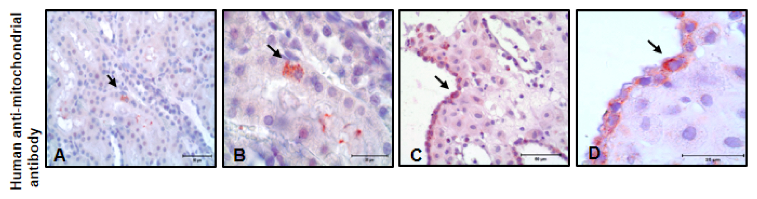
Fig. 4 At 30 day hAMSCs tracking in kidney tissue with immunolabeling of human anti-mitochondrial antibody (A, ×40, B, ×100). Positive control is human decidua tissue (C, ×40, D, ×100).
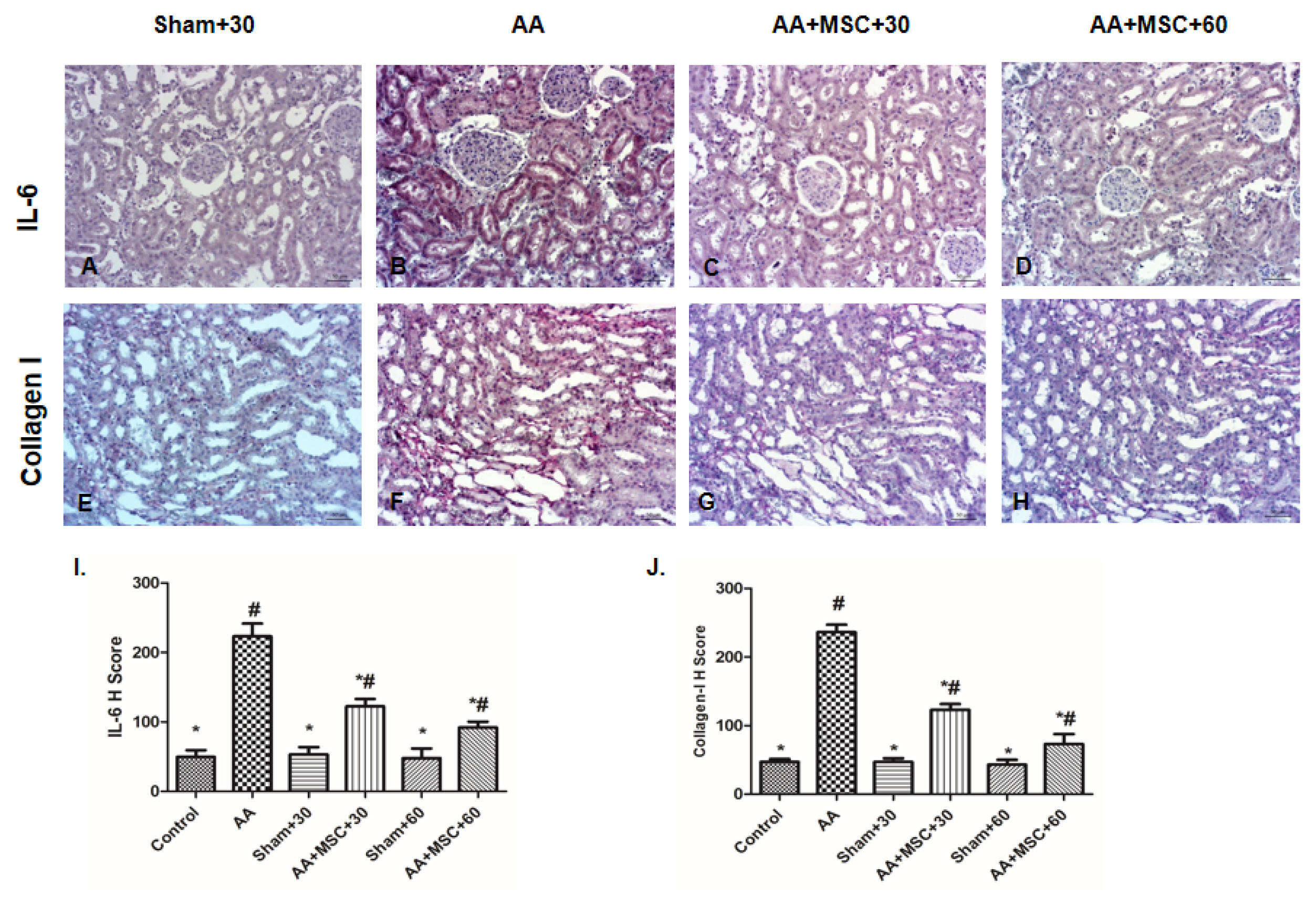
Fig. 5 Immunohistochemical staining of kidney tissue sections for the sham+30, AA, AA+hAMSC+30 and AA+hAMSC+60 groups using IL-6 (A–D), Collagen-I (E–H) I and J, H scores of IL-6 and Collagen-I. Values presented as mean ± SD. AA group compared with Sham, Control, AA+hAMSC+30, and AA+hAMSC +60 groups. Values statistically significant at: *p<0.05, Sham and Control groups compared with AA and hAMSC groups; #p<0.05.
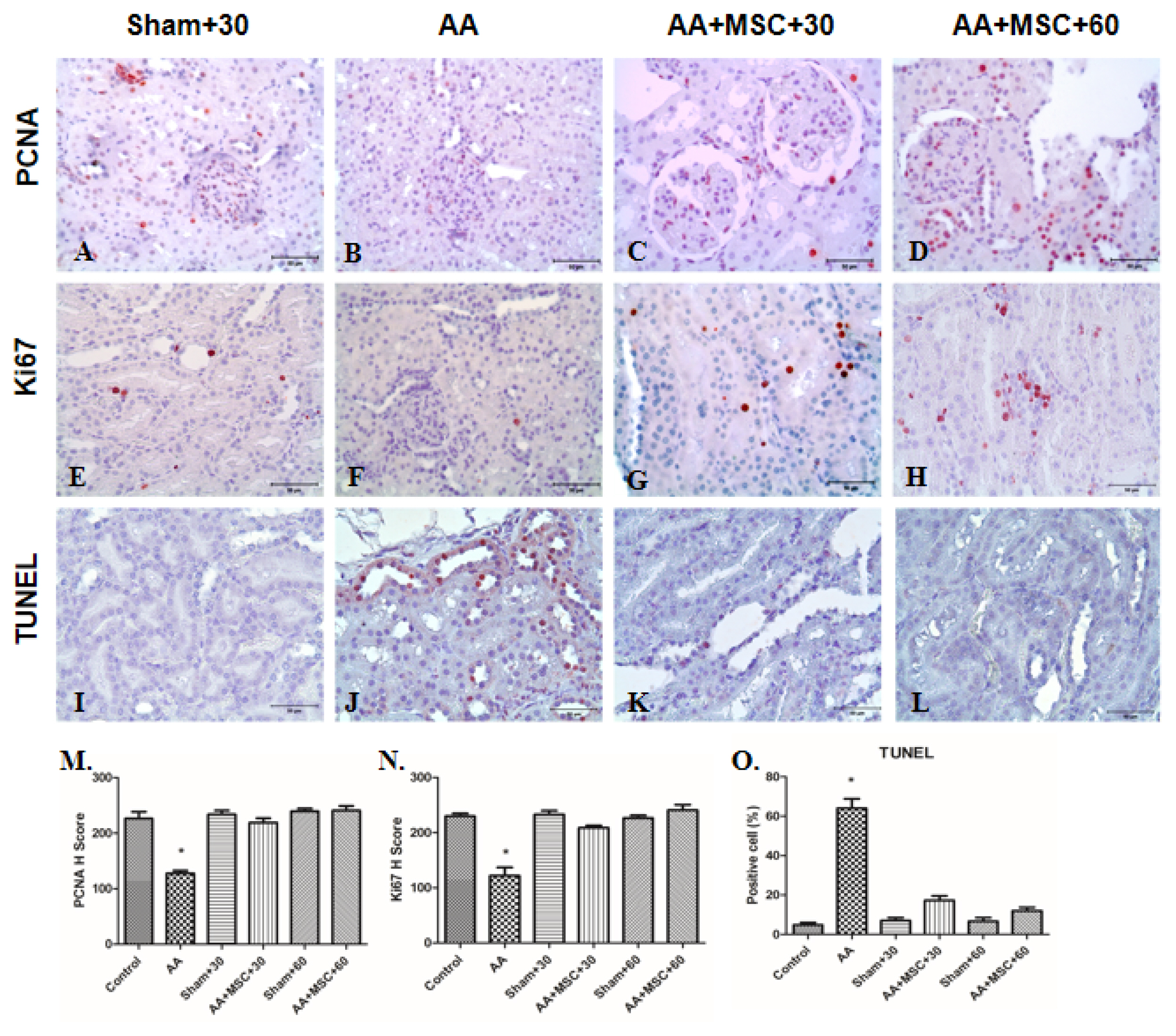
Fig. 6 PCNA, Ki67 and TUNEL immunostaining in kidneys tissue. For PCNA, sham+30 (A), AA (B), AA+ hAMSC+30 (C) and AA+hAMSC+ 60 (D) groups, for Ki67 sham+30 (E), AA (F), AA+hAMSC+30 (G) and AA+hAMSC+60 groups (H) and for TUNEL immunostaining sham+30 (I), AA (J), AA+hAMSC +30 (K) and AA+hAMSC+60 (L) groups. M and N, H scores of PCNA and Ki67. (O) Positive cells number for TUNEL. Values statistically significant at: *p<0.05.

Fig. 7 Western blot results of PCNA, p57, total PARP and cleaved-PARP. AA administration caused increased PARP and p57 expressions but decreased PCNA compared with control, sham+30 and sham+60. After hAMSC administration PARP and p57 expressions decreased and PCNA expression increased significantly. Protein levels between AA+hAMSC+30 and AA+hAMSC +60 were not statistically significant. All results were normalized to beta actin. Graphics represent means of optical densitometry measurements. *p<0.005.
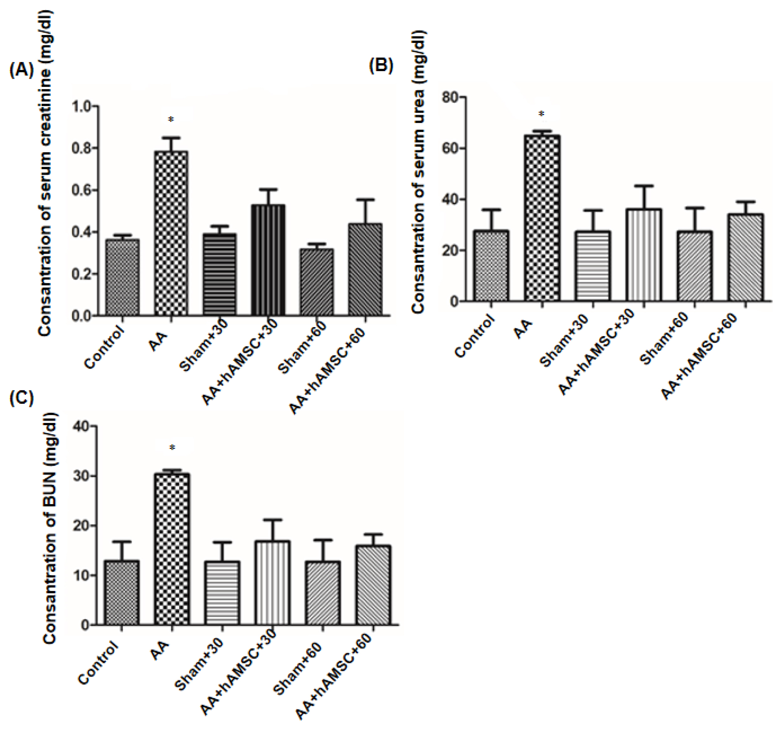
Fig. 8 Functional damage assessment. Serum creatinine (A), serum urea (B) and BUN (C) levels were determined. AA administration caused increased serum creatinine, serum urea and BUN compared to control and sham groups significantly. After hAMSC administration, serum creatinine, serum urea and BUN levels decreased significantly. Graphics represent means of optical densitometry measurements. *p< 0.005
Reference
-
References
1. Little MH. Regrow or repair: potential regenerative therapies for the kidney. J Am Soc Nephrol. 2006; 17:2390–2401. DOI: 10.1681/ASN.2006030218. PMID: 16870708.
Article2. Nangaku M. Mechanisms of tubulointerstitial injury in the kidney: final common pathways to end-stage renal failure. Intern Med. 2004; 43:9–17. DOI: 10.2169/internalmedicine.43.9. PMID: 14964574.
Article3. Vanhaelen M, Vanhaelen-Fastre R, But P, Vanherweghem JL. Identification of aristolochic acid in Chinese herbs. Lancet. 1994; 343:174. DOI: 10.1016/S0140-6736(94)90964-4. PMID: 7904018.
Article4. De Broe ME. Chinese herbs nephropathy and Balkan endemic nephropathy: toward a single entity, aristolochic acid nephropathy. Kidney Int. 2012; 81:513–515. DOI: 10.1038/ki.2011.428. PMID: 22373701.
Article5. Nortier JL, Deschodt-Lanckman MM, Simon S, Thielemans NO, de Prez EG, Depierreux MF, Tielemans CL, Richard C, Lauwerys RR, Bernard AM, Vanherweghem JL. Proximal tubular injury in Chinese herbs nephropathy: monitoring by neutral endopeptidase enzymuria. Kidney Int. 1997; 51:288–293. DOI: 10.1038/ki.1997.35. PMID: 8995745.
Article6. Vanherweghem JL, Depierreux M, Tielemans C, Abramowicz D, Dratwa M, Jadoul M, Richard C, Vandervelde D, Verbeelen D, Vanhaelen-Fastre R. Rapidly progressive interstitial renal fibrosis in young women: association with slimming regimen including Chinese herbs. Lancet. 1993; 341:387–391. DOI: 10.1016/0140-6736(93)92984-2. PMID: 8094166.
Article7. Schmeiser HH, Bieler CA, Wiessler M, van Ypersele de Strihou C, Cosyns JP. Detection of DNA adducts formed by aristolochic acid in renal tissue from patients with Chinese herbs nephropathy. Cancer Res. 1996; 56:2025–2028. PMID: 8616845.8. Nortier JL, Martinez MC, Schmeiser HH, Arlt VM, Bieler CA, Petein M, Depierreux MF, De Pauw L, Abramowicz D, Vereerstraeten P, Vanherweghem JL. Urothelial carcinoma associated with the use of a Chinese herb (Aristolochia fangchi). N Engl J Med. 2000; 342:1686–1692. DOI: 10.1056/NEJM200006083422301. PMID: 10841870.
Article9. Lebeau C, Arlt VM, Schmeiser HH, Boom A, Verroust PJ, Devuyst O, Beauwens R. Aristolochic acid impedes endocytosis and induces DNA adducts in proximal tubule cells. Kidney Int. 2001; 60:1332–1342. DOI: 10.1046/j.1523-1755.2001.00938.x. PMID: 11576347.
Article10. Lebeau C, Debelle FD, Arlt VM, Pozdzik A, De Prez EG, Phillips DH, Deschodt-Lanckman MM, Vanherweghem JL, Nortier JL. Early proximal tubule injury in experimental aristolochic acid nephropathy: functional and histological studies. Nephrol Dial Transplant. 2005; 20:2321–2332. DOI: 10.1093/ndt/gfi042. PMID: 16077141.
Article11. Pozdzik AA, Salmon IJ, Debelle FD, Decaestecker C, Van den Branden C, Verbeelen D, Deschodt-Lanckman MM, Vanherweghem JL, Nortier JL. Aristolochic acid induces proximal tubule apoptosis and epithelial to mesenchymal transformation. Kidney Int. 2008; 73:595–607. DOI: 10.1038/sj.ki.5002714. PMID: 18094681.
Article12. Soylemezoglu O, Wild G, Dalley AJ, MacNeil S, Milford-Ward A, Brown CB, el Nahas AM. Urinary and serum type III collagen: markers of renal fibrosis. Nephrol Dial Transplant. 1997; 12:1883–1889. DOI: 10.1093/ndt/12.9.1883. PMID: 9306339.13. Liu Y. Cellular and molecular mechanisms of renal fibrosis. Nat Rev Nephrol. 2011; 7:684–696. DOI: 10.1038/nrneph.2011.149. PMID: 22009250. PMCID: 4520424.
Article14. Sun D, Feng J, Dai C, Sun L, Jin T, Ma J, Wang L. Role of peritubular capillary loss and hypoxia in progressive tubulointerstitial fibrosis in a rat model of aristolochic acid nephropathy. Am J Nephrol. 2006; 26:363–371. DOI: 10.1159/000094778. PMID: 16873992.
Article15. Yang L, Li X, Wang H. Possible mechanisms explaining the tendency towards interstitial fibrosis in aristolochic acid-induced acute tubular necrosis. Nephrol Dial Transplant. 2007; 22:445–456. DOI: 10.1093/ndt/gfl556. PMID: 17124284.
Article16. Barry FP, Murphy JM. Mesenchymal stem cells: clinical applications and biological characterization. Int J Biochem Cell Biol. 2004; 36:568–584. DOI: 10.1016/j.biocel.2003.11.001. PMID: 15010324.
Article17. Lange C, Tögel F, Ittrich H, Clayton F, Nolte-Ernsting C, Zander AR, Westenfelder C. Administered mesenchymal stem cells enhance recovery from ischemia/reperfusion-induced acute renal failure in rats. Kidney Int. 2005; 68:1613–1617. DOI: 10.1111/j.1523-1755.2005.00573.x. PMID: 16164638.
Article18. Kunter U, Rong S, Djuric Z, Boor P, Müller-Newen G, Yu D, Floege J. Transplanted mesenchymal stem cells accelerate glomerular healing in experimental glomerulonephritis. J Am Soc Nephrol. 2006; 17:2202–2212. DOI: 10.1681/ASN.2005080815. PMID: 16790513.
Article19. Bi B, Schmitt R, Israilova M, Nishio H, Cantley LG. Stromal cells protect against acute tubular injury via an endocrine effect. J Am Soc Nephrol. 2007; 18:2486–2496. DOI: 10.1681/ASN.2007020140. PMID: 17656474.
Article20. Hopkins C, Li J, Rae F, Little MH. Stem cell options for kidney disease. J Pathol. 2009; 217:265–281. DOI: 10.1002/path.2477. PMID: 19058290.
Article21. Dominici M, Le Blanc K, Mueller I, Slaper-Cortenbach I, Marini F, Krause D, Deans R, Keating A, Prockop Dj, Horwitz E. Minimal criteria for defining multipotent mesenchymal stromal cells. The International Society for Cellular Therapy position statement. Cytotherapy. 2006; 8:315–317. DOI: 10.1080/14653240600855905. PMID: 16923606.
Article22. Jin CZ, Park SR, Choi BH, Lee KY, Kang CK, Min BH. Human amniotic membrane as a delivery matrix for articular cartilage repair. Tissue Eng. 2007; 13:693–702. DOI: 10.1089/ten.2006.0184. PMID: 17269856.
Article23. Díaz-Prado S, Muiños-López E, Hermida-Gómez T, Rendal-Vázquez ME, Fuentes-Boquete I, de Toro FJ, Blanco FJ. Multilineage differentiation potential of cells isolated from the human amniotic membrane. J Cell Biochem. 2010; 111:846–857. DOI: 10.1002/jcb.22769. PMID: 20665539.
Article24. Parolini O, Alviano F, Bagnara GP, Bilic G, Bühring HJ, Evangelista M, Hennerbichler S, Liu B, Magatti M, Mao N, Miki T, Marongiu F, Nakajima H, Nikaido T, Portmann-Lanz CB, Sankar V, Soncini M, Stadler G, Surbek D, Takahashi TA, Redl H, Sakuragawa N, Wolbank S, Zeisberger S, Zisch A, Strom SC. Concise review: isolation and characterization of cells from human term placenta: outcome of the first international Workshop on Placenta Derived Stem Cells. Stem Cells. 2008; 26:300–311. DOI: 10.1634/stemcells.2007-0594. PMID: 17975221.
Article25. Choi S, Park M, Kim J, Hwang S, Park S, Lee Y. The role of mesenchymal stem cells in the functional improvement of chronic renal failure. Stem Cells Dev. 2009; 18:521–529. DOI: 10.1089/scd.2008.0097. PMID: 18647091.
Article26. Semedo P, Correa-Costa M, Antonio Cenedeze M, Maria Avancini Costa Malheiros D, Antonia dos Reis M, Shimizu MH, Seguro AC, Pacheco-Silva A, Saraiva Camara NO. Mesenchymal stem cells attenuate renal fibrosis through immune modulation and remodeling properties in a rat remnant kidney model. Stem Cells. 2009; 27:3063–3073. DOI: 10.1002/stem.214. PMID: 19750536.
Article27. Villanueva S, Ewertz E, Carrión F, Tapia A, Vergara C, Céspedes C, Sáez PJ, Luz P, Irarrázabal C, Carreño JE, Figueroa F, Vio CP. Mesenchymal stem cell injection ameliorates chronic renal failure in a rat model. Clin Sci (Lond). 2011; 121:489–499. DOI: 10.1042/CS20110108. PMID: 21675962.
Article28. Klinkhammer BM, Kramann R, Mallau M, Makowska A, van Roeyen CR, Rong S, Buecher EB, Boor P, Kovacova K, Zok S, Denecke B, Stuettgen E, Otten S, Floege J, Kunter U. Mesenchymal stem cells from rats with chronic kidney disease exhibit premature senescence and loss of regenerative potential. PLoS One. 2014; 9:e92115. DOI: 10.1371/journal.pone.0092115. PMID: 24667162. PMCID: 3965415.
Article29. Soncini M, Vertua E, Gibelli L, Zorzi F, Denegri M, Albertini A, Wengler GS, Parolini O. Isolation and characterization of mesenchymal cells from human fetal membranes. J Tissue Eng Regen Med. 2007; 1:296–305. DOI: 10.1002/term.40. PMID: 18038420.
Article30. Korgun ET, Celik-Ozenci C, Seval Y, Desoye G, Demir R. Do glucose transporters have other roles in addition to placental glucose transport during early pregnancy? Histochem Cell Biol. 2005; 123:621–629. DOI: 10.1007/s00418-005-0792-3. PMID: 15965666.
Article31. Ozmen A, Unek G, Kipmen-Korgun D, Korgun ET. The expression of Akt and ERK1/2 proteins decreased in dexamethasone-induced intrauterine growth restricted rat placental development. J Mol Histol. 2011; 42:237–249. DOI: 10.1007/s10735-011-9328-4. PMID: 21512721.
Article32. Lin F, Cordes K, Li L, Hood L, Couser WG, Shankland SJ, Igarashi P. Hematopoietic stem cells contribute to the regeneration of renal tubules after renal ischemia-reperfusion injury in mice. J Am Soc Nephrol. 2003; 14:1188–1199. DOI: 10.1097/01.ASN.0000061595.28546.A0. PMID: 12707389.
Article33. Broekema M, Harmsen MC, Koerts JA, Petersen AH, van Luyn MJ, Navis G, Popa ER. Determinants of tubular bone marrow-derived cell engraftment after renal ischemia/reperfusion in rats. Kidney Int. 2005; 68:2572–2581. DOI: 10.1111/j.1523-1755.2005.00728.x. PMID: 16316332.
Article34. Fang TC, Otto WR, Rao J, Jeffery R, Hunt T, Alison MR, Cook HT, Wright NA, Poulsom R. Haematopoietic lineage-committed bone marrow cells, but not cloned cultured mesenchymal stem cells, contribute to regeneration of renal tubular epithelium after HgCl 2 -induced acute tubular injury. Cell Prolif. 2008; 41:575–591. DOI: 10.1111/j.1365-2184.2008.00545.x. PMID: 18616694.
Article35. Humphreys BD, Bonventre JV. Mesenchymal stem cells in acute kidney injury. Annu Rev Med. 2008; 59:311–325. DOI: 10.1146/annurev.med.59.061506.154239. PMID: 17914926.
Article36. Sun D, Bu L, Liu C, Yin Z, Zhou X, Li X, Xiao A. Therapeutic effects of human amniotic fluid-derived stem cells on renal interstitial fibrosis in a murine model of unilateral ureteral obstruction. PLoS One. 2013; 8:e65042. DOI: 10.1371/journal.pone.0065042. PMID: 23724119. PMCID: 3665750.
Article37. Cosyns JP, Dehoux JP, Guiot Y, Goebbels RM, Robert A, Bernard AM, van Ypersele de Strihou C. Chronic aristolochic acid toxicity in rabbits: a model of Chinese herbs nephropathy? Kidney Int. 2001; 59:2164–2173. DOI: 10.1046/j.1523-1755.2001.00731.x. PMID: 11380818.
Article38. Inumaru J, Nagano O, Takahashi E, Ishimoto T, Nakamura S, Suzuki Y, Niwa S, Umezawa K, Tanihara H, Saya H. Molecular mechanisms regulating dissociation of cell-cell junction of epithelial cells by oxidative stress. Genes Cells. 2009; 14:703–716. DOI: 10.1111/j.1365-2443.2009.01303.x. PMID: 19422420.
Article39. Huang L, Scarpellini A, Funck M, Verderio EA, Johnson TS. Development of a chronic kidney disease model in C57BL/6 mice with relevance to human pathology. Nephron Extra. 2013; 3:12–29. DOI: 10.1159/000346180. PMID: 23610565. PMCID: 3617971.
Article40. Grande MT, Pérez-Barriocanal F, López-Novoa JM. Role of inflammation in túbulo-interstitial damage associated to obstructive nephropathy. J Inflamm (Lond). 2010; 7:19. DOI: 10.1186/1476-9255-7-19. PMID: 20412564. PMCID: 2873503.
Article41. Gatti S, Bruno S, Deregibus MC, Sordi A, Cantaluppi V, Tetta C, Camussi G. Microvesicles derived from human adult mesenchymal stem cells protect against ischaemia-reperfusion-induced acute and chronic kidney injury. Nephrol Dial Transplant. 2011; 26:1474–1483. DOI: 10.1093/ndt/gfr015. PMID: 21324974.
Article42. Li Y, Liu Z, Guo X, Shu J, Chen Z, Li L. Aristolochic acid I-induced DNA damage and cell cycle arrest in renal tubular epithelial cells in vitro. Arch Toxicol. 2006; 80:524–532. DOI: 10.1007/s00204-006-0090-4. PMID: 16609888.
Article43. Cavaglieri RC, Martini D, Sogayar MC, Noronha IL. Mesenchymal stem cells delivered at the subcapsule of the kidney ameliorate renal disease in the rat remnant kidney model. Transplant Proc. 2009; 41:947–951. DOI: 10.1016/j.transproceed.2009.01.072. PMID: 19376395.
Article
- Full Text Links
-
- Actions
-
Cited
- CITED
-
- Close
- Share
-
- Similar articles
-
- Effect of amnion derived stem cells on inflammatory response in endotoxemic rats
- Comparison with human amniotic membrane- and adipose tissue-derived mesenchymal stem cells
- L-Theanine-Treated Adipose-Derived Mesenchymal Stem Cells Alleviate the Cytotoxicity Induced by N-Nitrosodiethylamine in Liver
- Effects of Mesenchymal Stem Cells Treatment on Radiation-Induced Proctitis in Rats
- Advanced Research on Stem Cell Therapy for Hepatic Diseases: Potential Implications of a Placenta-derived Mesenchymal Stem Cell-based Strategy
