J Korean Acad Prosthodont.
2019 Apr;57(2):142-149. 10.4047/jkap.2019.57.2.142.
Inhibition of Osteoclast differentiation based on precipitation time of titanium surfaces immersed in modified simulated body fluid
- Affiliations
-
- 1Department of Prosthodontics, School of Dentistry, Seoul National University, Seoul, Republic of Korea. young21c@snu.ac.kr
- KMID: 2444183
- DOI: http://doi.org/10.4047/jkap.2019.57.2.142
Abstract
- PURPOSE
The purpose of this study is to investigate the changes of osteoclast differentiation inhibition according to the period of precipitation when titanium disks were immersed in Modified simulated body fluid (mSBF).
MATERIALS AND METHODS
Titanium alloy (Ti grade III) disks with machined surfaces and anodized surfaces were immersed in distilled water and mSBF, respectively. The immersion periods were 7 days, 14 days, 21 days and 28 days, and the control group was immersed in distilled water for each period. RAW 264.7 cells capable of differentiating into osteoclasts were used to measure the number of adherent cells, the measurement of TRAP activity, and the expression pattern of NFATc1 by western blotting.
RESULTS
The degree of inhibition of osteoclast differentiation was found to be statistically significant when the disks were immersed in mSBF for more than 14 days on both machined surfaces and anodized surfaces. There was no correlation between immersion time and cell attachment. When the disks were immersed for more than 14 days, TRAP activity was decreased and NFATc1 expression was inhibited. Futhermore, the decrease in TRAP activity and the inhibition of NFATc1 expression remained unchanged.
CONCLUSION
Immersion of titanium disks in mSBF for more than 14 days can prevent RAW 264.7 cells from differentiating into osteoclasts. Inhibition activity does not change even if the immersion period is for more than 14 days.
Keyword
MeSH Terms
Figure
-
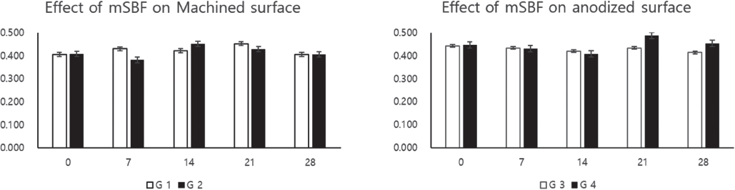
Fig. 1 Cell counting test. There is no significant difference among the groups. Deposition period dose not affect cell adhesion.
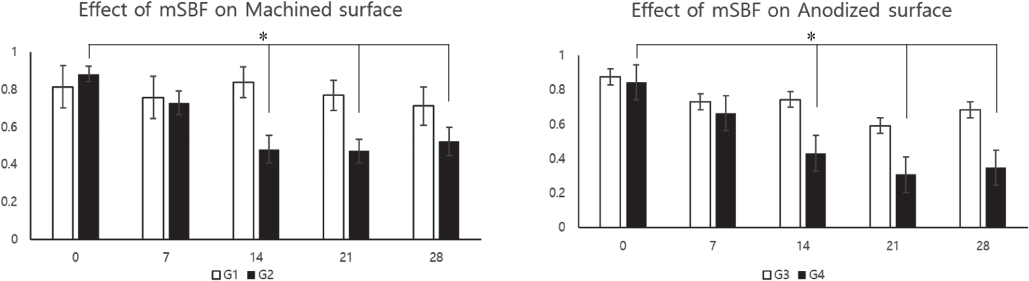
Fig. 2 TRAP activity on Ti disc. More than 14 days deposition period, TRAP activity was reduced regardless of surface roughness. *P < .05
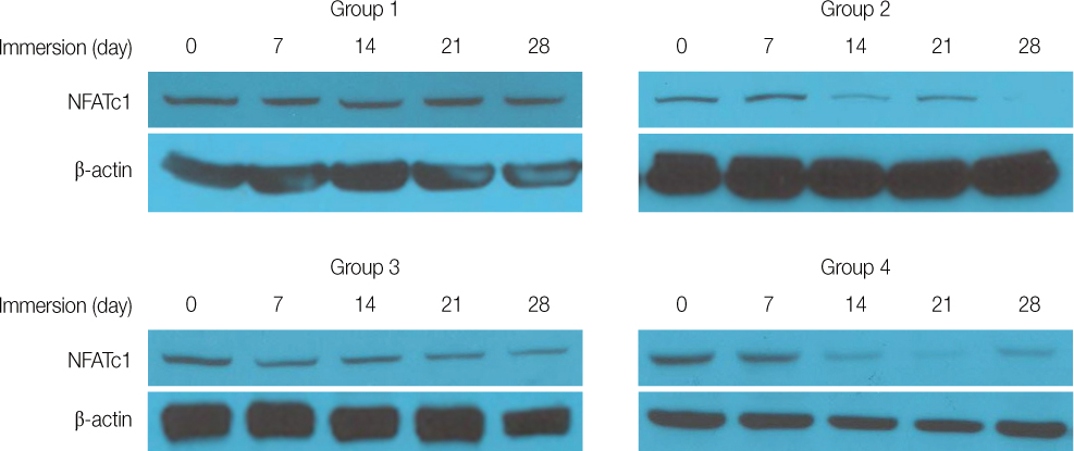
Fig. 3 Western blot of NFATc1, β-actin. Group 2, 4 show decreased expression of NFATc1 after 14 days deposition. NFATc1 expression of 0, 7 days deposition were not different among group 1 – 4. Group 1, 3 show constant expression of NFATc1. NFATc1 is a final signal transducer of osteoclast differentiation. β-actin is a control of protein expression.
Reference
-
1. Albrektsson T, Brånemark PI, Hansson HA, Kasemo B, Larsson K, Lundstrom I, McQueen DH, Skalak R. The interface zone of inorganic implantsIn vivo: Titanium implants in bone. Ann Biomed Eng. 1983; 11:1–27.
Article2. Brånemark PI, Adell R, Breine U, Hansson BO, Lindström J, Ohlsson A. Intra-osseous anchorage of dental prostheses. I. Experimental studies. Scand J Plast Reconstr Surg. 1969; 3:81–100.3. Albrektsson T, Zarb G, Worthington P, Eriksson AR. The long-term efficacy of currently used dental implants: a review and proposed criteria of success. Int J Oral Maxillofac Implants. 1986; 1:11–25.4. Le Guéhennec L, Soueidan A, Layrolle P, Amouriq Y. Surface treatments of titanium dental implants for rapid osseointegration. Dent Mater. 2007; 23:844–854.
Article5. de Jonge LT, Leeuwenburgh SC, Wolke JG, Jansen JA. Organic-inorganic surface modifications for titanium implant surfaces. Pharm Res. 2008; 25:2357–2369.
Article6. Lang NP, Salvi GE, Huynh-Ba G, Ivanovski S, Donos N, Bosshardt DD. Early osseointegration to hydrophilic and hydrophobic implant surfaces in humans. Clin Oral Implants Res. 2011; 22:349–356.
Article7. Zhao G, Schwartz Z, Wieland M, Rupp F, Geis-Gerstorfer J, Cochran DL, Boyan BD. High surface energy enhances cell response to titanium substrate microstructure. J Biomed Mater Res A. 2005; 74:49–58.
Article8. Hori N, Ueno T, Suzuki T, Yamada M, Att W, Okada S, Ohno A, Aita H, Kimoto K, Ogawa T. Ultraviolet light treatment for the restoration of age-related degradation of titanium bioactivity. Int J Oral Maxillofac Implants. 2010; 25:49–62.9. Junker R, Dimakis A, Thoneick M, Jansen JA. Effects of implant surface coatings and composition on bone integration: a systematic review. Clin Oral Implants Res. 2009; 20:185–206.
Article10. Kokubo T, Kushitani H, Sakka S, Kitsugi T, Yamamuro T. Solutions able to reproduce in vivo surface-structure changes in bioactive glass-ceramic A-W. J Biomed Mater Res. 1990; 24:721–734.
Article11. Bohner M, Lemaitre J. Can bioactivity be tested in vitro with SBF solution? Biomaterials. 2009; 30:2175–2179.
Article12. Kim MH, Lee SY, Kim MJ, Kim SK, Heo SJ, Koak JY. Effect of biomimetic deposition on anodized titanium surfaces. J Dent Res. 2011; 90:711–716.
Article13. Minkin C, Marinho VC. Role of the osteoclast at the bone-implant interface. Adv Dent Res. 1999; 13:49–56.
Article14. Kim MH, Lee SY, Heo SJ, Kim SK, Kim MJ, Koak JY. Osteoclastic response on titanium surfaces in modified simulated body fluid. Int J Oral Maxillofac Implants. 2017; 32:337–343.
Article15. Hsu H, Lacey DL, Dunstan CR, Solovyev I, Colombero A, Timms E, Tan HL, Elliott G, Kelley MJ, Sarosi I, Wang L, Xia XZ, Elliott R, Chiu L, Black T, Scully S, Capparelli C, Morony S, Shimamoto G, Bass MB, Boyle WJ. Tumor necrosis factor receptor family member RANK mediates osteoclast differentiation and activation induced by osteoprotegerin ligand. Proc Natl Acad Sci USA. 1999; 96:3540–3545.
Article16. Takayanagi H. Osteoimmunological insight into bone damage in rheumatoid arthritis. Mod Rheumatol. 2005; 15:225–231.
Article17. Asagiri M, Sato K, Usami T, Ochi S, Nishina H, Yoshida H, Morita I, Wagner EF, Mak TW, Serfling E, Takayanagi H. Autoamplification of NFATc1 expression determines its essential role in bone homeostasis. J Exp Med. 2005; 202:1261–1269.
Article
- Full Text Links
-
- Actions
-
Cited
- CITED
-
- Close
- Share
-
- Similar articles
-
- Comparison of alkaline phosphatase activity of MC3T3-E1 cells cultured on different Ti surfaces: modified sandblasted with large grit and acid-etched (MSLA), laser-treated, and laser and acid-treated Ti surfaces
- Magnesium vs. machined surfaced titanium - osteoblast and osteoclast differentiation
- Effect of Application of Tetracycline - HCl on Implant Surface - Scanning Electron Microscopic Study
- Surface characteristics and bioactivity of anodically oxidized titanium surfaces
- Study about Biodegradation of Hydroxyapatite and beta-tricalcium Phosphate Coating Layer
