Changes in Transepidermal Water Loss and Skin Hydration according to Expression of Aquaporin-3 in Psoriasis
- Affiliations
-
- 1Department of Dermatology, College of Medicine, Chungnam National University, Daejeon, Korea. jhoon@cnu.ac.kr
Abstract
- BACKGROUND
Aquaporins (AQPs) are a family of water transporting proteins present in many mammalian epithelial and endothelial cell types. Among the AQPs, AQP3 is known to be a water/glycerol transporter expressed in human skin.
OBJECTIVE
The relationship between the expression level of AQP3 and transpidermal water loss (TEWL) in the lesional and peri-lesional skin of psoriasis-affected patients, and skin hydration in the lesional and peri-lesional skin of psoriasis patients, was investigated.
METHODS
The expression of AQP3 in psoriasis-affected and healthy control skin was determined using immunohistochemical and immunofluroscence staining. TEWL and skin hydration were measured using a Tewameter(R) TM210 (Courage & Khazaka, Cologne, Germany) and a Corneometer(R) CM 820 (Courage & Khazaka), respectively.
RESULTS
AQP3 was mainly expressed in the plasma membrane of stratum corneum and the stratum spinosum in normal epidermis. Unlike the normal epidermis, AQP3 showed decreased expression in the lesional and peri-lesional epidermis of psoriasis. TEWL was increased, and skin hydration was decreased, in the lesional and peri-lesional skin of psoriasis patients, compared with the healthy control sample.
CONCLUSION
Although various factors contribute to reduced skin hydration in the lesional and peri-lesional skin of psoriasis, AQP3 appears to be a key factor in the skin dehydration of psoriasis-affected skin.
Keyword
MeSH Terms
Figure
-

Fig. 1 (A) Immunolocalization of aquaporins 3 (AQP3) uisng immunohistochemisty in the human epidermis. AQP3 is expressed in the stratum basale and the stratum spinosum. (B) AQP3 expression in calcium-induced keratinocyte differentiation. AQP3 from cultured keratinocyte extracts of 20 or 30 µg was loaded onto sodium dodecyl sulfate polyacrylamide gel electrophoresis and was detected using a peptide polyclonal anti-AQP3 antibody. Involucrin and filaggrin were used as control makers for differentiation. (C) The [3H]glycerol uptake ability in calcium differentiated human keratinocytes. Near confluent keratinocytes were incubated in Keratinocyte-basal medium containing 1 uCi/ml of [3H]glycerol with and without 1.2 mM Ca2+ for 0 min, 10 min, 30 min, 60 min, 4 hr, 12 hr, 24 hr, and 48 hr. NHEK: normal human epidermal keratinocyte, CPM: counting per minute.

Fig. 2 Aquaporins 3 (AQP3) immunoreactivity in (A) healthy control, (B) psoriatic lesional and (C) peri-lesional skin. AQP3 protein expression is decreased and mislocalized in lesional and peri-lesional skin, showing a diffuse cytoplasmic pattern. (D) AQP3 staining intensity between healthy control and psoriasis. The fluorescence intensity was analyzed using an image analyzer (i-solution TM, iMTechnology, Bucheon, Korea) *p<0.05 vs. healthy control.
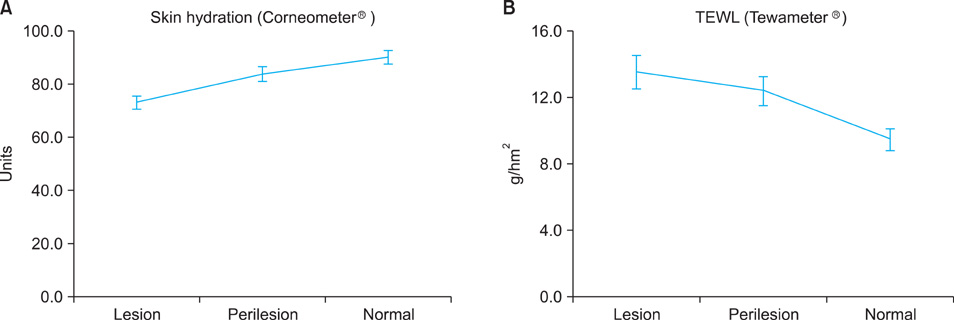
Fig. 3 (A) Differences in skin hydration among psoriatic lesional, peri-lesional, and normal non-psoriatic skin in psoriasis patients. Skin hydration was measured using a Corneometer® CM 820 (Courage & Khazaka, Cologne, Germany). (B) Transpidermal water loss (TEWL) differences among psoriatic lesional, peri-lesional, and normal non-psoriasis skin in psoriasis patients. TEWL was measured using a Tewameter® TM 210 (Courage & Khazaka).
Reference
-
1. Gudjonsson JE, Elder ST. Wolff K, Goldsmith LA, Kats SI, Gilchrest BA, Paller AS, Leffell DJ, editors. Psoriasis. Fitzpatrick's dermatology in general medicine. 2008. 7th ed. New York: McGraw-Hill;169–193.
Article2. Hara-Chikuma M, Verkman AS. Roles of aquaporin-3 in the epidermis. J Invest Dermatol. 2008. 128:2145–2151.
Article3. Lodén M. The clinical benefit of moisturizers. J Eur Acad Dermatol Venereol. 2005. 19:672–688.
Article4. Hara-Chikuma M, Verkman AS. Aquaporin-3 facilitates epidermal cell migration and proliferation during wound healing. J Mol Med (Berl). 2008. 86:221–231.
Article5. Hara-Chikuma M, Verkman AS. Prevention of skin tumorigenesis and impairment of epidermal cell proliferation by targeted aquaporin-3 gene disruption. Mol Cell Biol. 2008. 28:326–332.
Article6. Yoon HK, Sohn KC, Lee JS, Kim YJ, Bhak J, Yang JM, et al. Prediction and evaluation of protein-protein interaction in keratinocyte differentiation. Biochem Biophys Res Commun. 2008. 377:662–667.
Article7. Zheng X, Bollinger Bollag W. Aquaporin 3 colocates with phospholipase d2 in caveolin-rich membrane microdomains and is downregulated upon keratinocyte differentiation. J Invest Dermatol. 2003. 121:1487–1495.
Article8. Cho SH, Seo SJ, Hong CK. Expression of antimicrobial peptides according to changes of transepidermal water loss levels in patients with atopic dermatits. Ann Dermatol. 2007. 19:47–54.
Article9. Heinrich U, Koop U, Leneveu-Duchemin MC, Osterrieder K, Bielfeldt S, Chkarnat C, et al. Multicentre comparison of skin hydration in terms of physical-, physiological- and product-dependent parameters by the capacitive method (Corneometer CM 825). Int J Cosmet Sci. 2003. 25:45–53.
Article10. Suh DH, Kwon TE, Kim SD, Park SB, Kwon OS, Kim KH, et al. Changes of transepidermal water loss (TEWL) in psoriatic plaques during D-PUVA therapy. Ann Dermatol. 2001. 13:7–11.
Article11. Ma T, Hara M, Sougrat R, Verbavatz JM, Verkman AS. Impaired stratum corneum hydration in mice lacking epidermal water channel aquaporin-3. J Biol Chem. 2002. 277:17147–17153.
Article12. Hara M, Ma T, Verkman AS. Selectively reduced glycerol in skin of aquaporin-3-deficient mice may account for impaired skin hydration, elasticity, and barrier recovery. J Biol Chem. 2002. 277:46616–46621.
Article13. Li J, Tang H, Hu X, Chen M, Xie H. Aquaporin-3 gene and protein expression in sun-protected human skin decreases with skin ageing. Australas J Dermatol. 2010. 51:106–112.
Article14. Hara-Chikuma M, Verkman AS. Aquaporin-3 functions as a glycerol transporter in mammalian skin. Biol Cell. 2005. 97:479–486.
Article15. Voss KE, Bollag RJ, Fussell N, By C, Sheehan DJ, Bollag WB. Abnormal aquaporin-3 protein expression in hyperproliferative skin disorders. Arch Dermatol Res. 2011. 303:591–600.
Article16. Horie I, Maeda M, Yokoyama S, Hisatsune A, Katsuki H, Miyata T, et al. Tumor necrosis factor-alpha decreases aquaporin-3 expression in DJM-1 keratinocytes. Biochem Biophys Res Commun. 2009. 387:564–568.
Article17. Nakahigashi K, Kabashima K, Ikoma A, Verkman AS, Miyachi Y, Hara-Chikuma M. Upregulation of aquaporin-3 is involved in keratinocyte proliferation and epidermal hyperplasia. J Invest Dermatol. 2011. 131:865–873.
Article18. Bellemère G, Von Stetten O, Oddos T. Retinoic acid increases aquaporin 3 expression in normal human skin. J Invest Dermatol. 2008. 128:542–548.
Article
- Full Text Links
-
- Actions
-
Cited
- CITED
-
- Close
- Share
-
- Similar articles
-
- Variation in repeated measurements of transepidermal water loss, skin hydration, and sebum level in normal beagle dogs
- The Effects of Humidifier Generating Nano-sized Water Particles on Skin Hydration and Transepidermal Water Loss of the Normal Human Skin
- Changes of Transepidermal Water Loss (TEWL) in Psoriatic Plaques during D-PUVA Therapy
- Expression of Antimicrobial Peptides according to Changes of Transepidermal Water Loss Levels in Patients with Atopic Dermatits
- Effects of Topical N-Acetylcysteine on Skin Hydration/Transepidermal Water Loss in Healthy Volunteers and Atopic Dermatitis Patients
