Cyanidin-3-glucoside inhibits amyloid β₂₅₋₃₅-induced neuronal cell death in cultured rat hippocampal neurons
- Affiliations
-
- 1Department of Physiology, College of Medicine, Catholic Neuroscience Institute, The Catholic University of Korea, Seoul 06591, Korea. s-hyoon@catholic.ac.kr
- 2College of Pharmacy, The Catholic University of Korea, Bucheon 14662, Korea.
- KMID: 2430091
- DOI: http://doi.org/10.4196/kjpp.2018.22.6.689
Abstract
- Increasing evidence implicates changes in [Ca²âº]i and oxidative stress as causative factors in amyloid beta (Aβ)-induced neuronal cell death. Cyanidin-3-glucoside (C3G), a component of anthocyanin, has been reported to protect against glutamate-induced neuronal cell death by inhibiting Ca²âº and Zn²âº signaling. The present study aimed to determine whether C3G exerts a protective effect against Aβ₂₅₋₃₅-induced neuronal cell death in cultured rat hippocampal neurons from embryonic day 17 fetal Sprague-Dawley rats using MTT assay for cell survival, and caspase-3 assay and digital imaging methods for Ca²âº, Zn²âº, MMP and ROS. Treatment with Aβ25-35 (20 µM) for 48 h induced neuronal cell death in cultured rat pure hippocampal neurons. Treatment with C3G for 48 h significantly increased cell survival. Pretreatment with C3G for 30 min significantly inhibited Aβ₂₅₋₃₅-induced [Zn²âº]i increases as well as [Ca²âº]i increases in the cultured rat hippocampal neurons. C3G also significantly inhibited Aβ₂₅₋₃₅-induced mitochondrial depolarization. C3G also blocked the Aβ₂₅₋₃₅-induced formation of ROS. In addition, C3G significantly inhibited the Aβ₂₅₋₃₅-induced activation of caspase-3. These results suggest that cyanidin-3-glucoside protects against amyloid β-induced neuronal cell death by reducing multiple apoptotic signals.
Keyword
MeSH Terms
Figure
-
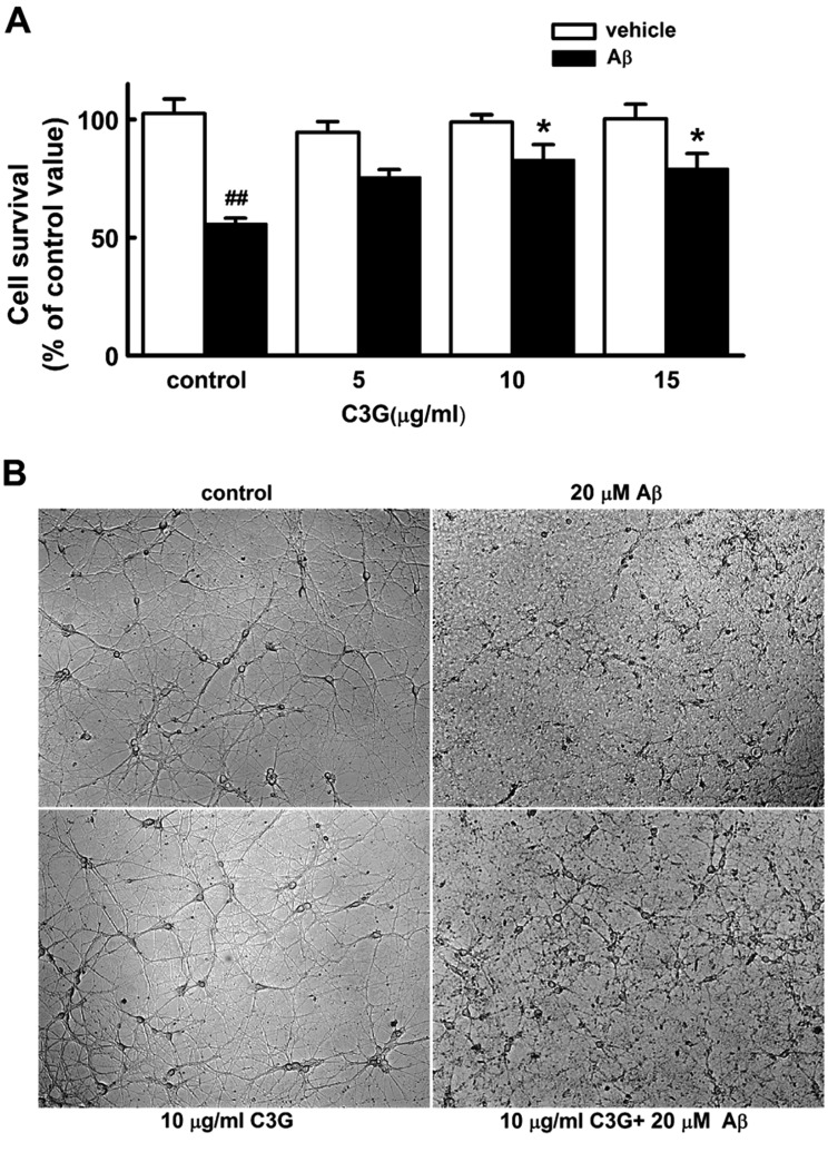
Fig. 1 Effects of C3G on Aβ25–35-induced cell death in cultured pure rat hippocampal neurons.Aβ25–35 (20 µM)-induced neuronal damage was measured through the reduction of MTT in viable cells at 11 days in culture. The bar graph shows the purple formazan product from pure hippocampal neurons after treatment. (A) Effects of C3G on Aβ25–35-induced cell death in the non-treated(control) (vehicle, n=5; Aβ, n=5), 5 µg/ml C3G (vehicle, n=4; Aβ, n=4), 10 µg/ml C3G (vehicle, n=4; Aβ, n=4), and 15 µg/ml C3G (vehicle, n=4; Aβ, n=4)-treated cells in the absence (vehicle) and presence of 20 µM Aβ25–35 for 48 h. (B) Representative phase contrast photomicrographs showing cultured pure rat hippocampal neurons 48 h following co-treatment of C3G with Aβ25–35 at 11 days in culture. Data are expressed as mean±S.E. of experiments. ##p<0.01 relative to control, *p<0.05 relative to control Aβ (ANOVA with Bonferroni test).
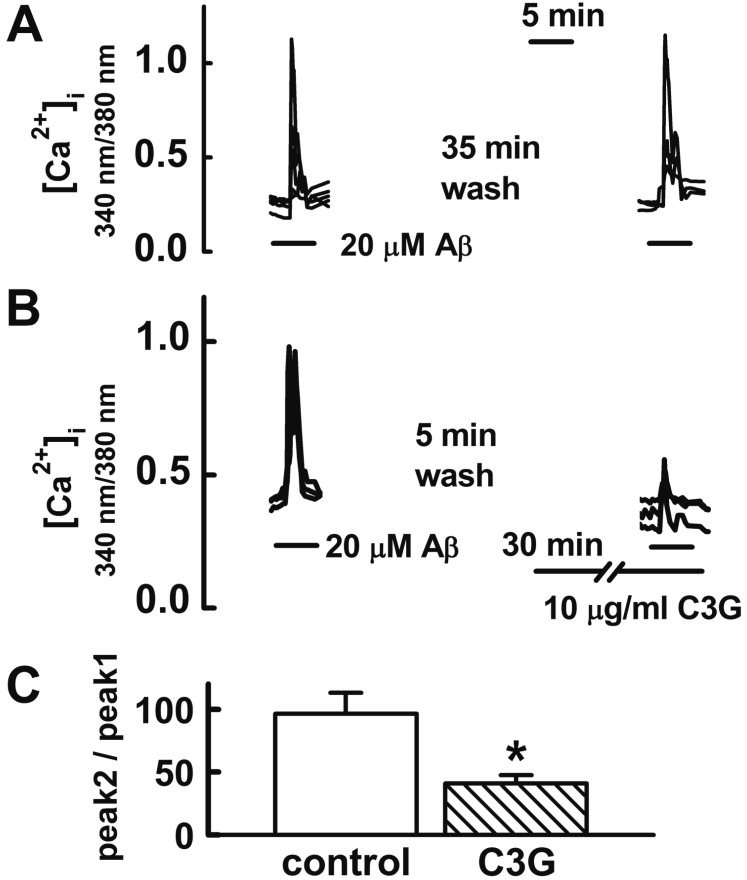
Fig. 2 Effects of C3G on Aβ25–35-induced [Ca2+]i increases.Reproducible [Ca2+]i increases were elicited by applying Aβ25–35 (20 µM) for 5 min at 35 min intervals (A). Pretreatment with C3G (10 µg/ml) for 30 min significantly inhibited Aβ25–35-induced [Ca2+]i responses (B). (C) Summary of the Aβ25–35-induced [Ca2+]i increases in non-treated (control, n=17) and C3G-pretreated (C3G, n=10) cells. Aβ25–35 induced response is presented as a percentage of the initial Aβ25–35 induced response (peak 2/peak 1). Data are expressed as mean±S.E. *p<0.05 relative to control (non-paired Student's t-test).
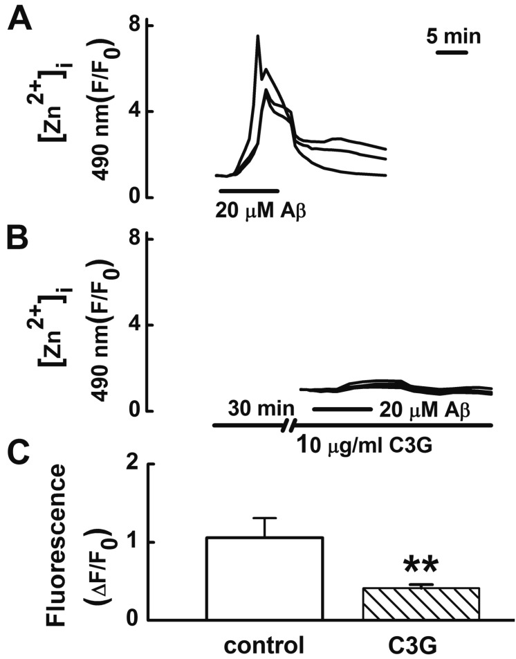
Fig. 3 Effects of C3G on Aβ25–35-induced [Zn2+]i increases.(A) Treatment with Aβ25–35 (20 µM) for 10 min increased [Zn2+]i increases (n=26). (B, C) Pretreatment with C3G (10 µg/ml) for 30 min significantly inhibited the Aβ25–35-induced [Zn2+]i responses (n=22). (C) Aβ25–35 induced response is normalized to the initial value measured before the addition of agonist. Data are expressed as mean±S.E. **p<0.01 relative to control (non-paired Student's t-test).
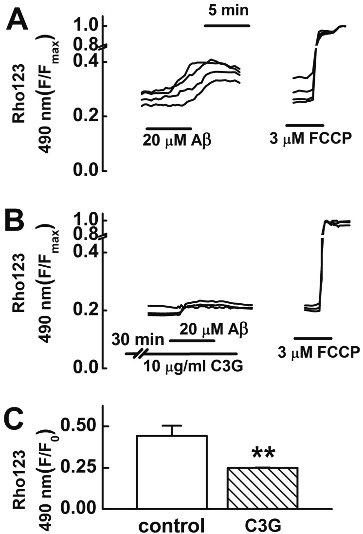
Fig. 4 Effects of C3G on Aβ25–35-induced mitochondrial depolarization.(A) Reproducible depolarization in mitochondrial membrane potential (MMP) was elicited by applying Aβ25–35 (20 µM) for 5 min at 35 min intervals. (B) Pretreatment with C3G (10 µg/ml) for 30 min significantly inhibited Aβ25–35-induced mitochondrial depolarization. (C) Summary of glutamate-induced mitochondrial depolarizations in non-treated (control, n=18) and C3G-pretreated (n=18) cells. Cells were preincubated with 10 µM rhodamine 123 for 15 min. Change in MMP was shown as a percentage of the maximal intensity of rhodamine 123 (3 µM FCCP). Data are expressed as mean±S.E. **p<0.01 relative to control (non-paired Student's t-test).
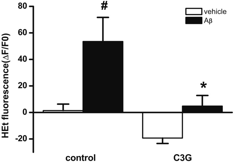
Fig. 5 Effects of C3G on the Aβ25–35-induced formation of intracelluar ROS.Treatment with Aβ25–35 (20 µM) for 10 min significantly increased superoxide formation (vehicle, n=19; Aβ, n=22). Pretreatment with C3G (10 µg/ml) for 30 min blocked Aβ25–35-induced superoxide formation (vehicle, n=16, Aβ, n=14). Superoxide formation was shown as a percentage of the initial intensity of dihydroxyehidine. Data are expressed as mean±S.E. #p<0.05 relative to vehicle (non-paired Student's t-test). *p<0.05 relative to respective control (non-paired Student t-test).
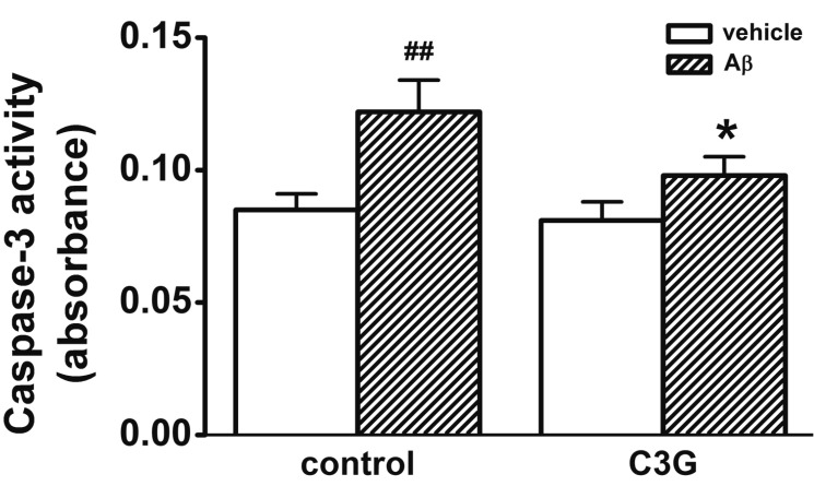
Fig. 6 Effects of C3G on Aβ25–35-induced activation of caspase-3.Treatment with Aβ25–35 (20 µM) for 24 hr increased the activation of caspase-3 (n=6). Co-treatment with C3G (10 µg/ml) for 24 hr significantly inhibited the Aβ25–35-induced activation of caspase-3 (n=4). Data are expressed as mean±S.E. ##p<0.01 relative to vehicle (non-paired Student's t-test) *p<0.05 relative to respective control (non-paired Student's t-test).
Reference
-
1. Terry RD, Masliah E, Salmon DP, Butters N, DeTeresa R, Hill R, Hansen LA, Katzman R. Physical basis of cognitive alterations in Alzheimer's disease: synapse loss is the major correlate of cognitive impairment. Ann Neurol. 1991; 30:572–580. PMID: 1789684.
Article2. Arispe N, Rojas E, Pollard HB. Alzheimer disease amyloid beta protein forms calcium channels in bilayer membranes: blockade by tromethamine and aluminum. Proc Natl Acad Sci U S A. 1993; 90:567–571. PMID: 8380642.
Article3. Mattson MP. Pathways towards and away from Alzheimer's disease. Nature. 2004; 430:631–639. PMID: 15295589.
Article4. Bezprozvanny I, Mattson MP. Neuronal calcium mishandling and the pathogenesis of Alzheimer's disease. Trends Neurosci. 2008; 31:454–463. PMID: 18675468.
Article5. Religa D, Strozyk D, Cherny RA, Volitakis I, Haroutunian V, Winblad B, Naslund J, Bush AI. Elevated cortical zinc in Alzheimer disease. Neurology. 2006; 67:69–75. PMID: 16832080.
Article6. Bush AI, Pettingell WH Jr, de Paradis M, Tanzi RE, Wasco W. The amyloid beta-protein precursor and its mammalian homologues. Evidence for a zinc-modulated heparin-binding superfamily. J Biol Chem. 1994; 269:26618–26621. PMID: 7929392.
Article7. Ahn SH, Kim HJ, Jeong I, Hong YJ, Kim MJ, Rhie DJ, Jo YH, Hahn SJ, Yoon SH. Grape seed proanthocyanidin extract inhibits glutamate-induced cell death through inhibition of calcium signals and nitric oxide formation in cultured rat hippocampal neurons. BMC Neurosci. 2011; 12:78. DOI: 10.1186/1471-2202-12-78. PMID: 21810275.
Article8. Bae JH, Mun KC, Park WK, Lee SR, Suh SI, Baek WK, Yim MB, Kwon TK, Song DK. EGCG attenuates AMPA-induced intracellular calcium increase in hippocampal neurons. Biochem Biophys Res Commun. 2002; 290:1506–1512. PMID: 11820792.9. Yang JS, Perveen S, Ha TJ, Kim SY, Yoon SH. Cyanidin-3-glucoside inhibits glutamate-induced Zn2+ signaling and neuronal cell death in cultured rat hippocampal neurons by inhibiting Ca2+-induced mitochondrial depolarization and formation of reactive oxygen species. Brain Res. 2015; 1606:9–20. PMID: 25721794.10. Bhuiyan MI, Kim HB, Kim SY, Cho KO. The Neuroprotective potential of cyanidin-3-glucoside fraction extracted from mulberry following oxygen-glucose deprivation. Korean J Physiol Pharmacol. 2011; 15:353–361. PMID: 22359473.
Article11. Kang TH, Hur JY, Kim HB, Ryu JH, Kim SY. Neuroprotective effects of the cyanidin-3-O-beta-d-glucopyranoside isolated from mulberry fruit against cerebral ischemia. Neurosci Lett. 2006; 391:122–126. PMID: 16181734.12. Ke Z, Liu Y, Wang X, Fan Z, Chen G, Xu M, Bower KA, Frank JA, Ou X, Shi X, Luo J. Cyanidin-3-glucoside ameliorates ethanol neurotoxicity in the developing brain. J Neurosci Res. 2011; 89:1676–1684. PMID: 21671257.
Article13. Tarozzi A, Morroni F, Hrelia S, Angeloni C, Marchesi A, Cantelli-Forti G, Hrelia P. Neuroprotective effects of anthocyanins and their in vivo metabolites in SH-SY5Y cells. Neurosci Lett. 2007; 424:36–40. PMID: 17709193.
Article14. Tarozzi A, Merlicco A, Morroni F, Franco F, Cantelli-Forti G, Teti G, Falconi M, Hrelia P. Cyanidin 3-O-glucopyranoside protects and rescues SH-SY5Y cells against amyloid-beta peptide-induced toxicity. Neuroreport. 2008; 19:1483–1486. PMID: 18797302.
Article15. Kubo T, Nishimura S, Kumagae Y, Kaneko I. In vivo conversion of racemized beta-amyloid ([D-Ser 26]A beta 1-40) to truncated and toxic fragments ([D-Ser 26]A beta 25-35/40) and fragment presence in the brains of Alzheimer's patients. J Neurosci Res. 2002; 70:474–483. PMID: 12391608.16. Kim HJ, Kim TH, Choi SJ, Hong YJ, Yang JS, Sung KW, Rhie DJ, Hahn SJ, Yoon SH. Fluoxetine suppresses synaptically induced [Ca2+]i spikes and excitotoxicity in cultured rat hippocampal neurons. Brain Res. 2013; 1490:23–34. PMID: 23131584.17. Kim HJ, Martemyanov KA, Thayer SA. Human immunodeficiency virus protein Tat induces synapse loss via a reversible process that is distinct from cell death. J Neurosci. 2008; 28:12604–12613. PMID: 19036954.
Article18. Dineley KE, Devinney MJ 2nd, Zeak JA, Rintoul GL, Reynolds IJ. Glutamate mobilizes [Zn2+] through Ca2+-dependent reactive oxygen species accumulation. J Neurochem. 2008; 106:2184–2193. PMID: 18624907.19. Agostinho P, Oliveira CR. Involvement of calcineurin in the neurotoxic effects induced by amyloid-beta and prion peptides. Eur J Neurosci. 2003; 17:1189–1196. PMID: 12670307.
Article20. Ferreiro E, Oliveira CR, Pereira C. Involvement of endoplasmic reticulum Ca2+ release through ryanodine and inositol 1,4,5-triphosphate receptors in the neurotoxic effects induced by the amyloidbeta peptide. J Neurosci Res. 2004; 76:872–880. PMID: 15160398.21. Zeng H, Chen Q, Zhao B. Genistein ameliorates beta-amyloid peptide (25-35)-induced hippocampal neuronal apoptosis. Free Radic Biol Med. 2004; 36:180–188. PMID: 14744630.22. Resende R, Pereira C, Agostinho P, Vieira AP, Malva JO, Oliveira CR. Susceptibility of hippocampal neurons to Abeta peptide toxicity is associated with perturbation of Ca2+ homeostasis. Brain Res. 2007; 1143:11–21. PMID: 17336275.23. Perveen S, Yang JS, Ha TJ, Yoon SH. Cyanidin-3-glucoside inhibits ATP-induced intracellular free Ca2+ concentration, ROS formation and mitochondrial depolarization in PC12 cells. Korean J Physiol Pharmacol. 2014; 18:297–305. PMID: 25177161.24. Nasr P, Gursahani HI, Pang Z, Bondada V, Lee J, Hadley RW, Geddes JW. Influence of cytosolic and mitochondrial Ca2+, ATP, mitochondrial membrane potential, and calpain activity on the mechanism of neuron death induced by 3-nitropropionic acid. Neurochem Int. 2003; 43:89–99. PMID: 12620277.25. Sensi SL, Yin HZ, Weiss JH. AMPA/kainate receptor-triggered Zn2+ entry into cortical neurons induces mitochondrial Zn2+ uptake and persistent mitochondrial dysfunction. Eur J Neurosci. 2000; 12:3813–3818. PMID: 11029652.26. Sensi SL, Ton-That D, Sullivan PG, Jonas EA, Gee KR, Kaczmarek LK, Weiss JH. Modulation of mitochondrial function by endogenous Zn2+ pools. Proc Natl Acad Sci U S A. 2003; 100:6157–6162. PMID: 12724524.27. Pereira C, Santos MS, Oliveira C. Mitochondrial function impairment induced by amyloid beta-peptide on PC12 cells. Neuroreport. 1998; 9:1749–1755. PMID: 9665595.28. Gottlieb E, Vander Heiden MG, Thompson CB. Bcl-x(L) prevents the initial decrease in mitochondrial membrane potential and subsequent reactive oxygen species production during tumor necrosis factor alpha-induced apoptosis. Mol Cell Biol. 2000; 20:5680–5689. PMID: 10891504.29. Schubert D, Behl C, Lesley R, Brack A, Dargusch R, Sagara Y, Kimura H. Amyloid peptides are toxic via a common oxidative mechanism. Proc Natl Acad Sci U S A. 1995; 92:1989–1993. PMID: 7892213.
Article30. Bisaglia M, Venezia V, Piccioli P, Stanzione S, Porcile C, Russo C, Mancini F, Milanese C, Schettini G. Acetaminophen protects hippocampal neurons and PC12 cultures from amyloid beta-peptides induced oxidative stress and reduces NF-kappaB activation. Neurochem Int. 2002; 41:43–54. PMID: 11918971.31. Brookes PS, Yoon Y, Robotham JL, Anders MW, Sheu SS. Calcium, ATP, and ROS: a mitochondrial love-hate triangle. Am J Physiol Cell Physiol. 2004; 287:C817–C833. PMID: 15355853.
Article32. Oyama Y, Furukawa K, Chikahisa L, Hatakeyama Y. Effect of N,N-diethyldithiocarbamate on ionomycin-induced increase in oxidation of cellular 2′,7′-dichlorofluorescin in dissociated cerebellar neurons. Brain Res. 1994; 660:158–161. PMID: 7827993.
Article33. Adam-Vizi V, Starkov AA. Calcium and mitochondrial reactive oxygen species generation: how to read the facts. J Alzheimers Dis. 2010; 20(Suppl 2):S413–S426. PMID: 20421693.
Article34. Thummayot S, Tocharus C, Pinkaew D, Viwatpinyo K, Sringarm K, Tocharus J. Neuroprotective effect of purple rice extract and its constituent against amyloid beta-induced neuronal cell death in SK-N-SH cells. Neurotoxicology. 2014; 45:149–158. PMID: 25451968.
Article35. Ullah I, Park HY, Kim MO. Anthocyanins protect against kainic acid-induced excitotoxicity and apoptosis via ROS-activated AMPK pathway in hippocampal neurons. CNS Neurosci Ther. 2014; 20:327–338. PMID: 24393263.
Article36. Mirzabekov T, Lin MC, Yuan WL, Marshall PJ, Carman M, Tomaselli K, Lieberburg I, Kagan BL. Channel formation in planar lipid bilayers by a neurotoxic fragment of the beta-amyloid peptide. Biochem Biophys Res Commun. 1994; 202:1142–1148. PMID: 7519420.37. Bondy SC, Guo-Ross SX, Truong AT. Promotion of transition metalinduced reactive oxygen species formation by beta-amyloid. Brain Res. 1998; 799:91–96. PMID: 9666089.38. Huang X, Atwood CS, Hartshorn MA, Multhaup G, Goldstein LE, Scarpa RC, Cuajungco MP, Gray DN, Lim J, Moir RD, Tanzi RE, Bush AI. The A beta peptide of Alzheimer's disease directly produces hydrogen peroxide through metal ion reduction. Biochemistry. 1999; 38:7609–7916. PMID: 10386999.39. Talavéra S, Felgines C, Texier O, Besson C, Gil-Izquierdo A, Lamaison JL, Rémésy C. Anthocyanin metabolism in rats and their distribution to digestive area, kidney, and brain. J Agric Food Chem. 2005; 53:3902–3908. PMID: 15884815.
Article
- Full Text Links
-
- Actions
-
Cited
- CITED
-
- Close
- Share
-
- Similar articles
-
- The Neuroprotective Potential of Cyanidin-3-glucoside Fraction Extracted from Mulberry Following Oxygen-glucose Deprivation
- Protective effect of the aerial parts of Silybum marianum against amyloid β protein (25-35)-induced neuronal death in cultured neurons
- Cyanidin-3-glucoside Inhibits ATP-induced Intracellular Free Ca2+ Concentration, ROS Formation and Mitochondrial Depolarization in PC12 Cells
- Inhibitory effect of the leaves and stems of Actinidia arguta on Aβ (25–35)-induced neuronal cell death and memory impairment
- A TUNEL and Electron Microscopic Study on the Delayed Neuronal Death of Rat CA1 Pyramidal Neurons after MCAO
