J Adv Prosthodont.
2018 Feb;10(1):73-78. 10.4047/jap.2018.10.1.73.
Comparison of removal torques between laser-etched and modified sandblasted acid-etched Ti implant surfaces in rabbit tibias
- Affiliations
-
- 1Department of Prosthodontics, School of Dentistry, Kyungpook National University, Daegu, Republic of Korea. sungamcho@gmail.com
- 2Department Oral and Maxillofacial Surgery in Al Hussein Hospital, Al Salt., Jordan.
- KMID: 2404001
- DOI: http://doi.org/10.4047/jap.2018.10.1.73
Abstract
- PURPOSE
The purpose of this study was to analyze the effects of two different implant surface treatments on initial bone connection by comparing the Removal Torque Values (RTQs) at 7 and 10 days after chemically modified, sandblasted, large-grit and acid-etched (modSLA), and Laser-etched (LE) Ti implant placements.
MATERIALS AND METHODS
Twenty modSLA and 20 LE implants were installed on the left and right tibias of 20 adult rabbits. RTQs were measured after 7 and 10 days in 10 rabbits each. Scanning electron microscope (SEM) photographs of the two implants were observed by using Quanta FEG 650 from the FEI company (Hillsboro, OR, USA). Analyses of surface elements and components were conducted using energy dispersive spectroscopy (EDS, Horiba, Kyoto, Japan).
RESULTS
The mean RTQs were 12.29 ± 0.830 and 12.19 ± 0.713 Ncm after 7 days (P=.928) and 16.47 ± 1.324 and 16.17 ± 1.165 Ncm after 10 days (P=.867) for LE and modSLA, respectively, indicating no significant inter-group differences. Pore sizes in the LE were 40 µm and consisted of numerous small pores, whereas pore sizes in the modSLA were 5 µm. In the EDS analysis, Ti, O, and C were the only three elements found in the LE surfaces. Na, Ca, Cl, and K were also observed in modSLA, in addition to Ti, O, and C.
CONCLUSION
The implants showed no significant difference in biomechanical bond strength to bone in early-stage osseointegration. LE implant can be considered an excellent surface treatment method in addition to the modSLA implant and can be applied to the early loading of the prosthesis clinically.
Keyword
MeSH Terms
Figure
-
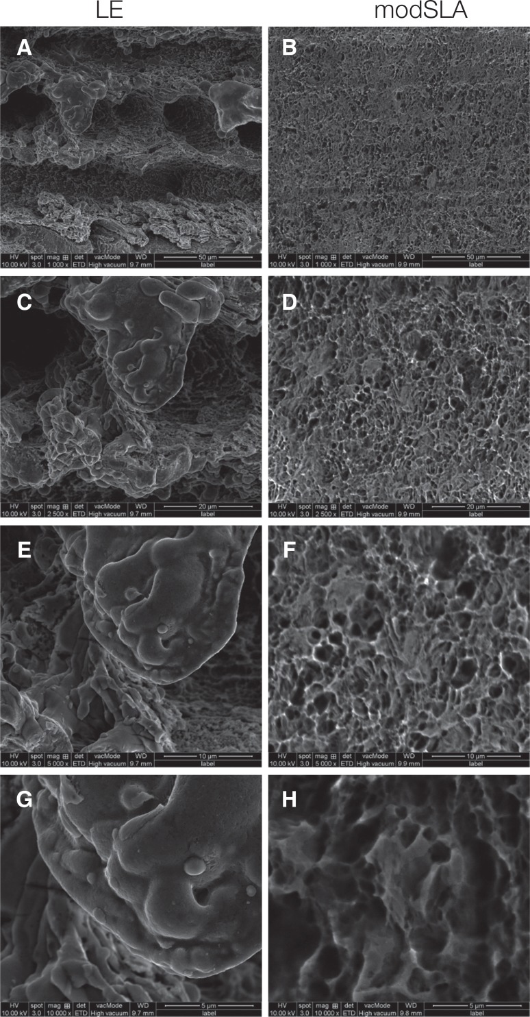
Fig. 1 Scanning electron microscopy image of LE and mod SLA implant surfaces. (A) ×1,000, (B) ×1,000, (C) ×2,500, (D) ×2,500, (E) ×5,000, (F) ×5,000, (G) ×10,000, (H) ×10,000.
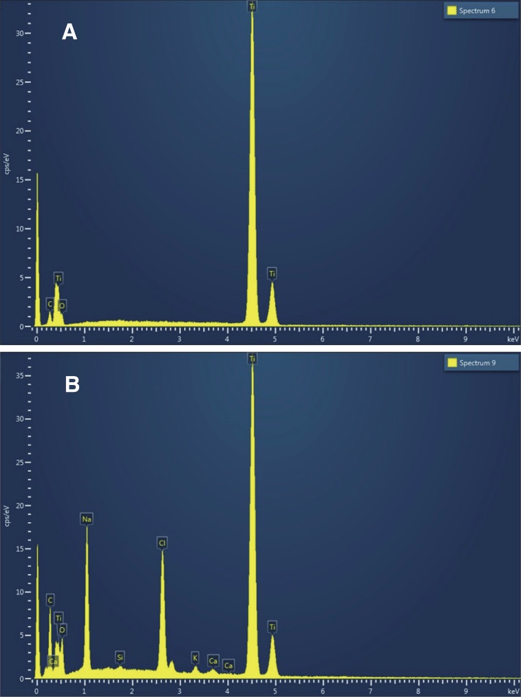
Fig. 2 (A) LE implant EDS spectrum. Ti, O, and C were the only three elements found in the LE implant surfaces. (B) modSLA implant EDS spectrum. Na, Ca, Cl, and K were also observed in modSLA, in addition to Ti, O, and C.
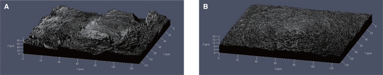
Fig. 3 Confocal laser scanning microscopy image: (A) LE implant surface; (B) modSLA implant surface.
Reference
-
1. Albrektsson T, Brånemark PI, Hansson HA, Lindström J. Osseointegrated titanium implants. Requirements for ensuring a long-lasting, direct bone-to-implant anchorage in man. Acta Orthop Scand. 1981; 52:155–170. PMID: 7246093.2. Brånemark PI, Adell R, Breine U, Hansson BO, Lindström J, Ohlsson A. Intra-osseous anchorage of dental prostheses. I. Experimental studies. Scand J Plast Reconstr Surg. 1969; 3:81–100. PMID: 4924041.3. Smeets R, Stadlinger B, Schwarz F, Beck-Broichsitter B, Jung O, Precht C, Kloss F, Gröbe A, Heiland M, Ebker T. Impact of dental implant surface modifications on osseointegration. Biomed Res Int. 2016; 2016:6285620. PMID: 27478833.
Article4. Chen CJ, Ding SJ, Chen CC. Effects of surface conditions of titanium dental implants on bacterial adhesion. Photomed Laser Surg. 2016; 34:379–388. PMID: 27454339.
Article5. Shah FA, Johansson ML, Omar O, Simonsson H, Palmquist A, Thomsen P. Laser-modified surface enhances osseointegration and biomechanical anchorage of commercially pure titanium implants for bone-anchored hearing systems. PLoS One. 2016; 11:e0157504. PMID: 27299883.
Article6. Park SH, Park KS, Cho SA. Comparison of removal torques of SLActive implant and blasted, laser-treated titanium implant in rabbit tibia bone healed with concentrated growth factor application. J Adv Prosthodont. 2016; 8:110–115. PMID: 27141254.
Article7. Junker R, Dimakis A, Thoneick M, Jansen JA. Effects of implant surface coatings and composition on bone integration: a systematic review. Clin Oral Implants Res. 2009; 20:185–206. PMID: 19663965.
Article8. Wong M, Eulenberger J, Schenk R, Hunziker E. Effect of surface topology on the osseointegration of implant materials in trabecular bone. J Biomed Mater Res. 1995; 29:1567–1575. PMID: 8600147.
Article9. von Wilmowsky C, Moest T, Nkenke E, Stelzle F, Schlegel KA. Implants in bone: part I. A current overview about tissue response, surface modifications and future perspectives. Oral Maxillofac Surg. 2014; 18:243–257. PMID: 23435578.
Article10. Dohan Ehrenfest DM, Coelho PG, Kang BS, Sul YT, Albrektsson T. Classification of osseointegrated implant surfaces: materials, chemistry and topography. Trends Biotechnol. 2010; 28:198–206. PMID: 20116873.
Article11. Zinelis S, Silikas N, Thomas A, Syres K, Eliades G. Surface characterization of SLActive dental implants. Eur J Esthet Dent. 2012; 7:72–92. PMID: 22319766.12. Buser D, Broggini N, Wieland M, Schenk RK, Denzer AJ, Cochran DL, Hoffmann B, Lussi A, Steinemann SG. Enhanced bone apposition to a chemically modified SLA titanium surface. J Dent Res. 2004; 83:529–533. PMID: 15218041.
Article13. Cochran DL, Buser D, ten Bruggenkate CM, Weingart D, Taylor TM, Bernard JP, Peters F, Simpson JP. The use of reduced healing times on ITI implants with a sandblasted and acid-etched (SLA) surface: early results from clinical trials on ITI SLA implants. Clin Oral Implants Res. 2002; 13:144–153. PMID: 11952734.14. Ferguson SJ, Broggini N, Wieland M, de Wild M, Rupp F, Geis-Gerstorfer J, Cochran DL, Buser D. Biomechanical evaluation of the interfacial strength of a chemically modified sandblasted and acid-etched titanium surface. J Biomed Mater Res A. 2006; 78:291–297. PMID: 16637025.
Article15. Marin C, Bonfante EA, Granato R, Suzuki M, Granjeiro JM, Coelho PG. The effect of alterations on resorbable blasting media processed implant surfaces on early bone healing: a study in rabbits. Implant Dent. 2011; 20:167–177. PMID: 21448026.
Article16. Lee JT, Cho SA. Biomechanical evaluation of laser-etched Ti implant surfaces vs. chemically modified SLA Ti implant surfaces: Removal torque and resonance frequency analysis in rabbit tibias. J Mech Behav Biomed Mater. 2016; 61:299–307. PMID: 27093590.
Article17. Gaggl A, Schultes G, Müller WD, Kärcher H. Scanning electron microscopical analysis of laser-treated titanium implant surfaces-a comparative study. Biomaterials. 2000; 21:1067–1073. PMID: 10768759.
Article18. Cho SA, Jung SK. A removal torque of the laser-treated titanium implants in rabbit tibia. Biomaterials. 2003; 24:4859–4863. PMID: 14530083.
Article19. Roberts WE, Smith RK, Zilberman Y, Mozsary PG, Smith RS. Osseous adaptation to continuous loading of rigid endosseous implants. Am J Orthod. 1984; 86:95–111. PMID: 6589962.
Article20. Johansson C, Albrektsson T. Integration of screw implants in the rabbit: a 1-year follow-up of removal torque of titanium implants. Int J Oral Maxillofac Implants. 1987; 2:69–75. PMID: 3481352.21. Johansson CB, Albrektsson T. A removal torque and histomorphometric study of commercially pure niobium and titanium implants in rabbit bone. Clin Oral Implants Res. 1991; 2:24–29. PMID: 1807419.
Article22. Johansson CB, Sennerby L, Albrektsson T. A removal torque and histomorphometric study of bone tissue reactions to commercially pure titanium and Vitallium implants. Int J Oral Maxillofac Implants. 1991; 6:437–441. PMID: 1820312.23. Ivanoff CJ, Sennerby L, Lekholm U. Influence of mono- and bicortical anchorage on the integration of titanium implants. A study in the rabbit tibia. Int J Oral Maxillofac Surg. 1996; 25:229–235. PMID: 8872230.24. Sennerby L, Thomsen P, Ericson LE. A morphometric and biomechanic comparison of titanium implants inserted in rabbit cortical and cancellous bone. Int J Oral Maxillofac Implants. 1992; 7:62–71. PMID: 1398826.25. Albrektsson T, Brånemark PI, Eriksson A, Lindström J. The preformed autologous bone graft. An experimental study in the rabbit. Scand J Plast Reconstr Surg. 1978; 12:215–223. PMID: 368971.
Article26. Raghavendra S, Wood MC, Taylor TD. Early wound healing around endosseous implants: a review of the literature. Int J Oral Maxillofac Implants. 2005; 20:425–431. PMID: 15973954.27. Brett PM, Harle J, Salih V, Mihoc R, Olsen I, Jones FH, Tonetti M. Roughness response genes in osteoblasts. Bone. 2004; 35:124–133. PMID: 15207748.
Article28. Bagno A, Di Bello C. Surface treatments and roughness properties of Ti-based biomaterials. J Mater Sci Mater Med. 2004; 15:935–949. PMID: 15448401.
Article29. Kang SH, Cho SA. Comparison of removal torques for lasertreated titanium implants with anodized implants. J Craniofac Surg. 2011; 22:1491–1495. PMID: 21778844.
Article30. Faeda RS, Tavares HS, Sartori R, Guastaldi AC, Marcantonio E Jr. Biological performance of chemical hydroxyapatite coating associated with implant surface modification by laser beam: biomechanical study in rabbit tibias. J Oral Maxillofac Surg. 2009; 67:1706–1715. PMID: 19615586.
Article
- Full Text Links
-
- Actions
-
Cited
- CITED
-
- Close
- Share
-
- Similar articles
-
- Comparison of removal torque of dual-acid etched and single-acid etched implants in rabbit tibias
- Comparison of removal torques between laser-treated and SLA-treated implant surfaces in rabbit tibiae
- Comparison of alkaline phosphatase activity of MC3T3-E1 cells cultured on different Ti surfaces: modified sandblasted with large grit and acid-etched (MSLA), laser-treated, and laser and acid-treated Ti surfaces
- Characteristics of contact and distance osteogenesis around modified implant surfaces in rabbit tibiae
- Survival of surface-modified short versus long implants in complete or partially edentulous patients with a follow-up of 1 year or more: a systematic review and meta-analysis
