Characteristics of contact and distance osteogenesis around modified implant surfaces in rabbit tibiae
- Affiliations
-
- 1Dental Research Institute, Seoul National University School of Dentistry, Seoul, Korea. pros53@snu.ac.kr
- 2Department of Prosthodontics, Seoul National University School of Dentistry, Seoul, Korea.
- KMID: 2383832
- DOI: http://doi.org/10.5051/jpis.2017.47.3.182
Abstract
- PURPOSE
Contact and distance osteogenesis occur around all endosseous dental implants. However, the mechanisms underlying these processes have not been fully elucidated. We hypothesized that these processes occur independently of each other. To test this, we used titanium (Ti) tubes to physically separate contact and distance osteogenesis, thus allowing contact osteogenesis to be measured in the absence of possible triggers from distance osteogenesis.
METHODS
Sandblasted and acid-etched (SLA) and modified SLA (modSLA) implants were used. Both types had been sandblasted with large grit and then etched with acid. The modSLA implants then underwent additional treatment to increase hydrophilicity. The implants were implanted into rabbit tibiae, and half were implanted within Ti tubes. The bone-to-implant contact (BIC) ratio was calculated for each implant. Immunohistochemical analyses of bone morphogenetic protein (BMP)-2 expression and new bone formation (Masson trichrome stain) were performed.
RESULTS
The implants outside of Ti tubes were associated with good bone formation along the implant surface. Implantation within a Ti tube significantly reduced the BIC ratio (P<0.001). Compared with the modSLA implants, the SLA implants were associated with significantly higher BIC ratios, regardless of the presence or absence of Ti tubes (P=0.043). In the absence of Ti tubes, the bone adjacent to the implant had areas of new bone formation that expressed BMP-2 at high levels.
CONCLUSIONS
This study disproved the null hypothesis and suggested that contact osteogenesis is initiated by signals from the old bone that undergoes distance osteogenesis after drilling. This signal may be BMP-2.
Keyword
MeSH Terms
Figure
-
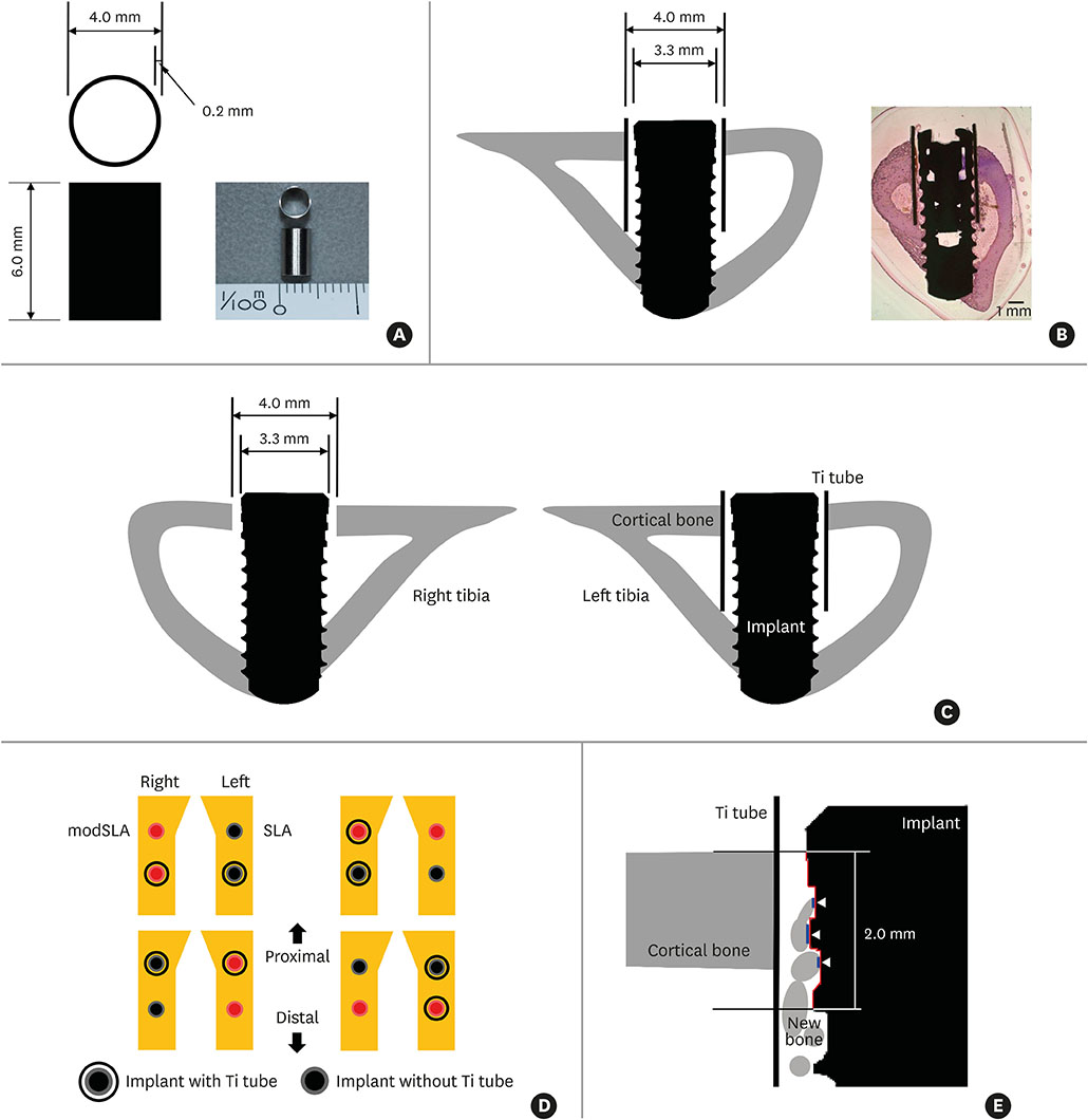
Figure 1 Key features of the in vivo experiment on rabbits. (A) The Ti tube that was used to obstruct the putative influence of distance osteogenesis on contact osteogenesis. The Ti tube is depicted schematically on the left. An actual image is shown on the right. (B) The Ti tube around the implant after both elements were inserted into a rabbit tibia. The resulting structure is depicted schematically on the left. An actual light microscopy image is shown on the right (H&E, bar=1 mm). Note that the implant is bicortically engaged in the tibial bone. (C) Schematic depiction of the implant after insertion with and without the Ti tube. Note that, in this image, there are gaps between the implant and the upper cortical bone in the right tibia and between the Ti tube and the implant inserted in the left tibia. During drilling, we noted that both gaps were filled with blood from the bone marrow and periosteum. This indicates that the gaps may contain various circulating factors such as platelet-derived growth factor. (D) Schematic depiction of the split-plot design with which implants with and without Ti tubes were placed in the 4 pairs of rabbit tibiae. The 4 rabbits used in this study were assumed to be identical. (E) Schematic depiction of how BIC was measured. A 2-mm-long stretch of the cross-sectional implant surface (indicated by the red line) was examined to determine how much of that length was in direct contact with bone: the white arrowheads and blue lines indicate where bone was in contact with the implant surface. The lengths of the blue lines were added up and divided by 2 mm to yield the BIC ratio. For each specimen, the BIC ratios of the right and left sides of the implant were calculated and the average ratio was used in the analyses. Ti: titanium, H&E: hematoxylin and eosin, BIC: bone-to-implant contact, modSLA: modified sandblasted and acid-etched, SLA: sandblasted and acid-etched.
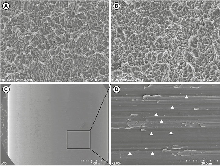
Figure 2 FE-SEM of the modSLA and SLA implants and the Ti tube. (A and B) FE-SEM images of the modSLA (A) and SLA (B) surfaces. Both implant types had been blasted with large-grit sand and acid-etched. The modSLA implant was further modified by being rinsed under nitrogen protection and then sealed in glass tubes containing isotonic NaCl. This procedure was conducted to increase the hydrophilicity of the implant. The surfaces exhibited similar micro-topographical features. The procedure intended to increase hydrophilicity had little effect on the modSLA surface topography. (C) FE-SEM images of the Ti tube. The left image shows a machined surface consisting of grade 4 commercially pure Ti. The right image shows a magnification of the left-hand FE-SEM image. It depicts many machined grooves that were etched by computer numerical control milling (white arrowheads). The bars in the left (A, B, C) and right (C) images indicate 20 μm, 20 μm, 1 mm, and 20 μm, respectively. FE-SEM: field emission scanning electron microscopy, modSLA: modified sandblasted and acid-etched, SLA: sandblasted and acid-etched, Ti: titanium, NaCl: sodium chloride.
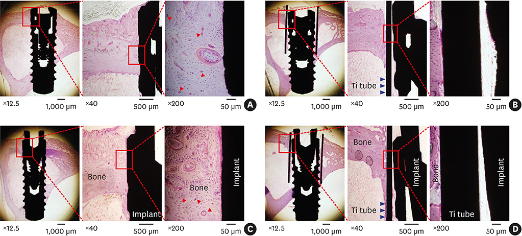
Figure 3 Light microscopic histological views of the modSLA and SLA implants with and without the Ti tube. Both implant types had been blasted with large-grit sand and acid-etched. The modSLA implant was further modified by being rinsed under nitrogen protection and then sealed in glass tubes containing isotonic NaCl. (A) modSLA without Ti tube. (B) modSLA with Ti tube. (C) SLA without Ti tube. (D) SLA with Ti tube. The red boxes indicate the area on the image that was further magnified on the right. The modSLA and SLA implants without Ti tubes showed good formation of cortical bone; Haversian systems (red arrowheads) are visible around the surfaces of both implants (A and C). By contrast, the modSLA and SLA implants with Ti tubes (blue arrowheads) exhibited little or no bone on their surfaces (B and D). Note that the implant surfaces in (A) and (C) were adjacent to a solid wall of bone. By contrast, the spaces between the implants and Ti tubes in (B) and (D) were empty, which indicates a lack of osteogenesis. In (A-D), the bars in the left, middle, and right images indicate 1,000 μm, 500 μm, and 50 μm, respectively (H&E). modSLA: modified sandblasted and acid-etched, SLA: sandblasted and acid-etched, Ti: titanium, NaCl: sodium chloride, H&E: hematoxylin and eosin.
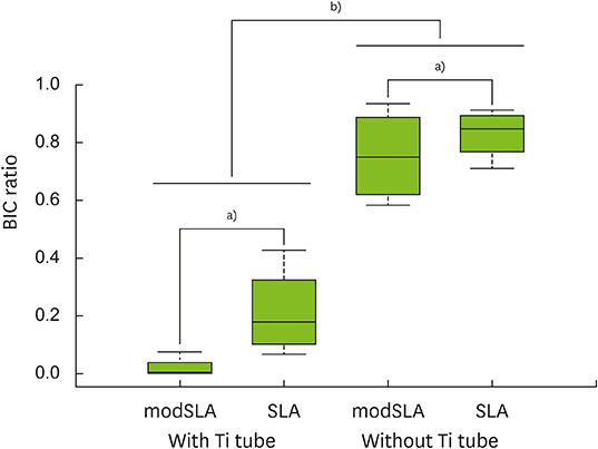
Figure 4 Box-plot analysis of the BIC ratios of each group. In terms of BIC ratios, the SLA surface was more biocompatible than the modSLA surface. However, this difference between the surface types was only marginally significant (i.e., the P values were close to 0.05). Notably, the Ti tubes slowed bone formation considerably regardless of the type of surface. BIC: bone-to-implant contact, modSLA: modified sandblasted and acid-etched, SLA: sandblasted and acid-etched, Ti: titanium. a)P<0.050, b) P<0.010.
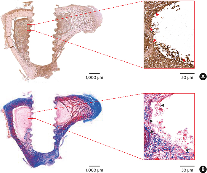
Figure 5 Immunohistochemistry of the tibial bone 10 days after implants were placed. Modified modSLA and SLA implants were placed in the tibiae of a rabbit. Ten days after surgery, the rabbit was sacrificed, the implants were removed, and tibial bone sections were generated. (A) The sections were stained for BMP-2 expression. BMP-2 staining is shown by the dark brown areas. Note the marked expression of BMP-2 (red arrowheads) at the bone-implant interface. This suggests that BMP-2 produced by old bone may initiate contact osteogenesis. (B) Other bone sections were stained with Masson trichrome to identify areas of new bone formation (blue color, black arrowheads) along the bone-implant interface. These areas correspond to the area of strong BMP-2 expression in (A). The red rectangular areas shown in the left-hand images (bar=1,000 μm) were magnified further in the right-hand images (bar=50 μm). modSLA: modified sandblasted and acid-etched, SLA: sandblasted and acid-etched, BMP: bone morphogenetic protein.
Reference
-
1. Osborn J, Newesely H. Dynamic aspects of the implant-bone interface. In : Heimke G, editor. Dental implants. Münche: Verlag;1980. p. 111–123.2. Berglundh T, Abrahamsson I, Lang NP, Lindhe J. De novo alveolar bone formation adjacent to endosseous implants. Clin Oral Implants Res. 2003; 14:251–262.3. Davies JE. In vitro modeling of the bone/implant interface. Anat Rec. 1996; 245:426–445.4. Moreo P, García-Aznar JM, Doblaré M. Bone ingrowth on the surface of endosseous implants. Part 1: mathematical model. J Theor Biol. 2009; 260:1–12.
Article5. Sela MN, Badihi L, Rosen G, Steinberg D, Kohavi D. Adsorption of human plasma proteins to modified titanium surfaces. Clin Oral Implants Res. 2007; 18:630–638.
Article6. Terheyden H, Lang NP, Bierbaum S, Stadlinger B. Osseointegration--communication of cells. Clin Oral Implants Res. 2012; 23:1127–1135.7. Kilkenny C, Browne WJ, Cuthill IC, Emerson M, Altman DG. Improving bioscience research reporting: the ARRIVE guidelines for reporting animal research. Osteoarthritis Cartilage. 2012; 20:256–260.
Article8. Yeo IS, Han JS, Yang JH. Biomechanical and histomorphometric study of dental implants with different surface characteristics. J Biomed Mater Res B Appl Biomater. 2008; 87:303–311.
Article9. Donath K, Breuner G. A method for the study of undecalcified bones and teeth with attached soft tissues. The Säge-Schliff (sawing and grinding) technique. J Oral Pathol. 1982; 11:318–326.
Article10. Berglundh T, Lindhe J, Jonsson K, Ericsson I. The topography of the vascular systems in the periodontal and peri-implant tissues in the dog. J Clin Periodontol. 1994; 21:189–193.
Article11. Nygren H, Tengvall P, Lundström I. The initial reactions of TiO2 with blood. J Biomed Mater Res. 1997; 34:487–492.
Article12. Park JY, Davies JE. Red blood cell and platelet interactions with titanium implant surfaces. Clin Oral Implants Res. 2000; 11:530–539.
Article13. Ammann AJ, Beck LS, DeGuzman L, Hirabayashi SE, Lee WP, McFatridge L, et al. Transforming growth factor-beta. Effect on soft tissue repair. Ann N Y Acad Sci. 1990; 593:124–134.14. Brighton CT. The biology of fracture repair. Instr Course Lect. 1984; 33:60–82.15. Brunski JB. In vivo bone response to biomechanical loading at the bone/dental-implant interface. Adv Dent Res. 1999; 13:99–119.
Article16. Cromack DT, Pierce GF, Mustoe TA. TGF-beta and PDGF mediated tissue repair: identifying mechanisms of action using impaired and normal models of wound healing. Prog Clin Biol Res. 1991; 365:359–373.17. Ikeda K, Takeshita S. Factors and mechanisms involved in the coupling from bone resorption to formation: how osteoclasts talk to osteoblasts. J Bone Metab. 2014; 21:163–167.
Article18. Mustoe TA, Pierce GF, Morishima C, Deuel TF. Growth factor-induced acceleration of tissue repair through direct and inductive activities in a rabbit dermal ulcer model. J Clin Invest. 1991; 87:694–703.
Article19. Pfeilschifter J, Wolf O, Naumann A, Minne HW, Mundy GR, Ziegler R. Chemotactic response of osteoblastlike cells to transforming growth factor beta. J Bone Miner Res. 1990; 5:825–830.
Article20. Postlethwaite AE, Keski-Oja J, Moses HL, Kang AH. Stimulation of the chemotactic migration of human fibroblasts by transforming growth factor beta. J Exp Med. 1987; 165:251–256.
Article21. Deuel TF, Senior RM, Huang JS, Griffin GL. Chemotaxis of monocytes and neutrophils to platelet-derived growth factor. J Clin Invest. 1982; 69:1046–1049.
Article22. Huang JS, Huang SS, Kennedy B, Deuel TF. Platelet-derived growth factor. Specific binding to target cells. J Biol Chem. 1982; 257:8130–8136.
Article23. Park JY, Gemmell CH, Davies JE. Platelet interactions with titanium: modulation of platelet activity by surface topography. Biomaterials. 2001; 22:2671–2682.
Article24. Pierce GF, Mustoe TA, Lingelbach J, Masakowski VR, Griffin GL, Senior RM, et al. Platelet-derived growth factor and transforming growth factor-beta enhance tissue repair activities by unique mechanisms. J Cell Biol. 1989; 109:429–440.
Article25. Seppä H, Grotendorst G, Seppä S, Schiffmann E, Martin GR. Platelet-derived growth factor in chemotactic for fibroblasts. J Cell Biol. 1982; 92:584–588.
Article26. Urist MR. Bone: formation by autoinduction. Science. 1965; 150:893–899.
Article27. El Bialy I, Jiskoot W, Reza Nejadnik M. Formulation, delivery and stability of bone morphogenetic proteins for effective bone regeneration. Pharm Res. 2017; 34:1152–1170.
Article
- Full Text Links
-
- Actions
-
Cited
- CITED
-
- Close
- Share
-
- Similar articles
-
- Stability and Efficacy of Titanium in Osteointegration in a Rabbit Model
- Effect of implant surface microtopography by hydroxyapatite grit-blasting on adhesion, proliferation, and differentiation of osteoblast-like cell line, MG-63
- Histomorphometric evaluation of osteogenesis with brushite implant surfaces in dogs
- Comparison between TiUniteTM and another oxidized implant using the rabbit tibia model
- Influence of the adjacent periodontium and inter-implant distance on bone resorption around non-submerged implants: A retrospective clinical and radiographic study
