Evaluation of the periodontal regenerative properties of patterned human periodontal ligament stem cell sheets
- Affiliations
-
- 1Department of Periodontology, Chonbuk National University School of Dentistry and Institute of Oral Bioscience, Jeonju, Korea. grayheron@hanmail.net
- 2Department of Bioengineering, University of Washington, Seattle, WA, USA.
- 3Institute for Stem Cell and Regenerative Medicine, University of Washington, Seattle, WA, USA.
- 4Center for Cardiovascular Biology, University of Washington, Seattle, WA, USA.
- 5Research Institute of Clinical Medicine, Chonbuk National University, Jeonju, Korea.
- 6Biomedical Research Institute, Chonbuk National University Hospital, Jeonju, Korea.
- KMID: 2399746
- DOI: http://doi.org/10.5051/jpis.2017.47.6.402
Abstract
- PURPOSE
The aim of this study was to determine the effects of patterned human periodontal ligament stem cell (hPDLSC) sheets fabricated using a thermoresponsive substratum.
METHODS
In this study, we fabricated patterned hPDLSC sheets using nanotopographical cues to modulate the alignment of the cell sheet.
RESULTS
The hPDLSCs showed rapid monolayer formation on various surface pattern widths. Compared to cell sheets grown on flat surfaces, there were no significant differences in cell attachment and growth on the nanopatterned substratum. However, the patterned hPDLSC sheets showed higher periodontal ligamentogenesis-related gene expression in early stages than the unpatterned cell sheets.
CONCLUSIONS
This experiment confirmed that patterned cell sheets provide flexibility in designing hPDLSC sheets, and that these stem cell sheets may be candidates for application in periodontal regenerative therapy.
MeSH Terms
Figure
-
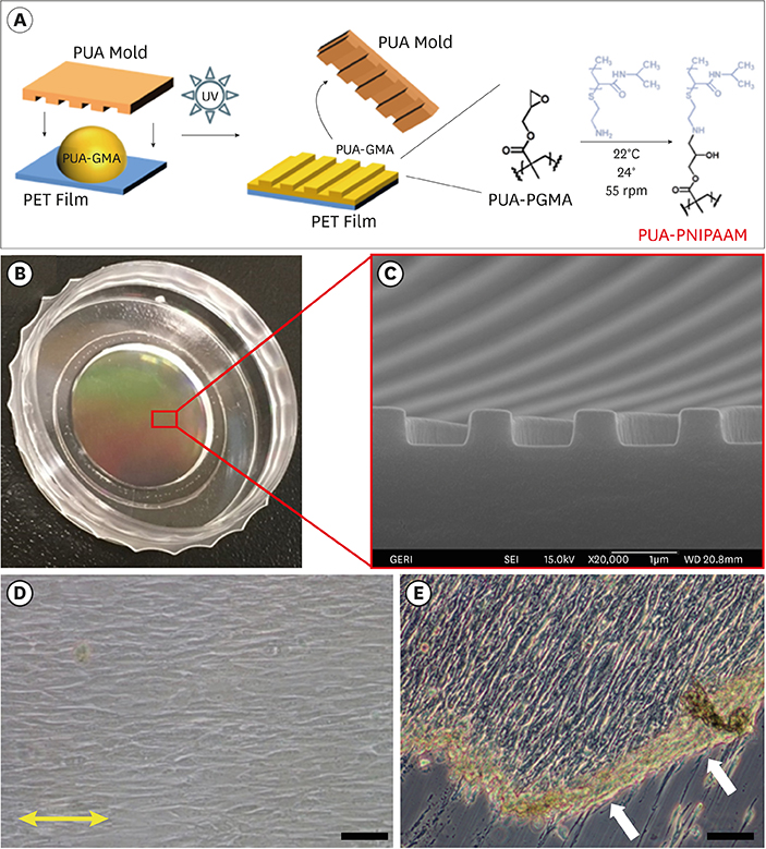
Figure 1 (A) The PNIPAM-functionalized thermoresponsive substratum with nanopatterns. Capillary force lithography was used for the fabrication of the substratum, with subsequent modification using amine-terminated PNIPAM on the PET film. (B) Representative macroscopic image of a large-area, scalable substratum, and (C) an SEM image of the 800-nm nanopatterned surface. (D) A microscopic view of hPDLSCs cultured on the 800-nm width nanopatterned surface incubated at 37°C for 48 hours following cell seeding. The yellow arrow designates the direction in which the hPDLSCs were aligned (scale bar=200 μm). (E) Cell sheet detachment from the substratum after low-temperature treatment. An aligned and interconnected hPDLSC sheet with intact cell-to-cell junctions was slowly detached from the substratum as a single monolayer sheet upon water penetration from the periphery after being maintained at 22°C for 30 minutes. The white arrow indicates the direction of detachment (scale bar=200 μm). PNIPAM: poly(N-isopropyl-acrylamide), SEM: scanning electron microscopy, hPDLSC: human periodontal ligament stem cell, PUA: poly(urethane acrylate), GMA: glycidyl methacrylate, PET: polyethylene terephthalate, PGMA, poly(glycidyl methacrylate).
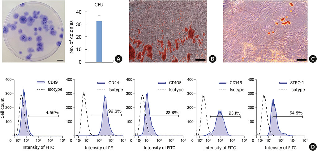
Figure 2 Characterization of primary cultured hPDLSCs. (A) Representative images of crystal violet staining of CFUs of primary cultured hPDLSCs at day 14 (scale bars=10 mm) and quantitative measurement of the CFUs (32.3±4.0, mean±SD). (B) Representative images of alizarin red staining of mineralized nodules after osteogenic differentiation (scale bar=100 μm). (C) Representative images of oil red O staining of lipid-rich vacuoles after adipogenic differentiation (scale bar=100 μm). (D) Immunophenotypical analyses of hPDLSCs. CD44, CD105, CD146, and STRO-1 were used as positive markers and CD19 was used as a negative marker. hPDLSC: human periodontal ligament stem cell, CFU: colony-forming unit, SD: standard deviation, STRO-1: stromal cell surface marker-1, FITC: fluorescein isothiocyanate, PE: phycoerythrin.
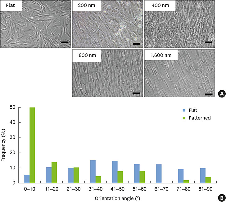
Figure 3 Microscopic images of hPDLSC sheet formation from the flat, 200-, 400-, 800-, and 1,600-nm nanopatterns at 48 hours. The hPDLSCs demonstrated varying degrees of confluent cell sheet formation depending on the culture plate surface condition. The 800-nm surface resulted in the optimal conditions for cell patterning (scale bars=200 μm). A histogram of the orientation of the hPDLSCs shows the quantified initial cellular adhesion angle on the flat surface and 800-nm width nanopatterned surface. The cells were patterned almost parallel to the direction of the grooves (n=4). hPDLSC: human periodontal ligament stem cell.

Figure 4 Cell proliferation was evaluated for up to 10 days (n=4, P<0.05). Microscopic image sets of cell growth on the flat and nanopatterned surfaces on days 1, 4, 7, and 10. On the nanopatterned surfaces, hPDLSCs extended and patterned in the direction of groove orientation with a spindle-like shape, and grew parallel to the direction of the grooves (scale bars=200 μm). hPDLSC: human periodontal ligament stem cell.
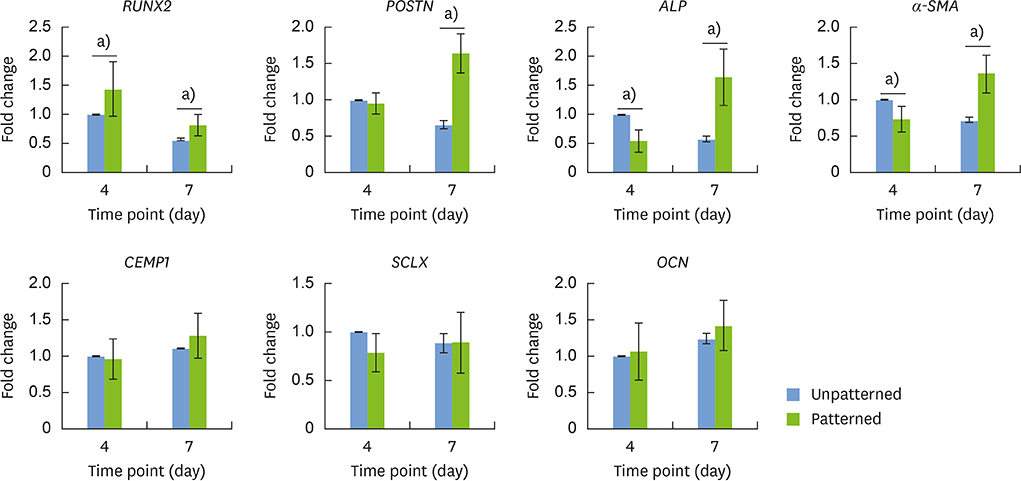
Figure 5 Differences in hPDLSC gene expression between patterned and unpatterned cell sheets as assessed by quantitative RT-PCR. The expression of the following genes was upregulated on the patterned cell sheet; RUNX2 on both days 4 and 7, and POSTN, ALP, and α-SMA on day 7. α-SMA and ALP showed decreased expression on day 4. There was no significant change in the expression of CEMP1, SCLX, and OCN. hPDLSC: human periodontal ligament stem cell, RT-PCR: real-time polymerase chain reaction, RUNX2: runt-related transcription factor 2, POSTN: periostin, ALP: alkaline phosphatase, α-SMA: α-smooth muscle actin, CEMP1: cementum protein 1, SCLX: scleraxis, OCN: osteocalcin. a)Statistical significance (P<0.05).
Reference
-
1. Armitage GC. Development of a classification system for periodontal diseases and conditions. Ann Periodontol. 1999; 4:1–6.
Article2. Apicella A, Heunemann P, Dejace L, Marascio M, Plummer CJ, Fischer P. Scaffold requirements for periodontal regeneration with enamel matrix derivative proteins. Colloids Surf B Biointerfaces. 2017; 156:221–226.
Article3. Reynolds MA, Kao RT, Camargo PM, Caton JG, Clem DS, Fiorellini JP, et al. Periodontal regeneration - intrabony defects: a consensus report from the AAP Regeneration Workshop. J Periodontol. 2015; 86:S105–S107.
Article4. Greenwell H. Committee on Research, Science and Therapy. American Academy of Periodontology. Position paper: guidelines for periodontal therapy. J Periodontol. 2001; 72:1624–1628.5. Hasegawa M, Yamato M, Kikuchi A, Okano T, Ishikawa I. Human periodontal ligament cell sheets can regenerate periodontal ligament tissue in an athymic rat model. Tissue Eng. 2005; 11:469–478.
Article6. Bright R, Hynes K, Gronthos S, Bartold PM. Periodontal ligament-derived cells for periodontal regeneration in animal models: a systematic review. J Periodontal Res. 2015; 50:160–172.
Article7. Gay IC, Chen S, MacDougall M. Isolation and characterization of multipotent human periodontal ligament stem cells. Orthod Craniofac Res. 2007; 10:149–160.
Article8. Ishikawa I, Iwata T, Washio K, Okano T, Nagasawa T, Iwasaki K, et al. Cell sheet engineering and other novel cell-based approaches to periodontal regeneration. Periodontol 2000. 2009; 51:220–238.
Article9. Xu Q, Li B, Yuan L, Dong Z, Zhang H, Wang H, et al. Combination of platelet-rich plasma within periodontal ligament stem cell sheets enhances cell differentiation and matrix production. J Tissue Eng Regen Med. 2017; 11:627–636.
Article10. Carter SD, Costa PF, Vaquette C, Ivanovski S, Hutmacher DW, Malda J. Additive biomanufacturing: an advanced approach for periodontal tissue regeneration. Ann Biomed Eng. 2017; 45:12–22.
Article11. Okano T, Bae YH, Jacobs H, Kim SW. Thermally on-off switching polymers for drug permeation and release. J Control Release. 1990; 11:255–265.
Article12. Li M, Feng C, Gu X, He Q, Wei F. Effect of cryopreservation on proliferation and differentiation of periodontal ligament stem cell sheets. Stem Cell Res Ther. 2017; 8:77.
Article13. Guo S, Kang J, Ji B, Guo W, Ding Y, Wu Y, et al. Periodontal-derived mesenchymal cell sheets promote periodontal regeneration in inflammatory microenvironment. Tissue Eng Part A. 2017; 23:585–596.
Article14. Wang Z, Feng Z, Wu G, Bai S, Dong Y, Zhao Y. In vitro studies on human periodontal ligament stem cell sheets enhanced by enamel matrix derivative. Colloids Surf B Biointerfaces. 2016; 141:102–111.
Article15. Dalby MJ, Gadegaard N, Tare R, Andar A, Riehle MO, Herzyk P, et al. The control of human mesenchymal cell differentiation using nanoscale symmetry and disorder. Nat Mater. 2007; 6:997–1003.
Article16. Kim DH, Provenzano PP, Smith CL, Levchenko A. Matrix nanotopography as a regulator of cell function. J Cell Biol. 2012; 197:351–360.
Article17. McMurray RJ, Gadegaard N, Tsimbouri PM, Burgess KV, McNamara LE, Tare R, et al. Nanoscale surfaces for the long-term maintenance of mesenchymal stem cell phenotype and multipotency. Nat Mater. 2011; 10:637–644.
Article18. Gao H, Li B, Zhao L, Jin Y. Influence of nanotopography on periodontal ligament stem cell functions and cell sheet based periodontal regeneration. Int J Nanomedicine. 2015; 10:4009–4027.19. Jiao A, Trosper NE, Yang HS, Kim J, Tsui JH, Frankel SD, et al. Thermoresponsive nanofabricated substratum for the engineering of three-dimensional tissues with layer-by-layer architectural control. ACS Nano. 2014; 8:4430–4439.
Article20. Kim K, Yi T, Yun JH. Maintained stemness of human periodontal ligament stem cells isolated after prolonged storage of extracted teeth. J Periodontol. 2016; 87:e148–e158.
Article21. Farag A, Vaquette C, Theodoropoulos C, Hamlet SM, Hutmacher DW, Ivanovski S. Decellularized periodontal ligament cell sheets with recellularization potential. J Dent Res. 2014; 93:1313–1319.
Article22. Hu J, Cao Y, Xie Y, Wang H, Fan Z, Wang J, et al. Periodontal regeneration in swine after cell injection and cell sheet transplantation of human dental pulp stem cells following good manufacturing practice. Stem Cell Res Ther. 2016; 7:130.
Article23. Dan H, Vaquette C, Fisher AG, Hamlet SM, Xiao Y, Hutmacher DW, et al. The influence of cellular source on periodontal regeneration using calcium phosphate coated polycaprolactone scaffold supported cell sheets. Biomaterials. 2014; 35:113–122.
Article24. Seo BM, Miura M, Gronthos S, Bartold PM, Batouli S, Brahim J, et al. Investigation of multipotent postnatal stem cells from human periodontal ligament. Lancet. 2004; 364:149–155.
Article25. Lemaitre M, Monsarrat P, Blasco-Baque V, Loubières P, Burcelin R, Casteilla L, et al. Periodontal tissue regeneration using syngeneic adipose-derived stromal cells in a mouse model. Stem Cells Transl Med. 2017; 6:656–665.
Article26. Wang ZS, Feng ZH, Wu GF, Bai SZ, Dong Y, Chen FM, et al. The use of platelet-rich fibrin combined with periodontal ligament and jaw bone mesenchymal stem cell sheets for periodontal tissue engineering. Sci Rep. 2016; 6:28126.
Article27. Feng R, Lengner C. Application of stem cell technology in dental regenerative medicine. Adv Wound Care (New Rochelle). 2013; 2:296–305.
Article28. Du J, Shan Z, Ma P, Wang S, Fan Z. Allogeneic bone marrow mesenchymal stem cell transplantation for periodontal regeneration. J Dent Res. 2014; 93:183–188.
Article29. Owaki T, Shimizu T, Yamato M, Okano T. Cell sheet engineering for regenerative medicine: current challenges and strategies. Biotechnol J. 2014; 9:904–914.
Article30. Kim JH, Kang MS, Eltohamy M, Kim TH, Kim HW. Dynamic mechanical and nanofibrous topological combinatory cues designed for periodontal ligament engineering. PLoS One. 2016; 11:e0149967.
Article31. Yin Z, Chen X, Chen JL, Shen WL, Hieu Nguyen TM, Gao L, et al. The regulation of tendon stem cell differentiation by the alignment of nanofibers. Biomaterials. 2010; 31:2163–2175.
Article32. Jiang W, Li L, Zhang D, Huang S, Jing Z, Wu Y, et al. Incorporation of aligned PCL-PEG nanofibers into porous chitosan scaffolds improved the orientation of collagen fibers in regenerated periodontium. Acta Biomater. 2015; 25:240–252.
Article33. Gauvin R, Parenteau-Bareil R, Larouche D, Marcoux H, Bisson F, Bonnet A, et al. Dynamic mechanical stimulations induce anisotropy and improve the tensile properties of engineered tissues produced without exogenous scaffolding. Acta Biomater. 2011; 7:3294–3301.
Article34. Yu N, Prodanov L, te Riet J, Yang F, Walboomers XF, Jansen JA. Regulation of periodontal ligament cell behavior by cyclic mechanical loading and substrate nanotexture. J Periodontol. 2013; 84:1504–1513.
Article35. San Miguel SM, Fatahi MR, Li H, Igwe JC, Aguila HL, Kalajzic I. Defining a visual marker of osteoprogenitor cells within the periodontium. J Periodontal Res. 2010; 45:60–70.
Article36. Kalajzic Z, Li H, Wang LP, Jiang X, Lamothe K, Adams DJ, et al. Use of an alpha-smooth muscle actin GFP reporter to identify an osteoprogenitor population. Bone. 2008; 43:501–510.
Article37. Jin H, Choung HW, Lim KT, Jin B, Jin C, Chung JH, et al. Recombinant human plasminogen activator inhibitor-1 promotes cementogenic differentiation of human periodontal ligament stem cells. Tissue Eng Part A. 2015; 21:2817–2828.
Article38. Zhu B, Liu W, Liu Y, Zhao X, Zhang H, Luo Z, et al. Jawbone microenvironment promotes periodontium regeneration by regulating the function of periodontal ligament stem cells. Sci Rep. 2017; 7:40088.
Article39. Kadkhoda Z, Safarpour A, Azmoodeh F, Adibi S, Khoshzaban A, Bahrami N. Histopathological comparison between bone marrow- and periodontium-derived stem cells for bone regeneration in rabbit calvaria. Int J Organ Transplant Med. 2016; 7:9–18.40. Scott A, Danielson P, Abraham T, Fong G, Sampaio AV, Underhill TM. Mechanical force modulates scleraxis expression in bioartificial tendons. J Musculoskelet Neuronal Interact. 2011; 11:124–132.
- Full Text Links
-
- Actions
-
Cited
- CITED
-
- Close
- Share
-
- Similar articles
-
- Autologous Stem Cell Application in Periodontal Regeneration Technique (SAI-PRT) Using PDLSCs Directly From an Extracted Tooth...An Insight
- Comparison of Gene Expression from Supernumerary Dental Pulp and Periodontal Ligament Stem Cells
- Stem cell properties of cells derived from canine periodontal ligament
- Immunomodulatory effect of canine periodontal ligament stem cells on allogenic and xenogenic peripheral blood mononuclear cells
- Effects of nitric oxide on the proliferation and differentiation of human periodontal ligament cells
