Dysembryoplastic Neuroepithelial Tumors
- Affiliations
-
- 1Department of Pathology, Samsung Medical Center, Sungkyunkwan University School of Medicine, Seoul, Korea. ylsuh76@skku.edu
- KMID: 2381403
- DOI: http://doi.org/10.4132/jptm.2015.10.05
Abstract
- Dysembryoplastic neuroepithelial tumor (DNT) is a benign glioneuronal neoplasm that most commonly occurs in children and young adults and may present with medically intractable, chronic seizures. Radiologically, this tumor is characterized by a cortical topography and lack of mass effect or perilesional edema. Partial complex seizures are the most common presentation. Three histologic subtypes of DNTs have been described. Histologically, the recognition of a unique, specific glioneuronal element in brain tumor samples from patients with medically intractable, chronic epilepsy serves as a diagnostic feature for complex or simple DNT types. However, nonspecific DNT has diagnostic difficulty because its histology is indistinguishable from conventional gliomas and because a specific glioneuronal element and/or multinodularity are absent. This review will focus on the clinical, radiographic, histopathological, and immunohistochemical features as well as the molecular genetics of all three variants of DNTs. The histological and cytological differential diagnoses for this lesion, especially the nonspecific variant, will be discussed.
Keyword
MeSH Terms
Figure
-
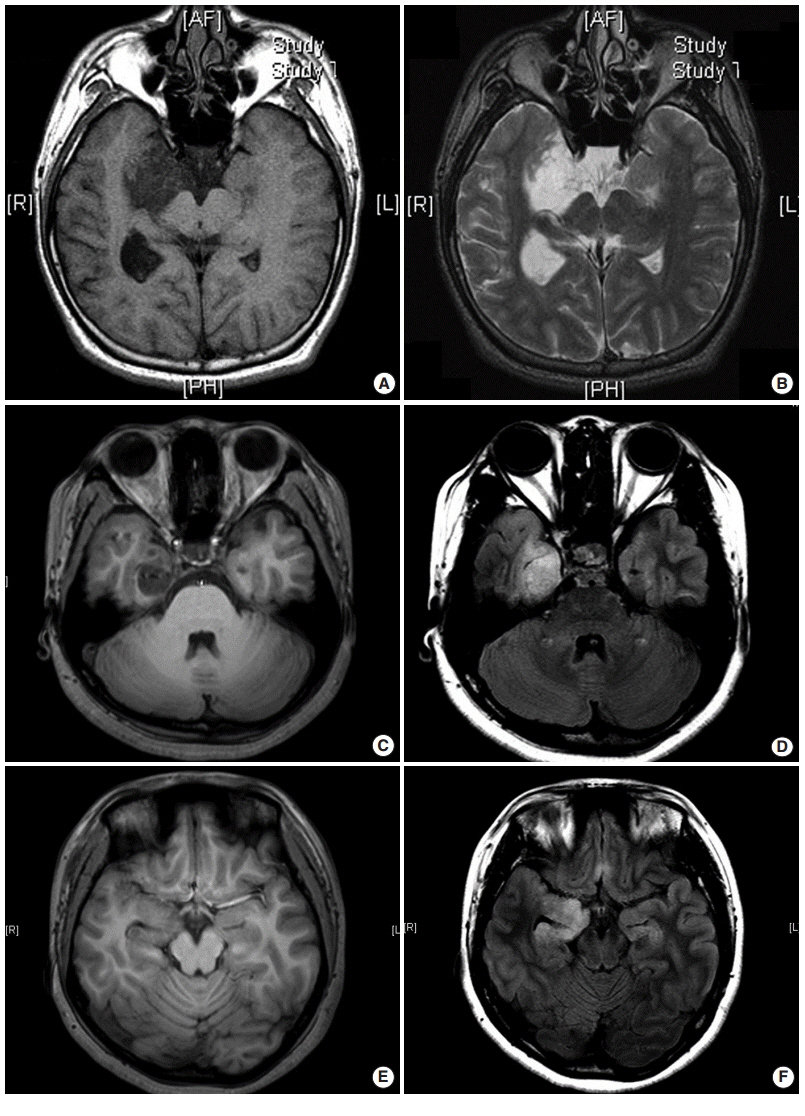
Fig. 1. Three different types of magnetic resonance imaging in dysembryoplastic neuroepithelial tumors. (A, B) Type 1 shows a well-delineated, polycystic-like tumor with strongly hypointense on T1- and hyperintense on T2-weighted images. (C, D) Type 2 shows a nodular-like, heterogeneous lesion. (E, F) Type 3 shows a poorly delineated, dysplastic-like, iso/hyposignal T1 with gray-white matter blurring. Type 1 is mainly found in simple or complex forms, and type 2 and 3 are observed in nonspecific forms. (A, C, E) T1-weighted images. (B, D, F) T2 FLARE images.
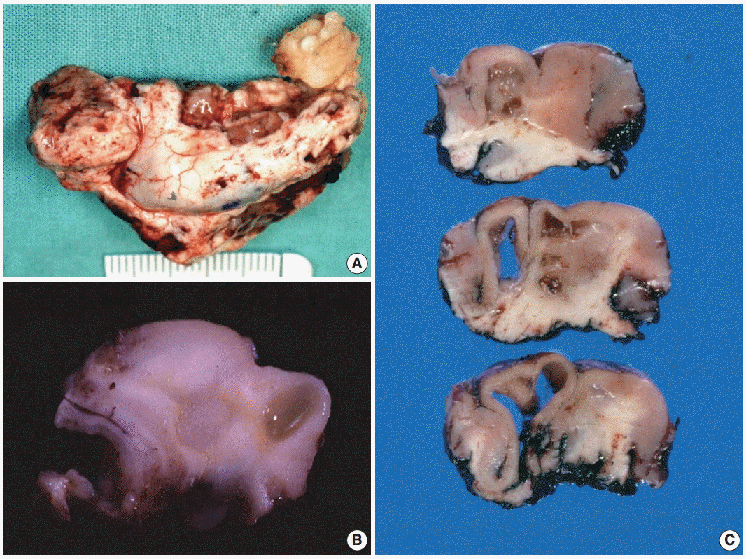
Fig. 2. Gross findings of dysembryoplastic neuroepithelial tumors (DNTs). (A) A hippocampectomy specimen shows a well circumscribed, gray white mass with two small satellite nodules. (B) On the cut section, a complex type of DNT shows multiple gray-white or gelatinous nodules affecting the cortex and white matter. (C) Nonspecific DNT shows a poorly demarcated, cortical thickening with underlying area of white matter rarefaction and cyst formation.
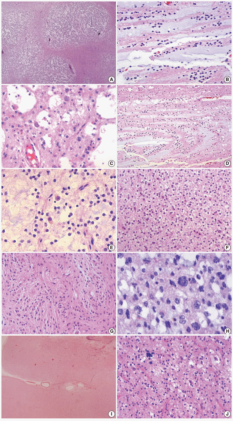
Fig. 3. Histopathological findings of dysembryoplastic neuroepithelial tumors (DNTs). (A) Multi-nodular appearance typical of complex DNTs. (B) Column arrangement of oligodendroglioma-like tumor cells (OLCs) in the specific glioneuronal element. (C) The characteristic appearance of DNTs with OLCs and mature neurons. (D) Targetoid structure of the specific glioneuronal element. (E) Floating neurons in the mucinous matrix. (F) The histology of glial nodules resembling oligodendroglioma. (G) This glial nodule with predominantly astrocytic differentiation. (H)Nuclear pleomorphism in glial component of DNTs. Histopathological findings of dysembryoplastic neuroepithelial tumors (DNTs). (I) A poorly demarcated cortical lesion of nonspecific DNT. (J) The similar histology of nonspecific DNTs to that observed within the glial nodules of complex DNTs.
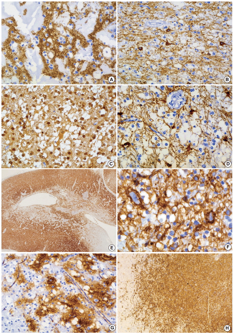
Fig. 4. Immunohistochemical findings of dysembryoplastic neuroepithelial tumors (DNTs). (A, B) Floating neurons are positive for synaptophysin (A) and phosphorylated neurofilament (B). (C, D) The oligodendroglioma-like cells (OLCs) are diffusely positive for S-100 (C) but negative for glial fibrillary acidic protein (D). (E) Nonspecific DNTs show slightly decreased synaptophysin granular staining compared with that seen in the adjacent normal cortex. (F) In the specific glioneuronal element, CD34 is expressed along the perikarya and in membrane of the floating neurons, pericellular stroma, and cytoplasm of OLCs. (G, H) The specific glioneuronal element shows cluster staining pattern of CD34 (G), while diffuse CD34 immunoreactivity in glial nodules of complex DNTs (H).
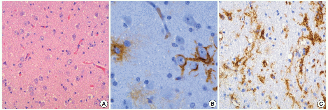
Fig. 5. The histology and CD34 immunoreactivity of focal cortical dysplasia (FCD) in the peritumoral cortex. (A) Dysplastic neurons and abnormalities in cortical lamination are observed. (B, C) CD34 immunoreactivity patterns of FCD are similar to that seen in dysembryoplastic neuroepithelial tumors.

Fig. 6. Ultrastructural findings of dysembryoplastic neuroepithelial tumors. (A) Oligodendroglioma-like cells (OLCs) show oval nuclei with small indentation, marginal aggregates of heterochromatin, and their cytoplasm with scanty organelles (×7,000). (B, C) Neuropil-like network of cellular process with a synaptic contact (arrow) (B, ×17,000) and scant dense core granules (arrow) (C, ×30,000) indicates neuronal differentiation of OLCs. (D) A few OLCs contain ribosome-lamellae complex inclusions (arrow) (×7,000).
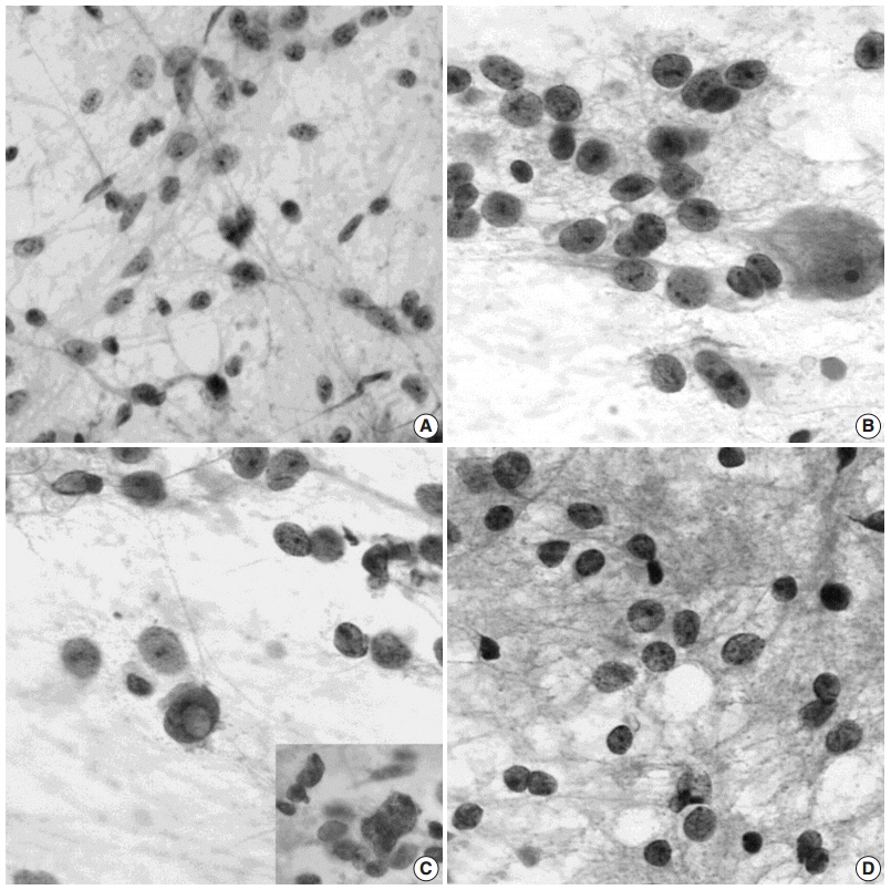
Fig. 7. Squash cytological findings of dysembryoplastic neuroepithelial tumors. (A) Squash preparation shows round, oval to elongated naked nuclei in the mucinous background. (B) The nuclei of oligodendroglioma-like cells (OLCs) are irregular with small indentations or deep grooves, and fine, granular chromatin and 1–4 small nucleoli. There is a large, normal-looking neuron in the mucinous background. (C) OLCs shows multinuclear giant cell formation (inset) and an intranuclear pseudoinclusion. (D) Squash preparation of oligodendroglioma shows smaller, dark nuclei without nucleoli and larger nuclei with granular chromatin and micronucleoli.
Reference
-
1. Burneo JG, Tellez-Zenteno J, Steven DA, et al. Adult-onset epilepsy associated with dysembryoplastic neuroepithelial tumors. Seizure. 2008; 17:498–504.
Article2. Daumas-Duport C, Scheithauer BW, Chodkiewicz JP, Laws ER Jr, Vedrenne C. Dysembryoplastic neuroepithelial tumor: a surgically curable tumor of young patients with intractable partial seizures. Report of thirty-nine cases. Neurosurgery. 1988; 23:545–56.3. Daumas-Duport C. Dysembryoplastic neuroepithelial tumours. Brain Pathol. 1993; 3:283–95.
Article4. Daumas-Duport C, Varlet P, Bacha S, Beuvon F, Cervera-Pierot P, Chodkiewicz JP. Dysembryoplastic neuroepithelial tumors: nonspecific histological forms: a study of 40 cases. J Neurooncol. 1999; 41:267–80.5. Louis DN, Ohgaki H, Wiestler OD, et al. The 2007 WHO classification of tumours of the central nervous system. Acta Neuropathol. 2007; 114:97–109.
Article6. Thom M, Toma A, An S, et al. One hundred and one dysembryoplastic neuroepithelial tumors: an adult epilepsy series with immunohistochemical, molecular genetic, and clinical correlations and a review of the literature. J Neuropathol Exp Neurol. 2011; 70:859–78.
Article7. Honavar M, Janota I, Polkey CE. Histological heterogeneity of dysembryoplastic neuroepithelial tumour: identification and differential diagnosis in a series of 74 cases. Histopathology. 1999; 34:342–56.
Article8. Bodi I, Selway R, Bannister P, et al. Diffuse form of dysembryoplastic neuroepithelial tumour: the histological and immunohistochemical features of a distinct entity showing transition to dysembryoplastic neuroepithelial tumour and ganglioglioma. Neuropathol Appl Neurobiol. 2012; 38:411–25.
Article9. Sung CO, Suh YL, Hong SC. CD34 and microtubule-associated protein 2 expression in dysembryoplastic neuroepithelial tumours with an emphasis on dual expression in non-specific types. Histopathology. 2011; 59:308–17.
Article10. Lee D, Cho YH, Kang SY, Yoon N, Sung CO, Suh YL. BRAF V600E mutations are frequent in dysembryoplastic neuroepithelial tumors and subependymal giant cell astrocytomas. J Surg Oncol. 2015; 111:359–64.11. Chassoux F, Landré E, Mellerio C, Laschet J, Devaux B, DaumasDuport C. Dysembryoplastic neuroepithelial tumors: epileptogenicity related to histologic subtypes. Clin Neurophysiol. 2013; 124:1068–78.
Article12. Devaux B, Chassoux F, Guenot M, et al. Epilepsy surgery in France. Neurochirurgie. 2008; 54:453–65.13. Blumcke I, Aronica E, Urbach H, Alexopoulos A, Gonzalez-Martinez JA. A neuropathology-based approach to epilepsy surgery in brain tumors and proposal for a new terminology use for long-term epilepsy-associated brain tumors. Acta Neuropathol. 2014; 128:39–54.
Article14. Piao YS, Lu DH, Chen L, et al. Neuropathological findings in intractable epilepsy: 435 Chinese cases. Brain Pathol. 2010; 20:902–8.
Article15. Kirkpatrick PJ, Honavar M, Janota I, Polkey CE. Control of temporal lobe epilepsy following en bloc resection of low-grade tumors. J Neurosurg. 1993; 78:19–25.
Article16. Rickert CH, Paulus W. Epidemiology of central nervous system tumors in childhood and adolescence based on the new WHO classification. Childs Nerv Syst. 2001; 17:503–11.
Article17. Spalice A, Ruggieri M, Grosso S, et al. Dysembryoplastic neuroepithelial tumors: a prospective clinicopathologic and outcome study of 13 children. Pediatr Neurol. 2010; 43:395–402.
Article18. Chang EF, Christie C, Sullivan JE, et al. Seizure control outcomes after resection of dysembryoplastic neuroepithelial tumor in 50 patients. J Neurosurg Pediatr. 2010; 5:123–130.
Article19. Lee J, Lee BL, Joo EY, et al. Dysembryoplastic neuroepithelial tumors in pediatric patients. Brain Dev. 2009; 31:671–81.
Article20. Leung SY, Gwi E, Ng HK, Fung CF, Yam KY. Dysembryoplastic neuroepithelial tumor: a tumor with small neuronal cells resembling oligodendroglioma. Am J Surg Pathol. 1994; 18:604–14.21. Cervera-Pierot P, Varlet P, Chodkiewicz JP, Daumas-Duport C. Dysembryoplastic neuroepithelial tumors located in the caudate nucleus area: report of four cases. Neurosurgery. 1997; 40:1065–9.
Article22. Fujimoto K, Ohnishi H, Tsujimoto M, Hoshida T, Nakazato Y. Dysembryoplastic neuroepithelial tumor of the cerebellum and brainstem: case report. J Neurosurg. 2000; 93:487–9.23. Ongürü O, Deveci S, Sirin S, Timurkaynak E, Günhan O. Dysembryoplastic neuroepithelial tumor in the left lateral ventricle. Minim Invasive Neurosurg. 2003; 46:306–9.
Article24. Hasselblatt M, Kurlemann G, Rickert CH, et al. Familial occurrence of dysembryoplastic neuroepithelial tumor. Neurology. 2004; 62:1020–1.
Article25. Ranger A, Diosy D. Seizures in children with dysembryoplastic neuroepithelial tumors of the brain: a review of surgical outcomes across several studies. Childs Nerv Syst. 2015; 31:847–55.26. Chassoux F, Daumas-Duport C. Dysembryoplastic neuroepithelial tumors: where are we now? Epilepsia. 2013; 54 Suppl 9:129–34.
Article27. Chassoux F, Rodrigo S, Mellerio C, et al. Dysembryoplastic neuroepithelial tumors: an MRI-based scheme for epilepsy surgery. Neurology. 2012; 79:1699–707.
Article28. Moazzam AA, Wagle N, Shiroishi MS. Malignant transformation of DNETs: a case report and literature review. Neuroreport. 2014; 25:894–9.29. Stanescu Cosson R, Varlet P, Beuvon F, et al. Dysembryoplastic neuroepithelial tumors: CT, MR findings and imaging follow-up: a study of 53 cases. J Neuroradiol. 2001; 28:230–40.30. Sung CO, Suh YL. Different pattern of expression of nestin in the non-specific form of dysembryoplastic neuroepithelial tumors compared to the simple and complex forms. J Neurooncol. 2009; 92:7–13.
Article31. Chappé C, Padovani L, Scavarda D, et al. Dysembryoplastic neuroepithelial tumors share with pleomorphic xanthoastrocytomas and gangliogliomas BRAF(V600E) mutation and expression. Brain Pathol. 2013; 23:574–83.32. Hirose T, Scheithauer BW, Lopes MB, VandenBerg SR. Dysembryoplastic neuroeptihelial tumor (DNT): an immunohistochemical and ultrastructural study. J Neuropathol Exp Neurol. 1994; 53:184–95.33. Biernat W, Liberski PP, Kordek R, Zakrzewski K, Polis L, Budka H. Dysembryoplastic neuroectodermal tumor: an ultrastructural study of six cases. Ultrastruct Pathol. 2001; 25:455–67.
Article34. Park JY, Suh YL, Han J. Dysembryoplastic neuroepithelial tumor: features distinguishing it from oligodendroglioma on cytologic squash preparations. Acta Cytol. 2003; 47:624–9.35. Blümcke I, Luyken C, Urbach H, Schramm J, Wiestler OD. An isomorphic subtype of long-term epilepsy-associated astrocytomas associated with benign prognosis. Acta Neuropathol. 2004; 107:381–8.36. Padovani L, Colin C, Fernandez C, et al. Search for distinctive markers in DNT and cortical grade II glioma in children: same clinicopathological and molecular entities? Curr Top Med Chem. 2012; 12:1683–92.
Article37. Prabowo AS, van Thuijl HF, Scheinin I, et al. Landscape of chromosomal copy number aberrations in gangliogliomas and dysembryoplastic neuroepithelial tumours. Neuropathol Appl Neurobiol. 2015; 41:743–55.
Article38. Prayson RA, Napekoski KM. Composite ganglioglioma/dysembryoplastic neuroepithelial tumor: a clinicopathologic study of 8 cases. Hum Pathol. 2012; 43:1113–8.
Article
- Full Text Links
-
- Actions
-
Cited
- CITED
-
- Close
- Share
-
- Similar articles
-
- Dysembryoplastic Neuroepithelial Tumor in the Cerebellum: Case Report
- Diffuse Ependymal Dysembryoplastic Neuroepithelial Tumor Causing Spinal Drop Metastases: A Case Report
- Dysembryoplastic Neuroepithelial Tumor: CT and MR Findings A Case Report
- Dysembryoplastic Neuroepithelial Tumor: A Case Report
- Dysembryoplastic Neuroepithelial Tumor in Young Patients with Temporal Lobe Epilepsy
