Diffuse Ependymal Dysembryoplastic Neuroepithelial Tumor Causing Spinal Drop Metastases: A Case Report
- Affiliations
-
- 1Department of Radiology, Chonnam National University Medical School, Chonnam National University Hospital, Gwangju 501-757, Korea. radyoon@jnu.ac.kr
- 2Department of Neurosurgery, Chonnam National University Hwasun Hospital, Hwasun 519-809, Korea.
- KMID: 1383862
- DOI: http://doi.org/10.3348/kjr.2012.13.4.492
Abstract
- Dysembryoplastic neuroepithelial tumors (DNETs) arise mostly in the supratentorial cerebral cortex. A very rare case of intraventricular DNET with diffuse ependymal involvement, which causes spinal drop metastasis, is presented.
MeSH Terms
Figure
-
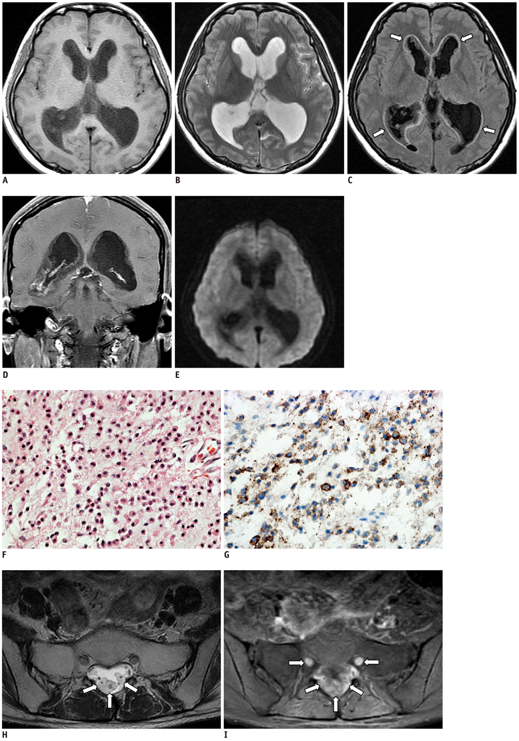
Fig. 1 Diffuse ependymal dysembryoplastic neuroepithelial tumor with spinal drop metastasis in 29-year-old female patient. A. Axial T1-weighted image shows diffuse and nodular low signal intensity lesions along ependymal surface of lateral ventricles and third ventricle. B. Lesions show bright signal intensity on axial T2-weighted image. C. Axial fluid attenuated inversion recovery image shows hyperintense rim (arrows) along wall of ventricles between mass lesions and underlying periventricular white matter. D. Coronal, contrast-enhanced T1-weighted image shows peripheral rim like or nodular enhancement along surface of mass lesions. E. Axial diffusion-weighted image shows no diffusion restriction within tumor. F. Photomicrograph shows oligodendrocyte-like cells in mucinous matrix (H&E, × 400). G. Immunohistochemical staining shows that synaptophysin is expressed in larger tumor cells, which are supposed to be neuronal component (IHC, × 400). H. Axial, T2-weighted lumbar spine magnetic resonance (MR) image obtained 29 months after initial presentation shows well defined, multiple intradural masses with high signal intensity surrounding cauda equina (arrows). I. Axial, contrast enhanced T1-weighted lumbar spine MR image shows extensive enhancement in intradural masses and bilateral S1 nerve roots (arrows).
Reference
-
1. Daumas-Duport C, Scheithauer BW, Chodkiewicz JP, Laws ER Jr, Vedrenne C. Dysembryoplastic neuroepithelial tumor: a surgically curable tumor of young patients with intractable partial seizures. Report of thirty-nine cases. Neurosurgery. 1988. 23:545–556.2. Cervera-Pierot P, Varlet P, Chodkiewicz JP, Daumas-Duport C. Dysembryoplastic neuroepithelial tumors located in the caudate nucleus area: report of four cases. Neurosurgery. 1997. 40:1065–1069. discussion 1069-1070.3. Guesmi H, Houtteville JP, Courthéoux P, Derlon JM, Chapon F. [Dysembryoplastic neuroepithelial tumors. Report of 8 cases including two with unusual localization]. Neurochirurgie. 1999. 45:190–200.4. Fujimoto K, Ohnishi H, Tsujimoto M, Hoshida T, Nakazato Y. Dysembryoplastic neuroepithelial tumor of the cerebellum and brainstem. Case report. J Neurosurg. 2000. 93:487–489.5. Cataltepe O, Marshall P, Smith TW. Dysembryoplastic neuroepithelial tumor located in pericallosal and intraventricular area in a child. Case report. J Neurosurg Pediatr. 2009. 3:456–460.6. Ongürü O, Deveci S, Sirin S, Timurkaynak E, Günhan O. Dysembryoplastic neuroepithelial tumor in the left lateral ventricle. Minim Invasive Neurosurg. 2003. 46:306–309.7. Harter DH, Omeis I, Forman S, Braun A. Endoscopic resection of an intraventricular dysembryoplastic neuroepithelial tumor of the septum pellucidum. Pediatr Neurosurg. 2006. 42:105–107.8. Altinörs N, Calisaneller T, Gülsşen S, Ozen O, Ongürü O. Intraventricular dysembryoplastic neuroepithelial tumor: case report. Neurosurgery. 2007. 61:E1332–E1333. discussion E1333.9. Bilginer B, Söylemezoğlu F, Cila A, Akalan N. Intraventricular dysembryoplastic neuroepithelial tumor-like neoplasm with disseminated spinal tumor. Turk Neurosurg. 2009. 19:69–72.10. Campos AR, Clusmann H, von Lehe M, Niehusmann P, Becker AJ, Schramm J, et al. Simple and complex dysembryoplastic neuroepithelial tumors (DNT) variants: clinical profile, MRI, and histopathology. Neuroradiology. 2009. 51:433–443.11. Stanescu Cosson R, Varlet P, Beuvon F, Daumas Duport C, Devaux B, Chassoux F, et al. Dysembryoplastic neuroepithelial tumors: CT, MR findings and imaging follow-up: a study of 53 cases. J Neuroradiol. 2001. 28:230–240.12. Parmar HA, Hawkins C, Ozelame R, Chuang S, Rutka J, Blaser S. Fluid-attenuated inversion recovery ring sign as a marker of dysembryoplastic neuroepithelial tumors. J Comput Assist Tomogr. 2007. 31:348–353.
