Effects of Gypenosides on Dopaminergic Neuronal Cell Death in 6-Hydroxydopamine-lesioned Rat Model of Parkinson's Disease with Long-term L-DOPA Treatment
- Affiliations
-
- 1College of Pharmacy and Research Center for Bioresource and Health, Chungbuk National University, Cheongju, Chungbuk 28644, Korea. myklee@chungbuk.ac.kr
- 2Department of Food and Nutrition, Chungcheong University, 38, Wuelgok-gil, Cheongju, Chungbuk 28171, Korea.
- KMID: 2355144
- DOI: http://doi.org/10.20307/nps.2016.22.3.187
Abstract
- The goal of this study was to determine whether gypenosides (GPS) exert protective effects against dopaminergic neuronal cell death in a 6-hydroxydopamine (OHDA)-lesioned rat model of Parkinson's disease (PD) with or without long-term 3,4-dihydroxyphenylalanine (L-DOPA) treatment. Rats were injected with 6-OHDA in the substantia nigra to induce PD-like symptoms; 14 days after injection, groups of 6-OHDA-lesioned animals were treated for 21 days with GPS (25 or 50 mg/kg) and/or L-DOPA (20 mg/kg). Dopaminergic neuronal cell death was assessed by counting tyrosine hydroxylase (TH)-immunopositive cells in the substantia nigra and measuring levels of dopamine, norepinephrine, 3,4-dihydroxyphenylacetic acid (DOPAC), and homovanillic acid (HVA) in the striatum. Dopaminergic neuronal cell death induced by 6-OHDA lesions was ameliorated by GPS treatment (50 mg/kg). L-DOPA treatment exacerbated 6-OHDA-induced dopaminergic neuronal cell death; however, these effects were partially reversed by GPS treatment (25 and 50 mg/kg). These results suggest that GPS treatment is protective against dopaminergic neuronal cell death in a 6-OHDA-lesioned rat model of PD with long-term L-DOPA treatment. Therefore, GPS may be useful as a phytotherapeutic agent for the treatment of PD.
Keyword
MeSH Terms
-
3,4-Dihydroxyphenylacetic Acid
Animals
Cell Death*
Dihydroxyphenylalanine
Dopamine
Dopaminergic Neurons*
Homovanillic Acid
Levodopa*
Models, Animal*
Norepinephrine
Oxidopamine
Parkinson Disease*
Rats*
Substantia Nigra
Tyrosine 3-Monooxygenase
3,4-Dihydroxyphenylacetic Acid
Dihydroxyphenylalanine
Dopamine
Homovanillic Acid
Levodopa
Norepinephrine
Oxidopamine
Tyrosine 3-Monooxygenase
Figure
-

Fig. 1. Representative photomicrographs of tyrosine hydroxylase (TH) immunoreactivity (A) and numbers of surviving TH-immu-nopositive cells (B) in the substantia nigra. Fourteen days after lesions were induced by 6-hydroxydopamine (6-OHDA) injection into the substantia nigra, rats were treated for 21 days with gypenosides (GPS) and/or a combination of L-DOPA (LD) and benserazide. (A) Arrows indicate 6-OHDA-lesioned areas. Scale bar = 100 µm. (B) Numbers of TH-immunopositive cells on the lesioned side (L) were analyzed as a percentage of the intact side (R). Results are presented as mean ± S.E.M.; n = 8 – 10 animals per group.∗P <0.05 compared with control group;#P <0.05 compared with 6-OHDA-lesioned group; §P < 0.05 compared with 6-OHDA-lesioned group treated with L-DOPA alone.
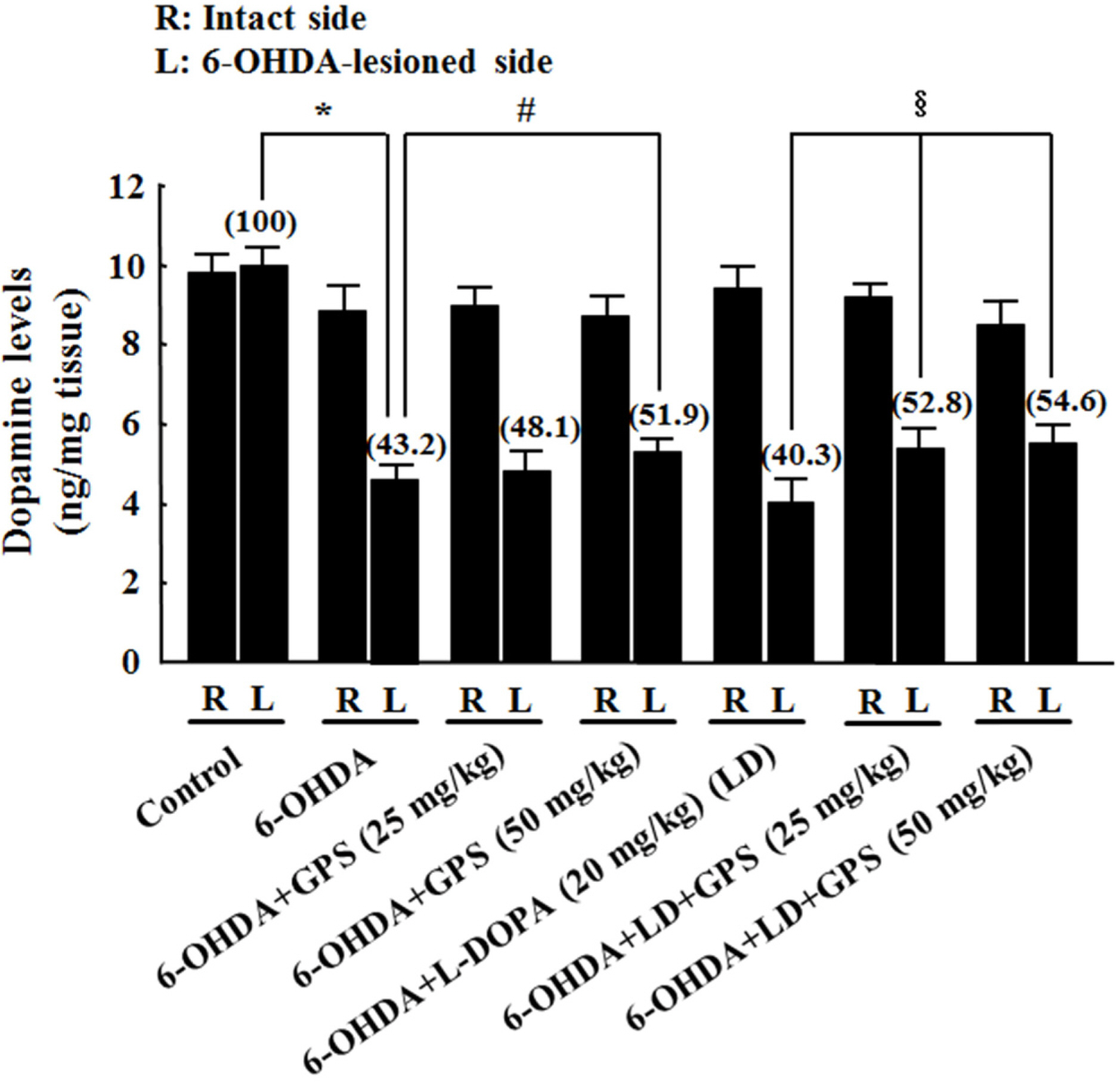
Fig. 2. Effects of GPS on striatal dopamine levels on the intact (R) and 6-OHDA-lesioned (L) sides. Following 6-OHDA lesion and L-DOPA and/or GPS treatment, levels of dopamine in the striatum were determined using HPLC. Numbers in parentheses represent dopamine levels expressed as percentages of the control group (L). Results are presented as mean ± S.E.M.; n = 8 – 10 animals per group.∗P < 0.05 compared with control group;#P <0.05 compared with 6-OHDA-lesioned group; §P <0.05 compared with 6-OHDA-lesioned group treated with L-DOPA alone.
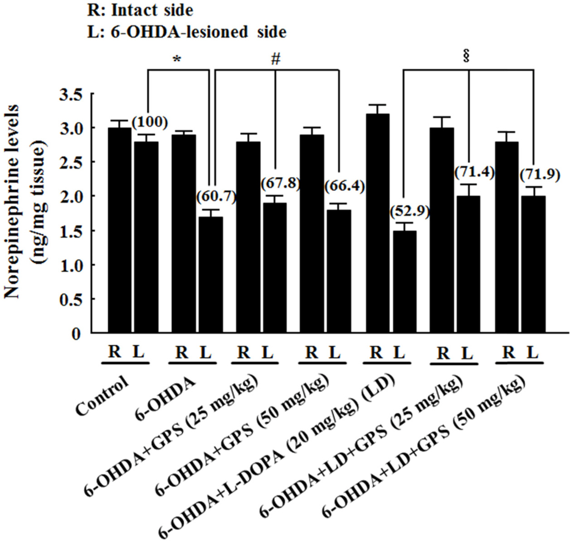
Fig. 3. Effects of GPS on striatal norepinephrine levels on the intact (R) and 6-OHDA-lesioned (L) sides. Numbers in parentheses represent norepinephrine levels expressed as percentages of the control group (L). Results are presented as mean ± S.E.M.; n =8–10 animals per group.∗P <0.05 compared with control group;#P < 0.05 compared with 6-OHDA-lesioned group;§P < 0.05 compared with 6-OHDA-lesioned group treated with L-DOPA alone.
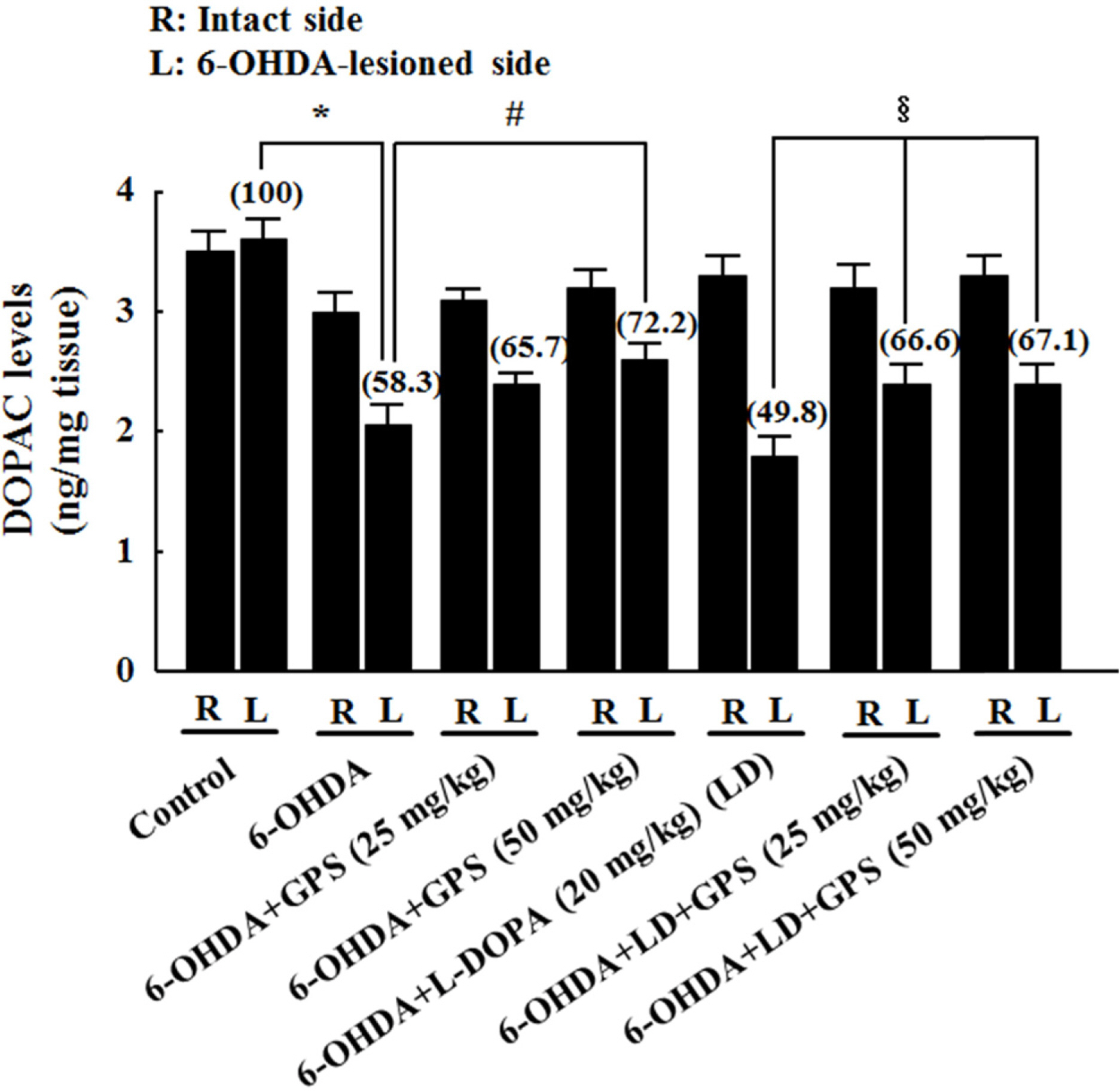
Fig. 4. Effects of GPS on striatal 3,4-dihydroxyphenylacetic acid (DOPAC) levels on the intact (R) and 6-OHDA-lesioned (L) sides. Numbers in parentheses represent DOPAC levels expressed as percentages of the control group (L). Results are presented as mean ± S.E.M.; n=8–10 animals per group.∗P <0.05 compared with control group;#P < 0.05 compared with 6-OHDA-lesioned group;§P < 0.05 compared with 6-OHDA-lesioned group treated with L-DOPA alone.
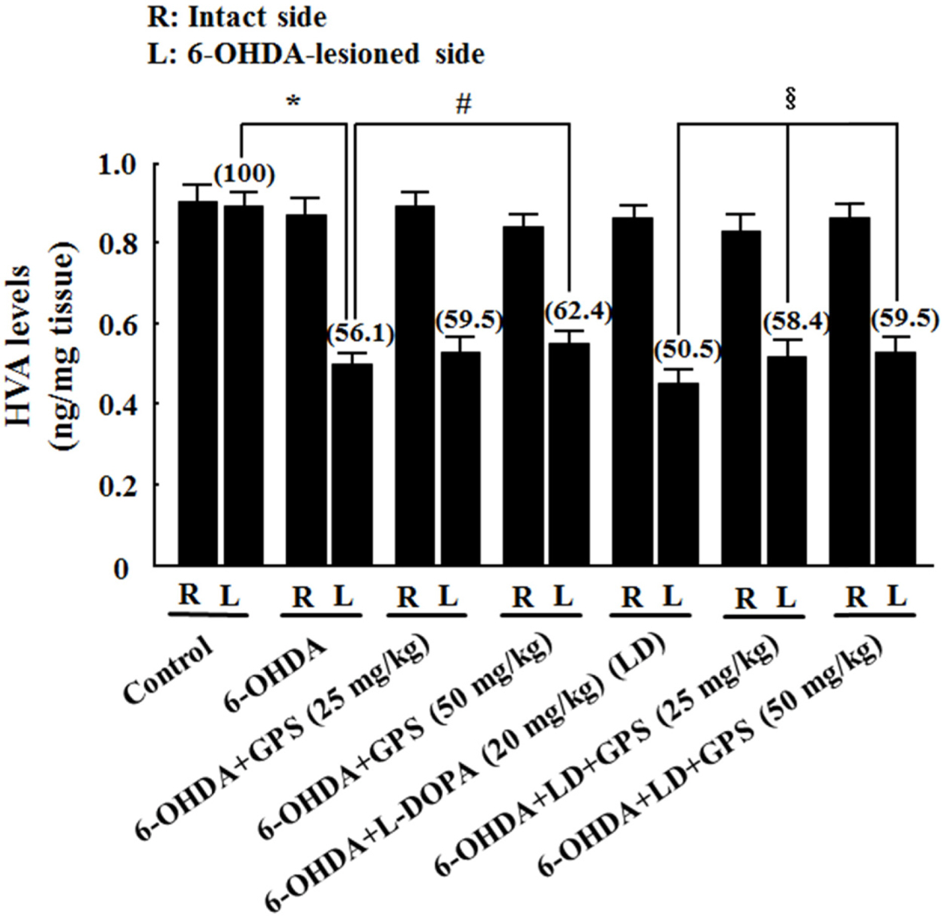
Fig. 5. Effects of GPS on striatal homovanillic acid (HVA) levels on the intact (R) and 6-OHDA-lesioned (L) sides. Numbers in parentheses represent HVA levels expressed as percentages of the control group (L). Results are presented as mean ± S.E.M.; n=8–10 animals per group.∗P < 0.05 compared with control group; #P < 0.05 compared with 6-OHDA-lesioned group; §P < 0.05 compared with 6-OHDA-lesioned group treated with L-DOPA alone.
Reference
-
(1). Fearnley J. M., Lees A. J.Brain. 1991; 114:2283–2301.(2). Fahn S. Ann. N. Y.Acad. Sci. 2003; 991:1–14.(3). Marsden C. D. J.Neurol. Neurosurg. Psychiatry. 1994; 57:672–681.(4). Jankovic J.Mov. Disord. 2005; 20:S11–S16.(5). Cheng N., Maeda T., Kume T., Kaneko S., Kochiyama H., Akaike A., Goshima Y., Misu Y.Brain Res. 1996; 16:278–283.(6). Walkinshaw G., Waters C. M. J.Clin. Invest. 1995; 95:2458–2464.(7). Shin K. S., Zhao T. T., Park K. H., Park H. J., Hwang B. Y., Lee C. K., Lee M. K.BMC Neurosci. 2015; 16:23.(8). Bové J., Perier C.Neuroscience. 2012; 211:51–76.(9). Jackson-Lewis V., Blesa J., Przedborski S.Parkinsonism Relat. Disord. 2012; 18:S183–S185.(10). Seidl S. E., Potashkin J. A.Front. Neurol. 2011; 2:68.(11). Razmovski-Naumovski V., Huang T. H. W., Tran V. H., Li G. Q., Duke C. C., Roufogalis B. D.Phytochem. Rev. 2005; 4:197–219.(12). Im S. A., Choi H. S., Hwang B. Y., Lee M. K., Lee C.K.Kor. J. Pharmacogn. 2009; 40:35–40.(13). Choi H. S., Zhao T. T., Shin K. S., Kim S. H., Hwang B. Y., Lee C. K., Lee M. K.Molecules. 2013; 18:4342–4356.(14). Choi H. S., Park M. S., Kim S. H., Hwang B. Y., Lee C. K., Lee M. K.Molecules. 2010; 15:2814–2824.(15). Shang L. S., Liu J. C., Zhu Q. J., Zhao L., Feng Y., Wang X., Cao W., Xin H.Brain Res. 2006; 1102:163–174.(16). Wang P., Niu L., Guo X. D., Gao L., Li W. X., Jia D., Wang X. L, Ma L. T., Gao G. D.Brain Res. Bull. 2010; 83:266–271.(17). Zhang G., Zhao Z., Gao L., Deng J., Wang B., Xu D., Liu B., Qu Y., Yu J., Li J., Gao G.Pharmacol. Biochem. Behav. 2011; 99:42–51.(18). Wang P., Niu L., Gao L., Li W. X., Jia D., Wang X. L., Gao G. D. J.Int. Med. Res. 2010; 38:1084–1092.(19). Paxinos G., Watson C.The rat brain in stereotaxic coordinates. 2nd ed. Academic Press;Australia: 1986.(20). Schwarting R. K. W., Huston J. P.Prog. Neurobiol. 1996; 50:275–331.(21). Mo J., Zhang H., Yu L. P., Sun P. H., Jin G. Z., Zhen X.Neurobiol. Aging. 2010; 31:926–936.(22). Izurieta-Sánchez P., Sarre S., Ebinger G., Michotte Y.Eur. J. Pharmacol. 1998; 353:33–42.(23). Ljungberg T., Ungerstedt U.Eur. J. Pharmacol. 1977; 46:147–151.(24). Blesa J., Phani S., Jackson-Lewis V., Przedborski S. J.BioMed. Biotechnol. 2012; 2012:845618.(25). Shin K. S., Zhao T. T., Choi H. S., Hwang B. Y., Lee C. K., Lee M. K.Brain Res. 2014; 1567:57–65.(26). Cenci M. A.Parkinsonism Relat. Disord. 2007; 13:S263–S267.(27). Cohen G.Neurotoxicology. 1984; 5:77–82.(28). Soto-Otero R., Méndez-Álvarez E., Hermida-Ameijeiras Á., Muñoz-Patiño A. M., Labandeira-Garcia J. L. J.Neurochem. 2000; 74:1605–1612.(29). Cadet J. L., Brannock C.Neurochem. Int. 1998; 32:117–131.(30). Fahn S., Cohen G.Ann. Neurol. 1992; 32:804–812. 2004.(31). Schober A.Cell Tissue Res. 2004; 318:215–224.(32). Blandini F., Armentero M. T., Martignoni E.Parkinsonism Relat. Disord. 2008; 14:S124–S129.(33). Severson J. A., Marcusson J., Winblad B., Finch C. E. J.Neurochem. 1982; 39:1623–1631.(34). Linert W., Herlinger E., Jameson R. F., Kienzl E., Jellinger K., Youdim M. B.Biochim. Biophys. Acta. 1996; 1316:160–168.(35). Maharaj H., Sukhdev Maharaj D., Scheepers M., Mokokong R., Daya S.Brain Res. 2005; 1063:180–186.(36). Basma A. N., Morris E. J., Nicklas W. J., Geller H. M. J.Neurochem. 1995; 64:825–832.(37). Migheli R., Godani C., Sciola L., Delogu M. R., Serra P. A., Zangani D., De Natale G., Miele E., Desole M. S. J.Neurochem. 1999; 73:1155–1163.(38). Zhang L., Dawson V. L., Dawson T. M.Pharmacol. Ther. 2006; 109:33–41.(39). Padovan-Neto F. E., Echeverry M. B., Tumas V., Del-Bel E. A.Neuroscience. 2009; 159:927–935.(40). Nicklas W. J., Vyas I., Heikkila R. E.Life Sci. 1985; 36:2503–2508.(41). Yacoubian T. A., Standaert D. G.Biochim. Biophys. Acta. 2009; 1792:676–687.(42). Cai T. S., Zhang S. F., Wang M. C.Chin. J. Clin. Rehabil. 2005; 9:106–107.(43). Tanner M. A., Bu X., Steimle J. A., Myers P. R.Nitric Oxide. 1999; 3:359–365.
- Full Text Links
-
- Actions
-
Cited
- CITED
-
- Close
- Share
-
- Similar articles
-
- Morphological Changes in Dopaminergic Neurons in Selective Parkinson's Rat Model
- Effects of Fetal Nondopaminergic Cortical Tissue Transplantation in the Rat Parkinsonian Model
- Neuroglial Reaction in the Substantia Nigra and Striatum of 6-Hydroxydopamine Induced Parkinson's Disease Rat Model
- The Behavioral Changes and the Patterns of Dopamine NeuronalDegeneration after Intrastriatal 6-hydroxydopamine Injection in Rat
- Tumor Necrosis Factor-Associated Protein 1 (TRAP1) is Released from the Mitochondria Following 6-hydroxydopamine Treatment
