The Effect of Autophagy to Cell Death in Nutrient-Deprived H460 Cells
- Affiliations
-
- 1Department of Internal Medicine, Wonkwang University College of Medicine, Iksan, Korea. yshpul@wonkwang.ac.kr
- 2Department of Pathology, Wonkwang University College of Medicine, Iksan, Korea.
- 3Department of Radiation Oncology, Wonkwang University College of Medicine, Iksan, Korea.
- 4Department of Thoracic Surgery, Wonkwang University College of Medicine, Iksan, Korea.
- 5Department of Nuclear Medicine, Wonkwang University College of Medicine, Iksan, Korea.
- 6Department of Biological Science, Wonkwang University College of Medicine, Iksan, Korea.
Abstract
- BACKGROUND
Autophagy is an important adaptive mechanism in normal development and in response to changing environmental stimuli in cancer. Previous papers have reported that different types of cancer underwent autophagy to obtain amino acids as energy source of dying cells in nutrient-deprived conditions. However, whether or not autophagy in the process of lung cancer causes death or survival is controversial. Therefore in this study, we investigated whether nutrient deprivation induces autophagy in human H460 lung cancer cells.
METHODS
H460, lung cancer cells were incubated in RPMI 1640 medium, and the starved media, which are BME and RPMI media without serum, including 2-deoxyl-D-glucose according to time dependence. To evaluate the viability and find out the mechanism of cell death under nutrient-deprived conditions, the MTT assay and flow cytometry were done and analyzed the apoptotic and autophagic related proteins. It is also measured the development of acidic vascular organelles by acridine orange.
RESULTS
The nutrient-deprived cancer cell is relatively sensitive to cell death rather than normal nutrition. Massive cytoplasmic vacuolization was seen under nutrient-deprived conditions. Autophagic vacuoles were visible at approximately 12 h and as time ran out, vacuoles became larger and denser with the increasing number of vacuoles. In addition, the proportion of acridine orange stain-positive cells increased according to time dependence. Localization of GFP-LC3 in cytoplasm and expression of LC-3II and Beclin 1 were increased according to time dependence on nutrient-deprived cells.
CONCLUSION
Nutrient deprivation induces cell death through autophagy in H460 lung cancer cells.
Keyword
MeSH Terms
Figure
-
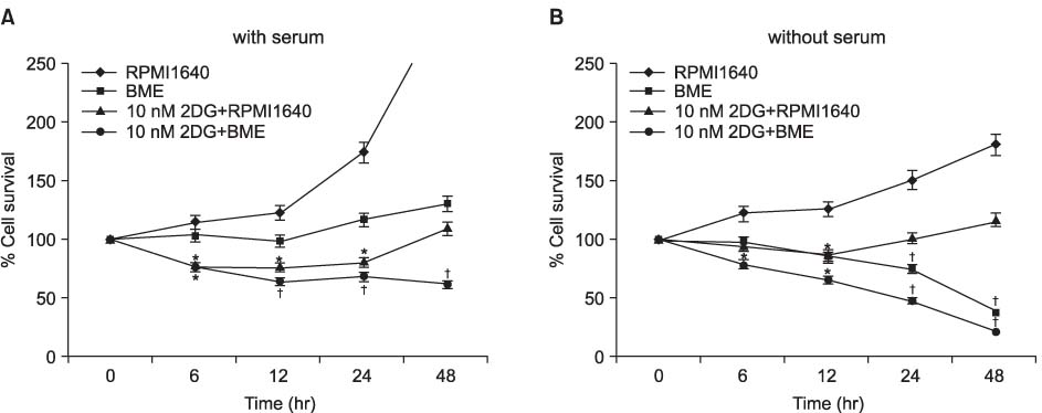
Figure 1 Effect of nutrient deprivation on cell survival in H460 cell line. Cells were seeded at a density of 1x10⁵ cells/mL in 48 well plates and incubated in completely fresh medium for 20 hours. The cells were washed with PBS and the medium was then changed to a medium without the nutrient factors. Basal medium eagle (BME) was deprived of each nutrient and prepared by adding 10 mM 2-deoxyl-D-glucose (2DG) or 10% of dialyzed fetal bovine serum to the basal medium. Cell survival in the presence (A) and absence (B) of serum were shown. Cell numbers at the start of starvation were considered to be 100%. The viability of cell was measured by MTT assay. Means±SD (n=4). *p≤0.05, †p≤0.01.

Figure 2 Apoptosis is not related with cell death induced by nutrient deprivation in NCI-H460 cells. Cells were incubated in RPMI 1640 medium, basal medium eagle (BME) medium or 10 mM 2-deoxyl-D-glucose (2DG) with BME medium (12 hours) assayed for apoptosis by FACS following annexin V-FITC staining. Control cells were contained RPMI 1640 Medium. 20 µM cisplatin was used as a positive control. The data shown are representative of three independent experiment.
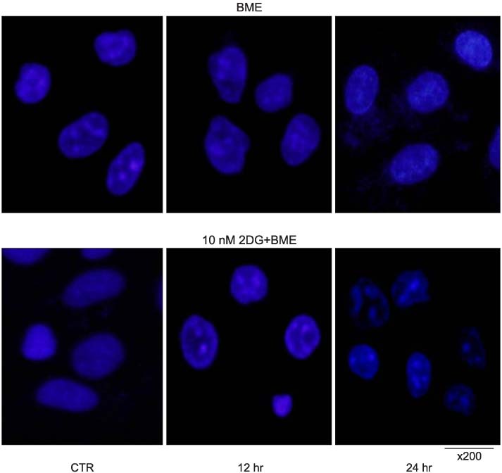
Figure 3 No apoptotic nuclear change occurred in nutrient-deprived H460 cells. H460 cells were plated on glass coverslips. Cells were incubated in RPMI 1640 medium, basal medium eagle (BME) medium (A) or 10 mM 2-deoxyl-D-glucose (2DG) with BME medium (B) for various periods. Hoechst staining was used to identify morphological features of cell death such as nuclear condensation and fragmentation. Scale bar, ×200. Samples were examined under a fluorescence microscope.

Figure 4 No apoptosis and necrosis is not associated with the nutrient-deprived H460 cell death. Cells probed with Western immunoblots were incubated in RPMI 1640 medium (CTR), basal medium eagle (BME) medium (A) or 10 mM 2-deoxyl-D-glucose (2DG) with BME medium (B) for various periods. The equal amounts of protein from cell lysate were subjected on 12% and 15% SDS-polyacrylamide gel electrophoresis (SDS-PAGE), transferred onto polyvinylidene fluoride (PVDF) membrane and immunoblotted with antibodies against Caspase 3 and cleaved caspase 3, PARP, cleaved PARP, HMGB1 and Actin. The immunoreactive signals were visualized by enhanced chemiluminescent kit (ECL). Actin was used as a roading control.

Figure 5 Cytoplasmic Vacuoles were increased according to time dependence in nutrient-deprived H460 cells. H460 cells were incubated in RPMI 1640 medium (CTR), basal medium eagle (BME) medium or 10 mM 2-deoxyl-D-glucose (2DG) with BME medium for 12 hours and 24 hours. Scale bar, ×200. Samples were examined under a light microscope.

Figure 6 Red acidic autophagic vacuoles were detected in cytoplasm of nutrient-deprived H460 cells according to time dependence. H460 cells were incubated in RPMI 1640 medium (CTR), basal medium eagle (BME) medium or 10 mM 2-deoxyl-D-glucose (2DG) with BME medium for 12 hours and 24 hours as shown. And then stained by 1 µg/mL acridine orange in medium for 15 minutes. The acidic autolysosomes induced by nutrition deprivation were stained orange-red.

Figure 7 Development of acidic vesicular organelles (AVO) in nutrient-deprived H460 cells and its quantification using FACS analysis. H460 cells were incubated in RPMI 1640 medium, basal medium eagle (BME) medium or 10 mM 2-deoxyl-D-glucose (2DG) with BME medidum for 6 hours, 12 hours, and 24 hours. AVOs were counted in cytoplasm of H460 cells. *p≤0.05, †p≤0.01.
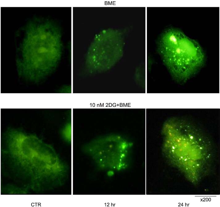
Figure 8 Fluorescence staining of GFP-LC3 in response to nutrient deprivation. H460 cells were plated on glass coverslips. Following transfection with GFP-LC3 plasmid, cells were incubated in RPMI 1640 medium, basal medium eagle (BME) medium, 10 mM 2-deoxyl-D-glucose (2DG) with BME medium for 12 hours and 24 hours as indicated. The transfected cells were observed using a single-photon fluorescence microscope.

Figure 9 Induction of autophagic protein LC3-I and formation of LC3-II and Beclin 1 in nutrient deprived condition. Lung cancer cells were incubated in basal medium eagle (BME) medium (A) and 10 mM 2-deoxyl-D-glucose (2DG) With BME medium (B) for various time periods as indicated. Beclin 1, the product of autophagy-promoting gene Beclin 1 (ATG6) expression was detected. LC3-I induction and conversion of LC3-I to LC3-II were determined by western blot analysis. GAPDH protein levels served as loading control.
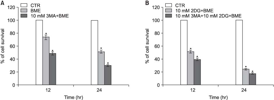
Figure 10 Effect of 3-Methyladenine (3-MA) on nutrient deprived cell death in H460 cells. Relative cell viability was determined 12 hours and 24 hours after nutrient-deprived medium cultured in the presence and absence of 10 mM 3-MA, respectively. Cell numbers at the start of starvation were considered to be 100%. The viability of cell was measured by MTT assay. Means±SD (n=4). *p≤0.01.
Reference
-
1. Jassem J. Combined chemotherapy and radiation in locally advanced non-small-cell lung cancer. Lancet Oncol. 2001. 2:335–342.2. Bae JM, Won YJ, Jung KW, Suh KA, Ahn DH, Park JG. Annual report of the Central Cancer Registry in Korea-1999: based on registered data from 128 hospitals. Cancer Res Treat. 2001. 33:367–372.3. Ferreira CG, Span SW, Peters GJ, Kruyt FA, Giaccone G. Chemotherapy triggers apoptosis in a caspase-8-dependent and mitochondria-controlled manner in the non-small cell lung cancer cell line NCI-H460. Cancer Res. 2000. 60:7133–7141.4. Fukuoka M, Yano S, Giaccone G, Tamura T, Nakagawa K, Douillard JY, et al. Multi-institutional randomized phase II trial of gefitinib for previously treated patients with advanced non-small-cell lung cancer (The IDEAL 1 Trial). J Clin Oncol. 2003. 21:2237–2246.5. Abraham MC, Shaham S. Death without caspases, caspases without death. Trends Cell Biol. 2004. 14:184–193.6. Bowen ID, Mullarkey K, Morgan SM. Programmed cell death during metamorphosis in the blow-fly Calliphora vomitoria. Microsc Res Tech. 1996. 34:202–217.7. Zakeri ZF, Ahuja HS. Apoptotic cell death in the limb and its relationship to pattern formation. Biochem Cell Biol. 1994. 72:603–613.8. Bursch W, Grasl-Kraupp B, Ellinger A, Török L, Kienzl H, Müllauer L, et al. Active cell death: role in hepatocarcinogenesis and subtypes. Biochem Cell Biol. 1994. 72:669–675.9. Dunn WA Jr. Studies on the mechanisms of autophagy: formation of the autophagic vacuole. J Cell Biol. 1990. 110:1923–1933.10. Bursch W, Ellinger A, Kienzl H, Török L, Pandey S, Sikorska M, et al. Active cell death induced by the anti-estrogens tamoxifen and ICI 164 384 in human mammary carcinoma cells (MCF-7) in culture: the role of autophagy. Carcinogenesis. 1996. 17:1595–1607.11. Kanzawa T, Kondo Y, Ito H, Kondo S, Germano I. Induction of autophagic cell death in malignant glioma cells by arsenic trioxide. Cancer Res. 2003. 63:2103–2108.12. Paglin S, Hollister T, Delohery T, Hackett N, McMahill M, Sphicas E, et al. A novel response of cancer cells to radiation involves autophagy and formation of acidic vesicles. Cancer Res. 2001. 61:439–444.13. Yao KC, Komata T, Kondo Y, Kanzawa T, Kondo S, Germano IM. Molecular response of human glioblastoma multiforme cells to ionizing radiation: cell cycle arrest, modulation of the expression of cyclin-dependent kinase inhibitors, and autophagy. J Neurosurg. 2003. 98:378–384.14. Butler R, Mitchell SH, Tindall DJ, Young CY. Nonapoptotic cell death associated with S-phase arrest of prostate cancer cells via the peroxisome proliferator-activated receptor gamma ligand, 15-deoxy-delta12,14-prostaglandin J2. Cell Growth Differ. 2000. 11:49–61.15. Dang CV, Semenza GL. Oncogenic alterations of metabolism. Trends Biochem Sci. 1999. 24:68–72.16. Sutherland RM. Cell and environment interactions in tumor microregions: the multicell spheroid model. Science. 1988. 240:177–184.17. Helmlinger G, Yuan F, Dellian M, Jain RK. Interstitial pH and pO2 gradients in solid tumors in vivo: high-resolution measurements reveal a lack of correlation. Nat Med. 1997. 3:177–182.18. Zhong D, Liu X, Schafer-Hales K, Marcus AI, Khuri FR, Sun SY, et al. 2-Deoxyglucose induces Akt phosphorylation via a mechanism independent of LKB1/AMP-activated protein kinase signaling activation or glycolysis inhibition. Mol Cancer Ther. 2008. 7:809–817.19. Lum JJ, Bauer DE, Kong M, Harris MH, Li C, Lindsten T, et al. Growth factor regulation of autophagy and cell survival in the absence of apoptosis. Cell. 2005. 120:237–248.20. Cummings BS, Schnellmann RG. Cisplatin-induced renal cell apoptosis: caspase 3-dependent and -independent pathways. J Pharmacol Exp Ther. 2002. 302:8–17.21. Kaushal GP, Kaushal V, Hong X, Shah SV. Role and regulation of activation of caspases in cisplatin-induced injury to renal tubular epithelial cells. Kidney Int. 2001. 60:1726–1736.22. Scaffidi P, Misteli T, Bianchi ME. Release of chromatin protein HMGB1 by necrotic cells triggers inflammation. Nature. 2002. 418:191–195.23. Kim J, Huang WP, Stromhaug PE, Klionsky DJ. Convergence of multiple autophagy and cytoplasm to vacuole targeting components to a perivacuolar membrane compartment prior to de novo vesicle formation. J Biol Chem. 2002. 277:763–773.24. Kabeya Y, Mizushima N, Ueno T, Yamamoto A, Kirisako T, Noda T, et al. LC3, a mammalian homologue of yeast Apg8p, is localized in autophagosome membranes after processing. EMBO J. 2000. 19:5720–5728.25. Asanuma K, Tanida I, Shirato I, Ueno T, Takahara H, Nishitani T, et al. MAP-LC3, a promising autophagosomal marker, is processed during the differentiation and recovery of podocytes from PAN nephrosis. FASEB J. 2003. 17:1165–1167.26. Pattingre S, Tassa A, Qu X, Garuti R, Liang XH, Mizushima N, et al. Bcl-2 antiapoptotic proteins inhibit Beclin 1-dependent autophagy. Cell. 2005. 122:927–939.27. Erlich S, Mizrachy L, Segev O, Lindenboim L, Zmira O, Adi-Harel S, et al. Differential interactions between Beclin 1 and Bcl-2 family members. Autophagy. 2007. 3:561–568.28. Bursch W, Ellinger A. Autophagy: a basic mechanism and a potential role for neurodegeneration. Folia Neuropathol. 2005. 43:297–310.29. Debnath J, Baehrecke EH, Kroemer G. Does autophagy contribute to cell death? Autophagy. 2005. 1:66–74.
- Full Text Links
-
- Actions
-
Cited
- CITED
-
- Close
- Share
-
- Similar articles
-
- Curcumin-Induced Autophagy Augments Its Antitumor Effect against A172 Human Glioblastoma Cells
- Induction of cytoprotective autophagy by morusin via AMP-activated protein kinase activation in human non-small cell lung cancer cells
- Dual Inhibition of PI3K/Akt/mTOR Pathway and Role of Autophagy in Non-Small Cell Lung Cancer Cells
- Role of Autophagy in the Control of Cell Death and Inflammation
- The role of autophagy in the placenta as a regulator of cell death
