Characterization of allergic response induced by repeated dermal exposure of IL-4/Luc/CNS-1 transgenic mice to low dose formaldehyde
- Affiliations
-
- 1Department of Biomaterials Science, College of Natural Resources & Life Science/Life and Industry Convergence Research Institute, Pusan National University, Miryang, Korea. dyhwang@pusan.ac.kr
- 2Department of Clinical Laboratory Science, Dong-Eui University, Busan, Korea.
- KMID: 2312120
- DOI: http://doi.org/10.5625/lar.2014.30.3.95
Abstract
- Although formaldehyde (FA) is known to be a major allergen responsible for allergic contact dermatitis, there are conflicting reports regarding correlation between FA exposure and interleukin (IL-4) expression. To investigate whether allergic responses including IL-4 expression were induced by repeated dermal exposure to low dose FA, alterations in the luciferase signal and allergic phenotypes were measured in IL-4/Luc/CNS-1 transgenic (Tg) mice containing luciferase cDNA under control of the IL-4 promoter after exposure to 4% FA for 2 weeks. High levels of luciferase were detected in the abdominal region of the whole body and submandibular lymph node (SLN) of FA treated mice. Additionally, the ear thickness and IgE concentration were significantly upregulated in the FA treated group when compared with the acetone olive oil (AOO) treated group. FA treated mice showed enhanced auricular lymph node (ALN) weight, epidermis and dermis thickness, and infiltration of inflammatory cells. Furthermore, the expression of IL-6 among T helper 2 cytokines was higher in the FA treated group than the AOO treated group, while vascular endothelial growth factor (VEGF) levels remained constant. Overall, the results presented herein provide additional evidence that various allergic responses may be successfully induced in IL-4/Luc/CNS-1 Tg mice after exposure to low dose FA for 2 weeks. The luciferase signal under the IL-4 promoter may reflect general indicators of the allergic response induced by exposure to low dose FA.
MeSH Terms
-
Acetone
Animals
Cytokines
Dermatitis, Allergic Contact
Dermis
DNA, Complementary
Ear
Epidermis
Formaldehyde*
Immunoglobulin E
Interleukin-4
Interleukin-6
Interleukins
Luciferases
Lymph Nodes
Mice
Mice, Transgenic*
Olea
Phenotype
Vascular Endothelial Growth Factor A
Olive Oil
Acetone
Cytokines
DNA, Complementary
Formaldehyde
Immunoglobulin E
Interleukin-4
Interleukin-6
Interleukins
Luciferases
Vascular Endothelial Growth Factor A
Figure
-
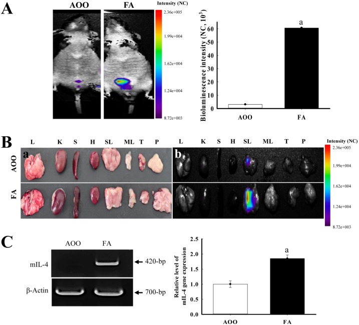
Figure 1 Detection of luciferase signal in the whole body (A) and each organ (B) of IL-4/Luc/CNS-1 Tg mice. Mice were treated with AOO and FA for 2 weeks and then imaged at 24 h after final treatment using the LivingImage software. The color overlay on the image represents the photons per second emitted from the organs in accordance with the pseudocolor scale shown next to the image. In this image, red indicates the highest number of photons per second, while blue indicates the lowest. L, lung; K, kidney; S, spleen; H, heart; SLN, submandibular lymph node; MLN, mesenteric lymph node; T, thymus; P, pancreas. (C) The expression of mouse IL-4 mRNA in SLN was measured by RT-PCR using a specific primer. Data shown are the means±SD (n=5). a, P<0.05 compared to the AOO treated group.
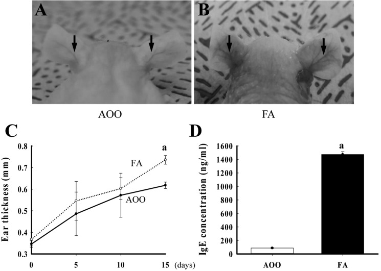
Figure 2 Alteration of ear phenotypes and IgE concentration. After repeated application of FA solution (4%) to the ear of IL-4/Luc/CNS-1 Tg mice for 2 weeks, the outline of the ear vein in two groups was analyzed using the picture image (A and B), and ear thickness of the mice was measured with a thickness gauge on each experimental day (C). Arrows indicate the vein in the ear. The serum used to measure the IgE concentration was prepared from blood samples collected from the abdominal vein of the mice. The IgE concentration in serum was quantified by an enzyme-linked immunosorbent assay (D). Data shown are the means±SD (n=5). a, P<0.05 compared to the AOO treated group.
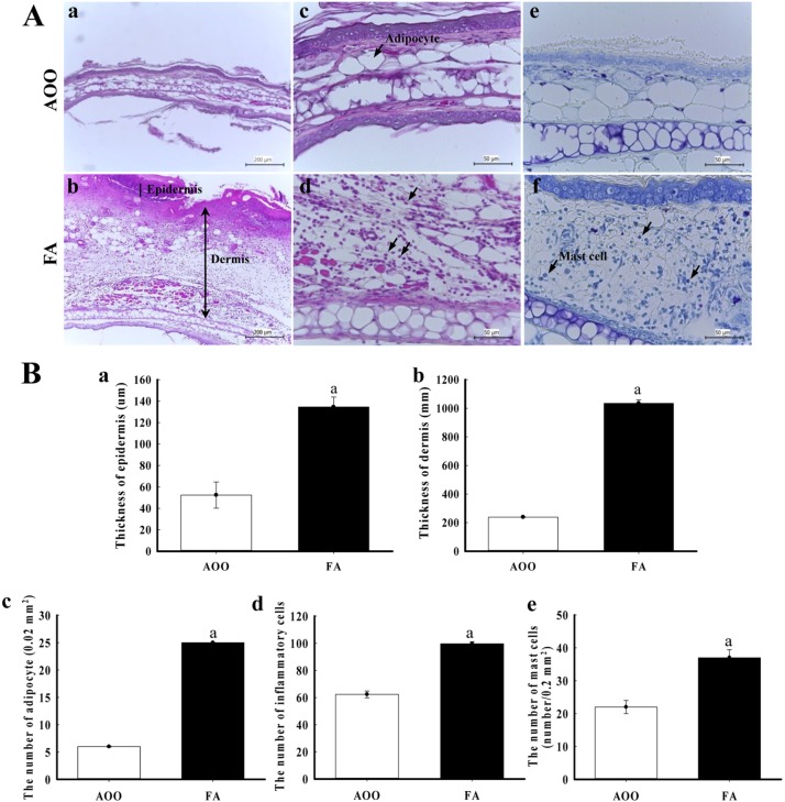
Figure 3 Histological alteration of ear skin. FA solution (4%) was repeatedly applied to the dorsum of the ear of IL-4/Luc/CNS-1 Tg mice. After 2 weeks, ear tissues were collected from AOO or FA treated IL-4/Luc/CNS-1 Tg mice. The histological changes were determined as described in the Materials and Methods. The slide sections of ear tissue were stained with hematoxylin & eosin and then observed at 100× or 400× magnification (Aa-d and Ba-d). Slide sections of ear tissue were stained with toluidine blue and then observed at 400× magnification (Ae and f, and Bc). Arrow heads indicate the infiltrated mast cells in the dermis of the ear. Data shown are the means±SD (n=5). a, P<0.05 compared to the AOO treated group.

Figure 4 IL-6 and VEGF expression in ear tissue. After detection of specific bands with two primary antibodies, the intensity of each band was determined using an imaging densitometer and the relative level of each protein was calculated based on the intensity of actin protein as an endogenous control. Data represent the mean±SD from three replicates. a, P<0.05 compared to the AOO-treated group.
Reference
-
1. Günther R, Walter D, Armin OG, Albrecht H. Formaldehyde. Ullmann's Encyclopedia of Industrial Chemistry. Weinheim: Wiley-VCH;2002.2. Marzulli FN, Maibach HI. The use of graded concentrations in studying skin sensitizers: experimental contact sensitization in man. Food Cosmet Toxicol. 1974; 12(2):219–227. PMID: 4459237.
Article3. Nethercott JR, Holness DL. Contact dermatitis in funeral service workers. Contact Dermatitis. 1988; 18(5):263–267. PMID: 2970932.
Article4. Dearman RJ, Basketter DA, Evans P, Kimber I. Comparison of cytokine secretion profiles provoked in mice by glutaraldehyde and formaldehyde. Clin Exp Allergy. 1999; 29(1):124–132. PMID: 10051711.
Article5. Saneyoshi K, Nohara O, Imai T, Shiraishi F, Moriyama H, Fujimaki H. IL-4 and IL-6 production of bone marrow-derived mast cells is enhanced by treatment with environmental pollutants. Int Arch Allergy Immunol. 1997; 114(3):237–245. PMID: 9363904.
Article6. Xu B, Aoyama K, Takeuchi M, Matsushita T, Takeuchi T. Expression of cytokine mRNAs in mice cutaneously exposed to formaldehyde. Immunol Lett. 2002; 84(1):49–55. PMID: 12161283.
Article7. Saito A, Tanaka H, Usuda H, Shibata T, Higashi S, Yamashita H, Inagaki N, Nagai H. Characterization of skin inflammation induced by repeated exposure of toluene, xylene, and formaldehyde in mice. Environ Toxicol. 2011; 26(3):224–232. PMID: 19904815.
Article8. De Jong WH, Arts JH, De Klerk A, Schijf MA, Ezendam J, Kuper CF, Van Loveren H. Contact and respiratory sensitizers can be identified by cytokine profiles following inhalation exposure. Toxicology. 2009; 261(3):103–111. PMID: 19422874.
Article9. Bae CJ, Shim SB, Jee SW, Lee SH, Kim MR, Lee JW, Lee CK, Hwang DY. IL-6, VEGF, KC and RANTES are a major cause of a high irritant dermatitis to phthalic anhydride in C57BL/6 inbred mice. Allergol Int. 2010; 59(4):389–397. PMID: 20864798.
Article10. Lee YJ, Kim JE, Kwak MH, Go J, Kim DS, Son HJ, Hwang DY. Quantitative evaluation of the therapeutic effect of fermented soybean products containing a high concentration of GABA on phthalic anhydride-induced atopic dermatitis in IL-4/Luc/CNS-1 Tg mice. Int J Mol Med. 2014; 33(5):1185–1194. PMID: 24604257.
Article11. Gao XK, Nakamura N, Fuseda K, Tanaka H, Inagaki N, Nagai H. Establishment of allergic dermatitis in NC/Nga mice as a model for severe atopic dermatitis. Biol Pharm Bull. 2004; 27(9):1376–1381. PMID: 15340222.
Article12. Leung DY. Infection in atopic dermatitis. Curr Opin Pediatr. 2003; 15(4):399–404. PMID: 12891053.
Article13. Umetsu DT, DeKruyff RH. The regulation of allergy and asthma. Immunol Rev. 2006; 212:238–255. PMID: 16903918.
Article14. Kapp A. The role of eosinophils in the pathogenesis of atopic dermatitis--eosinophil granule proteins as markers of disease activity. Allergy. 1993; 48(1):1–5. PMID: 8457021.15. Dubois GR, Bruijnzeel PL. IL-4-induced migration of eosinophils in allergic inflammation. Ann N Y Acad Sci. 1994; 725:268–273. PMID: 8030998.
Article
- Full Text Links
-
- Actions
-
Cited
- CITED
-
- Close
- Share
-
- Similar articles
-
- Therapeutic effect of ethyl acetate extract from Asparagus cochinchinensis on phthalic anhydride-induced skin inflammation
- IL-13 and STAT6 signaling involve in low dose lipopolysaccharide induced murine model of asthma
- Repeated Ozone Exposure Induces Th2 Immune Responses in Mice
- IL-4-deficient Mice Aggravate Hypersensitivity Pneumonitis
- Role of Mast Cells in a Aspergillus Murine Model of Allergic Rhinitis
