Histomorphometric Analysis of the Spine and Femur in Ovariectomized Rats Using Micro-Computed Tomographic Scan
- Affiliations
-
- 1Department of Neurosurgery, Wooridul Spine Hospital, Daegu, Korea.
- 2Department of Neurosurgery, School of Medicine, Kyungpook National University, Daegu, Korea. jksung@knu.ac.kr
- 3Department of Anatomy, School of Medicine, Kyungpook National University, Daegu, Korea.
- KMID: 2190515
- DOI: http://doi.org/10.3340/jkns.2012.52.1.1
Abstract
OBJECTIVE
The purpose of this study was to evaluate the different patterns of bone loss between the lumbar spine and the femur after ovariectomy in rats.
METHODS
Twenty-four female Sprague-Dawley rats underwent a sham operation (the sham group) or bilateral ovariectomy (the ovariectomized group). Four and eight weeks after operation, six rats from each of the two groups were euthanized. Serum biochemical markers of bone turnover including osteocalcin and alkaline phosphatase (ALP), which are sensitive biochemical markers of bone formation, and the telopeptide fragment of type I collagen C-terminus (CTX), which is a sensitive biochemical marker of bone resorption, were analyzed. Bone histomorphometric parameters of the 4th lumbar vertebrae and femur were determined by micro-computed tomography.
RESULTS
Ovariectomized rats were found to have higher osteocalcin, ALP and CTX levels than sham controls. Additionally, 8 weeks after ovariectomy in the OVX group, serum levels of osteocalcin, ALP and CTX were significantly higher than those of 4 weeks after ovariectomy. Bone loss after ovariectomy was more extensive in the 4th lumbar spine compared to the femur. Bone loss in the 4th lumbar spine was mainly caused by trabecular thinning, but in the femur, it was mainly caused by trabecular elimination.
CONCLUSION
The present study demonstrates different patterns of bone loss between the 4th lumbar spine and the femur in ovariectomized rats. Therefore, when considering animal models of osteoporosis, it is important that bone sites should be taken into account.
Keyword
MeSH Terms
Figure
-
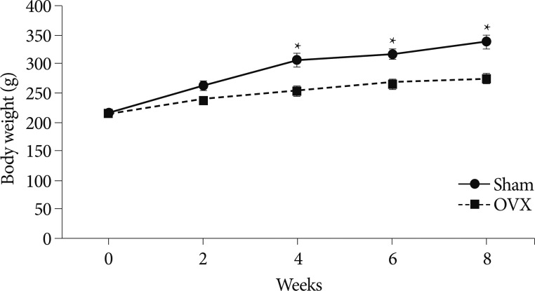
Fig. 1 Graph showing temporal changes of body weights of the twelve rats euthanized 8 weeks after ovariectomy or sham operations. Values represent mean±standard deviations (n=12). *indicates p<0.05 for OVX vs. sham group comparisons. OVX: ovariectomized.

Fig. 2 Serum estrogen levels in the two study groups at 4 and 8 weeks after ovariectomy or sham operations. *indicates p<0.05 for 4 weeks vs. 8 weeks in the OVX group. OVX: ovariectomized.
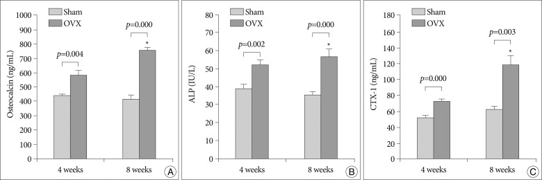
Fig. 3 The serum osteocalcin (A) and ALP (B) concentrations were used as markers of bone formation. Type I collagen C-telopeptide (CTX) (C) concentration was used as a marker of bone resorption. Values are mean±standard deviations. *indicates p<0.05 for 4 weeks vs. 8 weeks in the OVX group. ALP: alkaline phosphatase, OVX: ovariectomized.
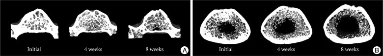
Fig. 4 Representative micro-CT images of the 4th lumbar spine (A) and the femur (B) in the OVX group. OVX rats 8 weeks after ovariectomy had less trabecular bone than at baseline and 4 weeks after ovariectomy. OVX: ovariectomized, CT: computed tomography.
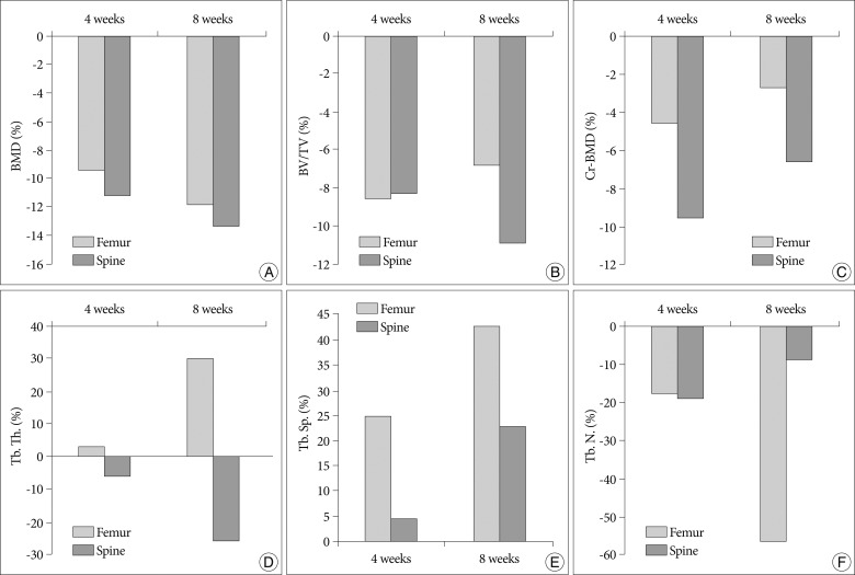
Fig. 5 Bar graph showing the result of histomorphometric analyses of 4th lumbar vertebrae in the sham and OVX groups at 4 and 8 weeks post-operation: bone mineral density (BMD) (A), trabecular bone volume fraction (BV/TV) (B), cortical bone mineral density (CrBMD) (C), trabecular thickness (Tb.Th.) (D), trabecular separation (Tb.Sp.) (E), and trabecular number (Tb.N.) (F). All values represent 100% minus the percentage of the value of the sham group/the value of the OVX group. OVX: ovariectomized.
Cited by 2 articles
-
Rutin Improves Bone Histomorphometric Values by Reduction of Osteoclastic Activity in Osteoporosis Mouse Model Induced by Bilateral Ovariectomy
Hye-Hwa Lee, Jae-Won Jang, Jung-Kil Lee, Choon-Keun Park
J Korean Neurosurg Soc. 2020;63(4):433-443. doi: 10.3340/jkns.2019.0097.Therapeutic Advantages of Treatment of High-Dose Curcumin in the Ovariectomized Rat
Dae-Chul Cho, Hyun-Sik Jung, Kyoung-Tae Kim, Younghoon Jeon, Joo-Kyung Sung, Jeong-Hyun Hwang
J Korean Neurosurg Soc. 2013;54(6):461-466. doi: 10.3340/jkns.2013.54.6.461.
Reference
-
1. Adachi JD, Loannidis G, Berger C, Joseph L, Papaioannou A, Pickard L, et al. The influence of osteoporotic fractures on health-related quality of life in community-dwelling men and women across Canada. Osteoporos Int. 2001; 12:903–908. PMID: 11804016.
Article2. Bauss F, Dempster DW. Effects of ibandronate on bone quality : preclinical studies. Bone. 2007; 40:265–273. PMID: 16996333.
Article3. Canpolat S, Tug N, Seyran AD, Kumru S, Yilmaz B. Effects of raloxifene and estradiol on bone turnover parameters in intact and ovariectomized rats. J Physiol Biochem. 2010; 66:23–28. PMID: 20428990.
Article4. Ferretti M, Bertoni L, Cavani F, Zavatti M, Resca E, Carnevale G, et al. Influence of ferutinin on bone metabolism in ovariectomized rats. II : role in recovering osteoporosis. J Anat. 2010; 217:48–56. PMID: 20492429.
Article5. French DL, Muir JM, Webber CE. The ovariectomized, mature rat model of postmenopausal osteoporosis : an assessment of the bone sparing effects of curcumin. Phytomedicine. 2008; 15:1069–1078. PMID: 18693096.
Article6. Kim TH, Jung JW, Ha BG, Hong JM, Park EK, Kim HJ, et al. The effects of luteolin on osteoclast differentiation, function in vitro and ovariectomy-induced bone loss. J Nutr Biochem. 2011; 22:8–15. PMID: 20233653.
Article7. Kleerekoper M, Villanueva AR, Stanciu J, Rao DS, Parfitt AM. The role of three-dimensional trabecular microstructure in the pathogenesis of vertebral compression fractures. Calcif Tissue Int. 1985; 37:594–597. PMID: 3937580.
Article8. Lei Z, Xiaoying Z, Xingguo L. Ovariectomy-associated changes in bone mineral density and bone marrow haematopoiesis in rats. Int J Exp Pathol. 2009; 90:512–519. PMID: 19765105.
Article9. Meli R, Pacilio M, Raso GM, Esposito E, Coppola A, Nasti A, et al. Estrogen and raloxifene modulate leptin and its receptor in hypothalamus and adipose tissue from ovariectomized rats. Endocrinology. 2004; 145:3115–3121. PMID: 15059958.
Article10. Miller SC, Bowman BM, Jee WS. Available animal models of osteopenia--small and large. Bone. 1995; 17:117S–123S. PMID: 8579907.11. Mosekilde L. Assessing bone quality--animal models in preclinical osteoporosis research. Bone. 1995; 17:343S–352S. PMID: 8579937.12. Omi N, Ezawa I. The effect of ovariectomy on bone metabolism in rats. Bone. 1995; 17:163S–168S. PMID: 8579912.
Article13. Palumbo C, Ferretti M, Bertoni L, Cavani F, Resca E, Casolari B, et al. Influence of ferutinin on bone metabolism in ovariectomized rats. I : role in preventing osteoporosis. J Bone Miner Metab. 2009; 27:538–545. PMID: 19333679.
Article14. Park SB, Lee YJ, Chung CK. Bone mineral density changes after ovariectomy in rats as an osteopenic model : stepwise description of double dorso-lateral approach. J Korean Neurosurg Soc. 2010; 48:309–312. PMID: 21113356.
Article15. Sheng ZF, Dai RC, Wu XP, Fang LN, Fan HJ, Liao EY. Regionally specific compensation for bone loss in the tibial trabeculae of estrogen-deficient rats. Acta Radiol. 2007; 48:531–539. PMID: 17520429.
Article16. Shiraishi A, Miyabe S, Nakano T, Umakoshi Y, Ito M, Mihara M. The combination therapy with alfacalcidol and risedronate improves the mechanical property in lumbar spine by affecting the material properties in an ovariectomized rat model of osteoporosis. BMC Musculoskelet Disord. 2009; 10:66. PMID: 19527501.
Article17. Szulc P, Delmas PD. Biochemical markers of bone turnover : potential use in the investigation and management of postmenopausal osteoporosis. Osteoporos Int. 2008; 19:1683–1704. PMID: 18629570.
Article18. Thompson DD, Simmons HA, Pirie CM, Ke HZ. FDA Guidelines and animal models for osteoporosis. Bone. 1995; 17:125S–133S. PMID: 8579908.
Article19. Turner AS. Animal models of osteoporosis--necessity and limitations. Eur Cell Mater. 2001; 1:66–81. PMID: 14562261.
- Full Text Links
-
- Actions
-
Cited
- CITED
-
- Close
- Share
-
- Similar articles
-
- The Therapeutic Effects of Combination Therapy with Curcumin and Alendronate on Spine Fusion Surgery in the Ovariectomized Rats
- The Change of Bone Metabolism in Ovariectomized Rats : Analyses of MicroCT Scan and Biochemical Markers of Bone Turnover
- Assessment of Bone Quality using Finite Element Analysis Based upon Micro-CT Images
- The evaluation of the correlation between histomorphometric analysis and micro-computed tomography analysis in AdBMP-2 induced bone regeneration in rat calvarial defects
- Effects of Caffeine on Bone Mineral Density and Bone Mineral Content in Ovariectomized Rats
