The Change of Bone Metabolism in Ovariectomized Rats : Analyses of MicroCT Scan and Biochemical Markers of Bone Turnover
- Affiliations
-
- 1Department of Neurosurgery, Daegu Veterans Hospital, Daegu, Korea.
- 2Department of Neurosurgery, School of Medicine, Kyungpook National University, Daegu, Korea. jksung@knu.ac.kr
- 3Department of Anesthesiology and Pain Medicine, School of Dentistry, Kyungpook National University, Daegu, Korea.
- KMID: 2190484
- DOI: http://doi.org/10.3340/jkns.2012.51.6.323
Abstract
OBJECTIVE
The purpose of this study was to verify the appropriateness of ovariectomized rats as the osteoporosis animal model.
METHODS
Twelve female Sprague-Dawley rats underwent a sham operation (the sham group) or bilateral ovariectomy [the ovariectomy (OVX) group]. Eight weeks after operations, serum biochemical markers of bone turnover were analyzed; osteocalcin and alkaline phosphatase, which are sensitive biochemical markers of bone formation, and C-terminal telopeptide fragment of type I collagen C-terminus (CTX), which is a sensitive biochemical marker of bone resorption. Bone histomorphometric parameters and microarchitectural properties of 4th lumbar vertebrae were determined by micro-computed tomographic (CT) scan.
RESULTS
The OVX group showed on average 75.4% higher osteocalcin and 72.5% higher CTX levels than the sham group, indicating increased bone turnover. Micro-CT analysis showed significantly lower bone mineral density (BMD) (p=0.005) and cortical BMD (p=0.021) in the OVX group. Furthermore, the OVX group was found to have a significantly lower trabecular bone volume fraction (p=0.002).
CONCLUSION
Our results showed that bone turnover was significantly increased and bone mass was significantly decreased 8 weeks after ovariectomy in rats. Thus, we propose that the ovariectomized rat model be considered a reproducible and reliable model of osteoporosis.
Keyword
MeSH Terms
Figure
-
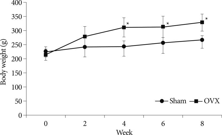
Fig. 1 Graph showing temporal changes of body weights in the two study groups. Values represent mean+standard deviations (n=12). *p<0.05 for OVX vs. sham group comparisons. OVX : ovariectomy.
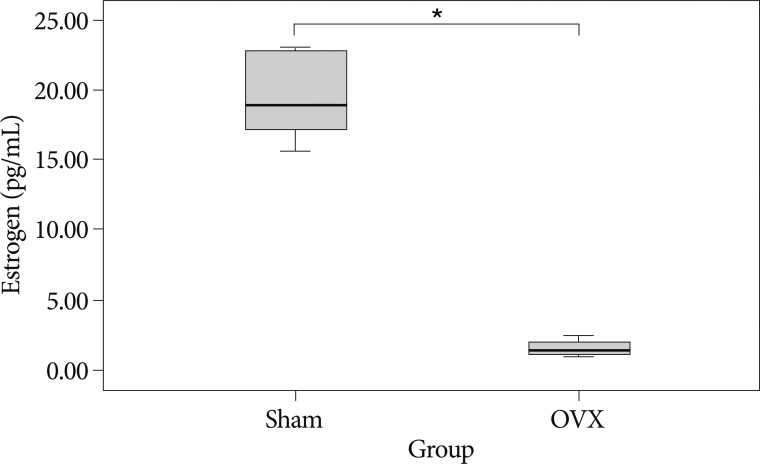
Fig. 2 Serum estrogen levels in the two study groups at 8 weeks after surgery. *p<0.05 for the OVX group compared to the sham group. OVX : ovariectomy.

Fig. 3 The serum osteocalcin (A) and ALP (B) concentrations were used as markers of bone formation. Type I collagen CTX (C) concentration was used as a marker of bone resorption. Values are means+standard deviations (n=12). *p<0.05 for the OVX vs. sham group comparisons. ALP : alkaline phosphatase, CTX : C-terminus, OVX : ovariectomy.
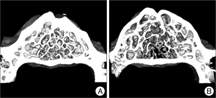
Fig. 4 Representative micro-CT images of the two groups; the sham group (A) and the OVX group (B). OVX rats had less trabecular bone than sham controls at 8 weeks after surgery. OVX : ovariectomy.
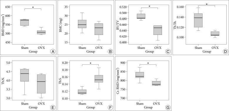
Fig. 5 Bar graph showing the result of histomorphometric analyses of 4th lumbar vertebrae in the sham and ovariectomy (OVX) groups; Bone mineral density (BMD) (A), bone mineral content (BMC) (B), trabecular bone volume fraction (BV/TV) (C), trabecular thickness (Tb.Th.) (D), trabecular number (Tb.N.) (E), trabecular separation (Tb.Sp.) (F), cortical bone mineral density (Cr.BMD) (G). *p<0.05 for OVX vs. sham group comparisons.
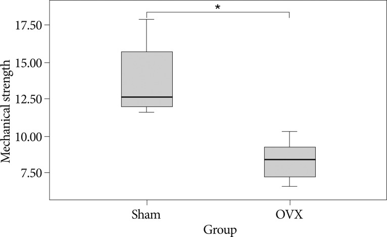
Fig. 6 Loading force to maximal load on the 4th lumbar vertebrae by using a three-point bending test. Ovariectomy significantly decreased maximal load as compared with Sham (p=0.001). *p<0.05 for OVX vs. sham group comparisons. OVX : ovariectomy.
Cited by 1 articles
-
Development of osteoporosis animal model using micropigs
Sang-Woo Kim, Kyoung-Shim Kim, Chester D. Solis, Myeong-Seop Lee, Byung-Hwa Hyun
Lab Anim Res. 2013;29(3):174-177. doi: 10.5625/lar.2013.29.3.174.
Reference
-
1. Canpolat S, Tug N, Seyran AD, Kumru S, Yilmaz B. Effects of raloxifene and estradiol on bone turnover parameters in intact and ovariectomized rats. J Physiol Biochem. 2010; 66:23–28. PMID: 20428990.
Article2. Ferretti M, Bertoni L, Cavani F, Zavatti M, Resca E, Carnevale G, et al. Influence of ferutinin on bone metabolism in ovariectomized rats. II: role in recovering osteoporosis. J Anat. 2010; 217:48–56. PMID: 20492429.
Article3. Kim TH, Jung JW, Ha BG, Hong JM, Park EK, Kim HJ, et al. The effects of luteolin on osteoclast differentiation, function in vitro and ovariectomy-induced bone loss. J Nutr Biochem. 2011; 22:8–15. PMID: 20233653.
Article4. Kleerekoper M, Villanueva AR, Stanciu J, Rao DS, Parfitt AM. The role of three-dimensional trabecular microstructure in the pathogenesis of vertebral compression fractures. Calcif Tissue Int. 1985; 37:594–597. PMID: 3937580.
Article5. Lei Z, Xiaoying Z, Xingguo L. Ovariectomy-associated changes in bone mineral density and bone marrow haematopoiesis in rats. Int J Exp Pathol. 2009; 90:512–519. PMID: 19765105.
Article6. Meli R, Pacilio M, Raso GM, Esposito E, Coppola A, Nasti A, et al. Estrogen and raloxifene modulate leptin and its receptor in hypothalamus and adipose tissue from ovariectomized rats. Endocrinology. 2004; 145:3115–3121. PMID: 15059958.
Article7. Miller SC, Bowman BM, Jee WS. Available animal models of osteopenia--small and large. Bone. 1995; 17:117S–123S. PMID: 8579907.8. Mosekilde L. Assessing bone quality--animal models in preclinical osteoporosis research. Bone. 1995; 17:343S–352S. PMID: 8579937.9. Omi N, Ezawa I. The effect of ovariectomy on bone metabolism in rats. Bone. 1995; 17:163S–168S. PMID: 8579912.
Article10. Palumbo C, Ferretti M, Bertoni L, Cavani F, Resca E, Casolari B, et al. Influence of ferutinin on bone metabolism in ovariectomized rats. I : role in preventing osteoporosis. J Bone Miner Metab. 2009; 27:538–545. PMID: 19333679.
Article11. Park SB, Lee YJ, Chung CK. Bone mineral density changes after ovariectomy in rats as an osteopenic model : stepwise description of double dorso-lateral approach. J Korean Neurosurg Soc. 2010; 48:309–312. PMID: 21113356.
Article12. Riggs BL, Khosla S, Melton LJ 3rd. Sex steroids and the construction and conservation of the adult skeleton. Endocr Rev. 2002; 23:279–302. PMID: 12050121.
Article13. Sheng ZF, Dai RC, Wu XP, Fang LN, Fan HJ, Liao EY. Regionally specific compensation for bone loss in the tibial trabeculae of estrogen-deficient rats. Acta Radiol. 2007; 48:531–539. PMID: 17520429.
Article14. Shiraishi A, Miyabe S, Nakano T, Umakoshi Y, Ito M, Mihara M. The combination therapy with alfacalcidol and risedronate improves the mechanical property in lumbar spine by affecting the material properties in an ovariectomized rat model of osteoporosis. BMC Musculoskelet Disord. 2009; 10:66. PMID: 19527501.
Article15. Szulc P, Delmas PD. Biochemical markers of bone turnover : potential use in the investigation and management of postmenopausal osteoporosis. Osteoporos Int. 2008; 19:1683–1704. PMID: 18629570.
Article16. Thompson DD, Simmons HA, Pirie CM, Ke HZ. FDA Guidelines and animal models for osteoporosis. Bone. 1995; 17:125S–133S. PMID: 8579908.
Article17. Tommasini SM, Morgan TG, van der Meulen MCh, Jepsen KJ. Genetic variation in structure-function relationships for the inbred mouse lumbar vertebral body. J Bone Miner Res. 2005; 20:817–827. PMID: 15824855.
Article18. Turner AS. Animal models of osteoporosis--necessity and limitations. Eur Cell Mater. 2001; 1:66–81. PMID: 14562261.
- Full Text Links
-
- Actions
-
Cited
- CITED
-
- Close
- Share
-
- Similar articles
-
- Changes of Biochemical Markers of Bone turnover in Pre-, Peri-and Postmenopausal Women
- Biochemical Markers of Bone Turnover
- Histomorphometric Analysis of the Spine and Femur in Ovariectomized Rats Using Micro-Computed Tomographic Scan
- Study for Usefulness of Total Alkaline Phosphatase as a Biochemical Markers of Bone Turnover in Healthy Menopausal Women
- Comparison on New Bone Formation Between Ovariectomized Rats and Normal Rats After Graft of Alloplastic bone material
