Effect of IL-4 on the Development and Function of Memory-like CD8 T Cells in the Peripheral Lymphoid Tissues
- Affiliations
-
- 1Graduate Course of Translational medicine, Seoul National University College of Medicine, Seoul 03080, Korea. jungkc66@snu.ac.kr
- 2Department of Biochemistry, College of Life Science & Biotechnology, Yonsei University, Seoul 03722, Korea. sjha@yonsei.ac.kr
- 3Transplantation Research Institute, Seoul National University Medical Research Center, Seoul 03080, Korea.
- 4Department of Pathology, Seoul National University College of Medicine, Seoul 03080, Korea.
- KMID: 2168053
- DOI: http://doi.org/10.4110/in.2016.16.2.126
Abstract
- Unlike conventional T cells, innate CD8 T cells develop a memory-like phenotype in the thymus and immediately respond upon antigen stimulation, similar to memory T cells. The development of innate CD8 T cells in the thymus is known to require IL-4, which upregulates Eomesodermin (Eomes). These features are similar to that of virtual memory CD8 T cells and IL-4-induced memory-like CD8 T cells generated in the peripheral tissues. However, the relationship between these cell types has not been clearly documented. In the present study, IL-4-induced memory-like CD8 T cells generated in the peripheral tissues were compared with innate CD8 T cells in terms of phenotype and function. When an IL-4/anti-IL-4 antibody complex (IL-4C) was injected into C57BL/6 mice daily for 7 days, the Eomes(hi)CXCR3+ CD8 T cell population was markedly increased in the peripheral lymphoid organs and blood. These cells were generated from naïve CD8 T cells or accumulated via the expansion of pre-existing CD44(hi)CXCR3+ CD8 T cells. Initially, the majority of these CXCR3+ CD8 T cells expressed low levels of CD44, which was followed by the conversion to the CD44(hi) phenotype. This conversion was associated with the acquisition of enhanced effector function. After discontinuation of IL-4C treatment, Eomes expression levels gradually decreased in CXCR3+ CD8 T cells. Taken together, the results of this study demonstrate that IL-4-induced memory-like CD8 T cells generated in the peripheral lymphoid tissues are phenotypically and functionally similar to the innate CD8 T cells generated in the thymus.
Keyword
MeSH Terms
Figure
-

Figure 1 Generation of a memory-like CD8 T cell population by IL-4 and anti-IL-4 antibody complex injections. (A & B) Comparison of memory-like CD8 T cells in CIITA-transgenic (CIITATg) mice and IL-4C treated B6 mice. WT B6 mice were injected with IL-4C daily for 7 days. On day 8, CD4 and CD8 T cell populations among CD24low mature thymocytes and total splenocytes (A) and the expression levels of CD44 and CXCR3 on CD8 T cells from the thymus and spleen (B) were compared with those of WT and CIITATg mice. Representative flow cytometry data (left panel) and summarized graph (n=3, right panel) from two independent experiments are shown. Numbers in the plots indicate the percentages of cells in each quadrant. The bars represent mean±SD. *p<0.05; **p<0.01; ***p <0.001. (C) Thymus-independent accumulation of memory-like CD8 T cells in IL-4C-treated mice. WT and thymectomized B6 mice were injected with IL-4C daily for 7 days. CD8 T cells were isolated from the spleen to compare CD44 and CXCR3 expression levels with those of untreated mice. Representative flow cytometry data from two independent experiments are shown. Numbers in the plots indicate the percentages of cells in each quadrant. (D) Generation of memory-like CD8 T cells in monoclonal TCR transgenic mice. OT-1 TCR transgenic mice were treated with IL-4C for 7 days. CD8 T cells were isolated from the spleen, and then labeled with anti-CD44 and anti-CXCR3 antibodies. Representative flow cytometry data from three independent experiments are shown. Numbers in the plots indicate the percentages of cells in each quadrant.
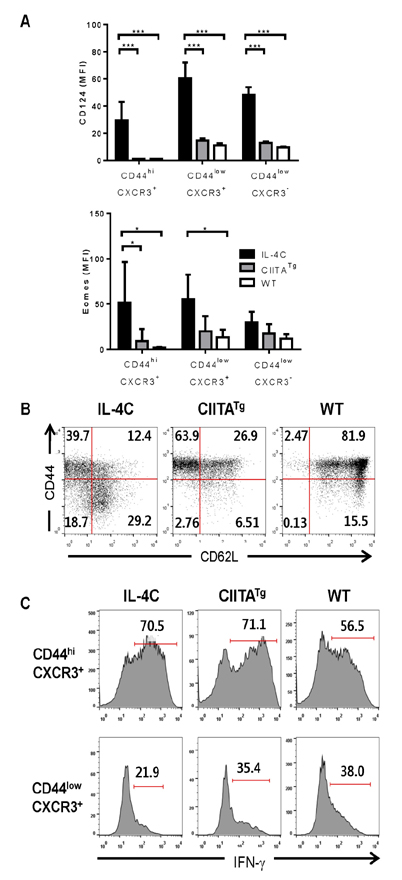
Figure 2 Phenotype and function of memory-like CD8 T cells in IL-4C-treated mice. Splenic CD8 T cells were isolated from IL-4C-treated mice, and the expression levels of the indicated markers (A & B) or IFN-γ production (C) were compared with those of CIITA-transgenic (CIITATg) and untreated WT mice. Summarized graph (n=3, A) and representative data from three independent experiments (B & C) are shown. The bars represent mean±SD. Numbers in the plots indicate the percentages of cells producing IFN-γ. *p< 0.05; ***p<0.001.
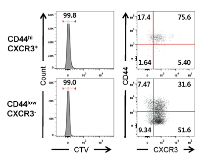
Figure 3 Generation of memory-like CD8 T cells from naïve CD8 T cells CD44hiCXCR3 + (memory phenotype) and CD44lowCXCR3 - (naïve) cells sorted from CD45.1 + B6 splenocytes were labeled with CTV and transferred into CD45.2 + B6 mice via intravenous injection. The recipient mice were injected with IL-4C. The phenotypic changes and proliferation of transferred CD8 T cells in the spleen were analyzed. Numbers in the plots indicate the percentages of cells in each population or quadrant.
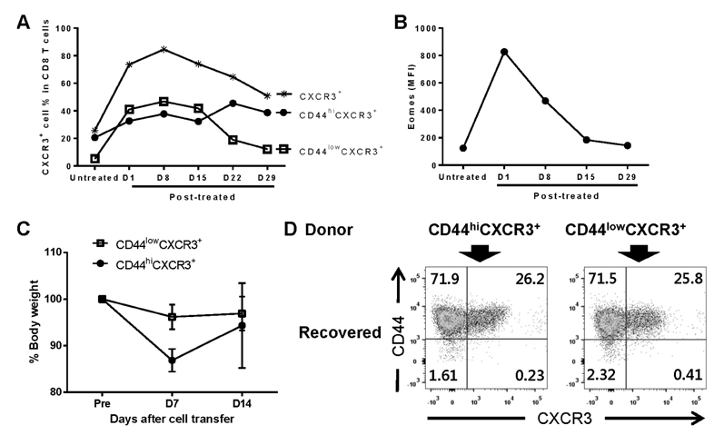
Figure 4 Conversion of CD44lowCXCR3 + CD8 T cells to the CD44hi phenotype. (A & B) Effect that discontinuing IL-4C treatment has on the CXCR3 + CD8 T cell population. B6 mice were treated with IL-4C for 7 days, and the percentage of total CXCR3 + , CD44hiCXCR3 + , and CD44lowCXCR3 + cell populations in peripheral blood CD8 T cells (A) and Eomes expression levels in CXCR3 + CD8 T cells (B) were analyzed by flow cytometry on the indicated days after treatment. Data summarized from two experiments are shown. (C & D) Conversion of CD44lowCXCR3 + CD8 T cells to the CD44hi phenotype by allo-stimulation. Splenic CD8 T cells from IL-4C-treated B6 mice were sorted into CD44hiCXCR3 + and CD44lowCXCR3 + populations by FACSAria and labeled with CTV. A total of 2×10⁵ CTV-labeled cells from each population were mixed with 3×10⁶ T cell-depleted bone marrow cells from untreated B6 mice, and transferred into irradiated BALB/c mice through intravenous injection. After the transfer, body weight was monitored every week (C). On day 14 after the cell transfer, splenocytes from the recipient BALB/c mice were labeled with antibodies against H-2Kb, CD8, CD44 and CXCR3, and the expression levels of CD44 and CXCR3 in CTVlow H-2Kb+ donor CD8 T cells that had proliferated were analyzed by flow cytometry (D). Numbers in the plots indicate the percentage of cells in each quadrant.
Cited by 2 articles
-
Serosal Cavities Contain Two Populations of Innate-like Integrin α4highCD4+ T Cells, Integrin α4β1+α6β1+α4β7− and α4β1+α6β1−α4β7+ Cells
Jeong In Yang, Chanho Park, Inseong Kho, Sujin Lee, Kyung-Suk Suh, Tae Jin Kim
Immune Netw. 2017;17(6):392-401. doi: 10.4110/in.2017.17.6.392.Aquatic Exercise at Thermoneutral Water Temperature Enhances Antitumor Immune Responses
Boae Lee, Geona Kim, Yuna Jo, Byunghyuk Lee, Yong-Il Shin, Changwan Hong
Immune Netw. 2019;19(2):. doi: 10.4110/in.2019.19.e10.
Reference
-
1. Berg LJ. Signalling through TEC kinases regulates conventional versus innate CD8+ T-cell development. Nat Rev Immunol. 2007; 7:479–485.
Article2. Veillette A, Dong Z, Latour S. Consequence of the SLAM-SAP signaling pathway in innate-like and conventional lymphocytes. Immunity. 2007; 27:698–710.
Article3. Lee YJ, Jameson SC, Hogquist KA. Alternative memory in the CD8 T cell lineage. Trends Immunol. 2011; 32:50–56.
Article4. Atherly LO, Lucas JA, Felices M, Yin CC, Reiner SL, Berg LJ. The Tec family tyrosine kinases Itk and Rlk regulate the development of conventional CD8+ T cells. Immunity. 2006; 25:79–91.
Article5. Broussard C, Fleischacker C, Horai R, Chetana M, Venegas AM, Sharp LL, Hedrick SM, Fowlkes BJ, Schwartzberg PL. Altered development of CD8+ T cell lineages in mice deficient for the Tec kinases Itk and Rlk. Immunity. 2006; 25:93–104.
Article6. Weinreich MA, Odumade OA, Jameson SC, Hogquist KA. T cells expressing the transcription factor PLZF regulate the development of memory-like CD8+ T cells. Nat Immunol. 2010; 11:709–716.
Article7. Lee A, Park SP, Park CH, Kang BH, Park SH, Ha SJ, Jung KC. IL-4 induced innate CD8+ T cells control persistent viral infection. PLoS Pathog. 2015; 11:e1005193.8. Choi EY, Jung KC, Park HJ, Chung DH, Song JS, Yang SD, Simpson E, Park SP. Thymocyte-thymocyte interaction for efficient positive selection and maturation of CD4 T cells. Immunity. 2005; 23:387–396.
Article9. Min HS, Lee YJ, Jeon YK, Kim EJ, Kang BH, Jung KC, Chang CH, Park SH. MHC class II-restricted interaction between thymocytes plays an essential role in the production of innate CD8+ T cells. J Immunol. 2011; 186:5749–5757.
Article10. Haluszczak C, Akue AD, Hamilton SE, Johnson LD, Pujanauski L, Teodorovic L, Jameson SC, Kedl RM. The antigen-specific CD8+ T cell repertoire in unimmunized mice includes memory phenotype cells bearing markers of homeostatic expansion. J Exp Med. 2009; 206:435–448.
Article11. Akue AD, Lee JY, Jameson SC. Derivation and maintenance of virtual memory CD8 T cells. J Immunol. 2012; 188:2516–2523.
Article12. Lee JY, Hamilton SE, Akue AD, Hogquist KA, Jameson SC. Virtual memory CD8 T cells display unique functional properties. Proc Natl Acad Sci USA. 2013; 110:13498–13503.
Article13. Ventre E, Brinza L, Schicklin S, Mafille J, Coupet CA, Marcais A, Djebali S, Jubin V, Walzer T, Marvel J. Negative regulation of NKG2D expression by IL-4 in memory CD8 T cells. J Immunol. 2012; 189:3480–3489.
Article14. Morris SC, Heidorn SM, Herbert DR, Perkins C, Hildeman DA, Khodoun MV, Finkelman FD. Endogenously produced IL-4 nonredundantly stimulates CD8+ T cell proliferation. J Immunol. 2009; 182:1429–1438.
Article15. Kurzweil V, LaRoche A, Oliver PM. Increased peripheral IL-4 leads to an expanded virtual memory CD8+ population. J Immunol. 2014; 192:5643–5651.
Article16. Kopf M, Legros G, Bachmann M, Lamers MC, Bluethmann H, Kohler G. Disruption of the murine Il-4 gene blocks Th2 cytokine tesponses. Nature. 1993; 362:245–248.
Article17. Ouyang W, Ranganath SH, Weindel K, Bhattacharya D, Murphy TL, Sha WC, Murphy KM. Inhibition of Th1 development mediated by GATA-3 through an IL-4-independent mechanism. Immunity. 1998; 9:745–755.
Article18. Carty SA, Koretzky GA, Jordan MS. Interleukin-4 regulates eomesodermin in CD8 T cell development and differentiation. Plos One. 2014; 9:e106659.
Article19. Oghumu S, Terrazas CA, Varikuti S, Kimble J, Vadia S, Yu L, Seveau S, Satoskar AR. CXCR3 expression defines a novel subset of innate CD8+ T cells that enhance immunity against bacterial infection and cancer upon stimulation with IL-15. FASEB J. 2015; 29:1019–1028.
Article
- Full Text Links
-
- Actions
-
Cited
- CITED
-
- Close
- Share
-
- Similar articles
-
- Heterogeneity of IL-22-producing Lymphoid Tissue Inducer-like Cells in Human and Mouse
- CD43 Expression Regulated by IL-12 Signaling Is Associated with Survival of CD8 T Cells
- The Roles of CCR7 for the Homing of Memory CD8+ T Cells into Their Survival Niches
- The Role of CD4 T Cell Help in CD8 T Cell Differentiation and Function During Chronic Infection and Cancer
- IL‑4/IL‑4 Ab complex enhances the accumulation of both antigen‑specific and bystander CD8 T cells in mouse lungs infected with influenza A virus
