Resveratrol at High Doses Acts as an Apoptotic Inducer in Endothelial Cells
- Affiliations
-
- 1Department of Molecular Biology & the Institute of Nanosensor and Biotechnology, Dankook Univiersity, Seoul, Korea. heonyong@dankook.ac.kr
Abstract
- PURPOSE
S: Resveratrol is a phenolic compound found in grapes and other food products. In order to assess the availability of resveratrol as an angio-inhibiting drug, we examined whether resveratrol plays an important role in bovine aortic endothelial cells (BAECs) for cell apoptosis and cell migration.
METHODS
AND MATERIALS: Endothelial cell apoptosis was observed as detected by the Hoechst staining and the caspase-3 activity. Additionally, Western blotting was performed for monitoring the activities of various cell signaling molecules.
RESULTS
Resveratrol was shown to act as a pro-apoptotic agent. The pro-apoptotic effect of resveratrol was as great as that of etoposide, a well-known anti-cancer drug. In addition, resveratrol had an inhibitory effect on endothelial cell migration. The demonstrated efficacy of resveratrol suggests that resveratrol may be utilized as an anti-angiogenic drug. To determine the underlying mechanisms, we further investigated which signaling molecules are activated by resveratrol. Extracellular signal-regulated kinase (ERK) was activated by the treatment with resveratrol in BAECs, whereas endothelial nitric oxide synthetase (eNOS), Akt, and Jun N-terminal kinase (JNK) were inhibited. The pretreatment with PD compound, an ERK inhibitor, had no effect on the pro-apoptosis induced by resveratrol.
CONCLUSION
Resveratrol plays an important role in endothelial cell apoptosis, indicating that resveratrol can be utilized as a potent anti-angiogenic drug.
Keyword
MeSH Terms
Figure
-

Fig. 1 Resveratrol induces endothelial cell apoptosis. Confluent BAECs were starved for more than 4 h and then the cells were incubated for additional hours (0, 4, 8, 12, 24 or 72 h) with the indicated doses of resveratrol (RES). Consecutively, the cells were incubated for additional hours (0, 4, 8, 12, 24 or 72 h) We then counted the apoptotic cells (round shrunken cells) under the microscope. The line graphs represent the percentages of apoptotic cells (means±S.E.). The experiments were independently performed at least three times.
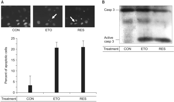
Fig. 2 The resveratrol-induced apoptosis is mediated by activation of caspase-3. Confluent cells were incubated with 0.5% FBS-DMEM containing none (CON), 100 µM etoposide (ETO) or 100 µM resveratrol (RES) for 36 h. The cell were then stained with Hoechst 33258 and we observed the nuclei under a fluorescence microscope (A). Arrows indicate the fragmented nuclei. In panel B, after the cells were lysed, proteins in the cell lysates were resolved by SDS-PAGE, electrotransferred to PVDF membranes and immunoblotted with caspase-3 antibodies.

Fig. 3 Resveratrol blocks the endothelial cell migration. Confluent endothelial cells were serum-starved and then scraped with a razor blade after 24 h starvation. The cells were subsequently incubated in media containing 20% fetal bovine serum (CON) or various concentration of resveratrol (RES). After 26 h, we then observed the migrated cells under the microscope. The line on each photograph indicates the boundary lines immediately after scraping. The photographs are representative of at least three different observations. We then counted the cells that migrated over the lines. Quantification was performed by counting the number of cells that migrated in the same field. The bar graphs show the mean number of migrated cells±S.E. (n=3).
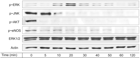
Fig. 4 Resveratrol modulates a variety of cell signaling molecules. Serum-depleted cells (grown in 0.5% FBS-DMEM) were stimulated with 100 µM resveratrol treatment as a function of time. After the cells were lysed, the proteins in cell lysates were resolved by SDS-PAGE, electrotransferred to PVDF membranes and immunoblotted with the appropriate antibodies.
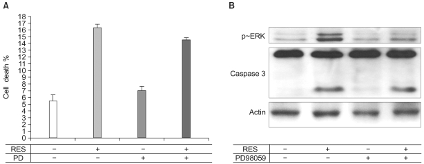
Fig. 5 ERK is not associated with the resveratrol-induced apoptosis. (A) Apoptosis was observed as described in Fig. 1. Interestingly, 10 µM of PD compound had no effect on apoptosis. The bar graphs represent means±S.E. (n=3). (B) Serum-depleted cells (grown in 0.5% FBS-DMEM) were stimulated with 100 µM resveratrol treatment for 24 h. After the cells were lysed, proteins in the cell lysates were resolved by SDS-PAGE, electrotransferred to PVDF membranes and immunoblotted with caspase-3 antibodies.
Reference
-
1. Gasparini G. The rationale and future potential of angiogenesis inhibitors in neoplasia. Drugs. 1999; 58:17–38. PMID: 10439927.
Article2. Jang M, Cai L, Udeani GO, Slowing KV, Thomas CF, Beecher CW, et al. Cancer Chemopreventive activity of resveratrol, a natural product derived from grapes. Science. 1997; 275:218–220. PMID: 8985016.
Article3. Signorelli P, Ghidoni R. Resveratrol as an anticancer nutrient: molecular basis, open questions and promises. J Nutr Biochem. 2005; 16:449–466. PMID: 16043028.
Article4. Klinge CM, Blankenship KA, Resinger KE, Bhatnagar S, Noisin EL, Sumanasekera WK, et al. Resveratrol and estradiol rapidly activate MAPK signaling through estrogen receptors α and β in endothelial cells. J Biol Chem. 2005; 280:7460–7468. PMID: 15615701.
Article5. Jo H, Sipos K, Go YM, Law R, Rong J, McDonald JM. Differential effect of shear stress on extracellular signal-regulated kinase and N-terminal Jun kinase in endothelial cells. Gi2- and Gbeta/gamma-dependent signaling pathways. J Biol Chem. 1997; 272:1395–1401. PMID: 8995450.6. Park H, Go YM, St John PL, Maland MC, Lisanti MP, Abrahamson DR, et al. Plasma membrane cholesterol is a key molecule in shear stress-dependent activation of extracellular signal-regulated kinase. J Biol Chem. 1998; 273:32304–32311. PMID: 9822710.
Article7. Park SG, Kang YS, Ahn YH, Lee SH, Kim KR, Kim KW, et al. Dose-dependent biphasic activity of tRNA synthetase-associating factor, p43, in angiogenesis. J Biol Chem. 2002; 277:45243–45248. PMID: 12237313.
Article8. Ou HC, Chou FP, Sheen HM, Lin TM, Yang CH, Huey-Herng Sheu W. Resveratrol, a polyphenolic compound in red wine, protects against oxidized LDL-induced cytotoxicity in endothelial cells. Clin Chim Acta. 2006; 364:196–204. PMID: 16095586.
Article9. Rodriguez-Lopez AM, Xenaki D, Eden TO, Hickman JA, Chresta CM. MDM2 mediated nuclear exclusion of p53 attenuates ectoposide-induced apoptosis in neuroblastoma cells. Mol Pharmacol. 2001; 59:135–143. PMID: 11125034.10. Li J, Zhang YP, Kirsner RS. Angiogenesis in wound repair: angiogenic growth factors and the extracellular matrix. Microsc Res Tech. 2003; 60:107–114. PMID: 12500267.
Article11. Oak MH, Bedoui J, Schini-Kerth VB. Antiangiogenic properties of natural polyphenols from red wine and green tea. J Nutr Biochem. 2005; 16:1–8. PMID: 15629234.
Article12. Karin M. The regulation of AP-1 activity by mitogen-activated protein kinases. Philos Trans R Soc Lond B Biol Sci. 1996; 351:127–134. PMID: 8650258.
Article13. Zuckerbraun BS, McCloskey CA, Mahidhara RS, Kim PK, Taylor BS, Tzeng E. Overexpression of mutated IkappaBalpha inhibits vascular smooth muscle cell proliferation and intimal hyperplasia formation. J Vasc Surg. 2003; 38:812–819. PMID: 14560235.14. Park H, Park SG, Kim J, Ko YG, Kim S. Signaling pathways for TNF production induced by human aminoacyl-tRNA synthetase-associating factor, p43. Cytokine. 2002; 20:148–153. PMID: 12543078.
Article15. She QB, Huang C, Zhang Y, Dong Z. Involvment of c-jun NH(2)-terminal kinase in resveratrol-induced activation of p53 and apoptosis. Mol Carcinog. 2002; 33:244–250. PMID: 11933078.16. Sanna B, Debidda M, Pintus G, Tandolini B, Posadino AM, Bennardini F, et al. The anti-metastatic agent imidazolium trans-imidazoledimethylsulfoxide-tetrachlororuthenate induces endothelial cell apoptosis by inhibiting the mitogen-activated protein kinase/extracellular signal-regulated kinase signaling pathway. Arch Biochem Biophys. 2002; 403:209–218. PMID: 12139970.
Article17. Ranieri G, Gasparini G. Angiogenesis and angiogenesis inhibitors: a new potential anticancer therapeutic strategy. Curr Drug Targets Immune Endocr Metabol Disord. 2001; 1:241–253. PMID: 12477290.18. Iyer S, Chaplin DJ, Rosenthal DS, Boulares AH, Li Ly, Smulson ME. Induction of apoptosis in proliferating human endothelial cells by the tumor-specific antiangiogenesis agent combretastatin A-4. Cancer Res. 1998; 58:4510–4514. PMID: 9788591.19. Lenz HJ. Antiangiogenic agents in cancer therapy. Oncology. 2005; 19(4):Suppl 3. 17–25. PMID: 15934499.
- Full Text Links
-
- Actions
-
Cited
- CITED
-
- Close
- Share
-
- Similar articles
-
- Dose-dependent effect of resveratrol on proliferation and apoptosis in endothelial and tumor cell cultures
- Effect of Resveratrol on Cell Differentiation and Mineralization in Cultured Odontoblasts
- Resveratrol blunts tumor necrosis factor-alpha-induced monocyte adhesion and transmigration
- Antichemosensitizing effect of resveratrol in cotreatment with oxaliplatin in HCT116 colon cancer cell
- Neuroprotective effects of resveratrol on 6-hydroxydopamine-induced damage of SH-SY5Y cell line
