Immune Netw.
2012 Dec;12(6):223-229. 10.4110/in.2012.12.6.223.
In Vivo Non Invasive Molecular Imaging for Immune Cell Tracking in Small Animals
- Affiliations
-
- 1Department of Nuclear Medicine, Cancer Research Institute, Seoul National University College of Medicine, Seoul 110-799, Korea. hwyoun@snu.ac.kr
- 2Laboratory of Molecular Imaging and Therapy, Cancer Research Institute, Seoul National University College of Medicine, Seoul 110-799, Korea.
- 3Cancer Imaging Center, Seoul National University Cancer Hospital Tumor Biology, Seoul 110-799, Korea.
- 4Division of High-Risk Pathogen Research, Center for Infectious Diseases, Korea National Research of Health, Osong 363-951, Korea.
- KMID: 2150753
- DOI: http://doi.org/10.4110/in.2012.12.6.223
Abstract
- Clinical and preclinical in vivo immune cell imaging approaches have been used to study immune cell proliferation, apoptosis and interaction at the microscopic (intra-vital imaging) and macroscopic (whole-body imaging) level by use of ex vivo or in vivo labeling method. A series of imaging techniques ranging from non-radiation based techniques such as optical imaging, MRI, and ultrasound to radiation based CT/nuclear imaging can be used for in vivo immune cell tracking. These imaging modalities highlight the intrinsic behavior of different immune cell populations in physiological context. Fluorescent, radioactive or paramagnetic probes can be used in direct labeling protocols to monitor the specific cell population. Reporter genes can also be used for genetic, indirect labeling protocols to track the fate of a given cell subpopulation in vivo. In this review, we summarized several methods dealing with dendritic cell, macrophage, and T lymphocyte specifically labeled for different macroscopic wholebody imaging techniques both for the study of their physiological function and in the context of immunotherapy to exploit imaging-derived information and immune-based treatments.
MeSH Terms
Figure
-
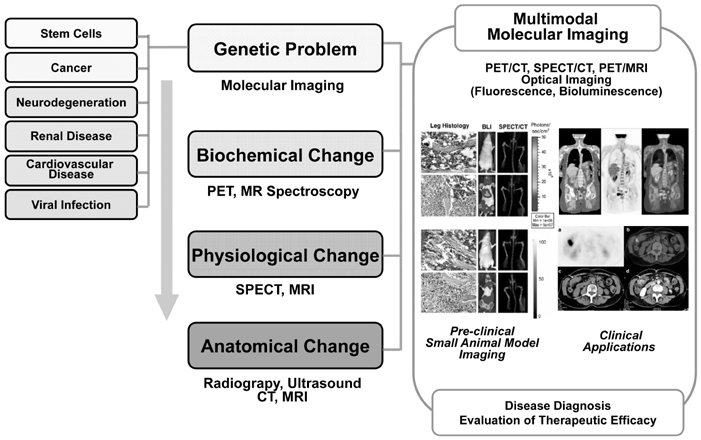
Figure 1 Molecular imaging techniques for disease diagnosis and evaluation of therapeutic efficacy.
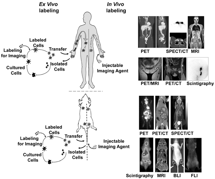
Figure 2 Ex vivo vs. In vivo labeling for immune cell tracking.
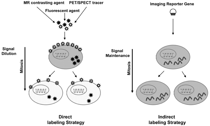
Figure 3 Direct vs. indirect cell labeling strategies.
Cited by 2 articles
-
Non-invasive molecular imaging of immune cell dynamics for vaccine research
Hyewon Youn, Kee-Jong Hong
Clin Exp Vaccine Res. 2019;8(2):89-93. doi: 10.7774/cevr.2019.8.2.89.In Vivo Stem Cell Imaging Principles and Applications
Seongje Hong, Dong-Sung Lee, Geun-Woo Bae, Juhyeong Jeon, Hak Kyun Kim, Siyeon Rhee, Kyung Oh Jung
Int J Stem Cells. 2023;16(4):363-375. doi: 10.15283/ijsc23045.
Reference
-
1. Blasberg RG, Gelovani-Tjuvajev J. In vivo molecular-genetic imaging. J Cell Biochem Suppl. 2002. 39:172–183.
Article2. Forss-Petter S, Danielson PE, Catsicas S, Battenberg E, Price J, Nerenberg M, Sutcliffe JG. Transgenic mice expressing beta-galactosidase in mature neurons under neuron-specific enolase promoter control. Neuron. 1990. 5:187–197.
Article3. Yu YA, Timiryasova T, Zhang Q, Beltz R, Szalay AA. Optical imaging: bacteria, viruses, and mammalian cells encoding light-emitting proteins reveal the locations of primary tumors and metastases in animals. Anal Bioanal Chem. 2003. 377:964–972.
Article4. Shaner NC, Steinbach PA, Tsien RY. A guide to choosing fluorescent proteins. Nat Methods. 2005. 2:905–909.
Article5. Tjuvajev JG, Stockhammer G, Desai R, Uehara H, Watanabe K, Gansbacher B, Blasberg RG. Imaging the expression of transfected genes in vivo. Cancer Res. 1995. 55:6126–6132.6. Gambhir SS, Barrio JR, Phelps ME, Iyer M, Namavari M, Satyamurthy N, Wu L, Green LA, Bauer E, MacLaren DC, Nguyen K, Berk AJ, Cherry SR, Herschman HR. Imaging adenoviral-directed reporter gene expression in living animals with positron emission tomography. Proc Natl Acad Sci U S A. 1999. 96:2333–2338.
Article7. Brown RS, Leung JY, Fisher SJ, Frey KA, Ethier SP, Wahl RL. Intratumoral distribution of tritiated-FDG in breast carcinoma: correlation between Glut-1 expression and FDG uptake. J Nucl Med. 1996. 37:1042–1047.8. Ichikawa T, Högemann D, Saeki Y, Tyminski E, Terada K, Weissleder R, Chiocca EA, Basilion JP. MRI of transgene expression: correlation to therapeutic gene expression. Neoplasia. 2002. 4:523–530.
Article9. Blasberg RG, Tjuvajev JG. Molecular-genetic imaging: current and future perspectives. J Clin Invest. 2003. 111:1620–1629.
Article10. Massoud TF, Gambhir SS. Molecular imaging in living subjects: seeing fundamental biological processes in a new light. Genes Dev. 2003. 17:545–580.
Article11. Serganova I, Mayer-Kukuck P, Huang R, Blasberg R. Molecular imaging: reporter gene imaging. Handb Exp Pharmacol. 2008. (185 Pt 2):167–223.
Article12. Hoffman JM, Gambhir SS. Molecular imaging: the vision and opportunity for radiology in the future. Radiology. 2007. 244:39–47.
Article13. Townsend DW, Carney JP, Yap JT, Hall NC. PET/CT today and tomorrow. J Nucl Med. 2004. 45:Suppl 1. 4S–14S.14. Kang JH, Chung JK. Molecular-genetic imaging based on reporter gene expression. J Nucl Med. 2008. 49(Suppl 2):164S–179S.
Article15. Pichler BJ, Judenhofer MS, Catana C, Walton JH, Kneilling M, Nutt RE, Siegel SB, Claussen CD, Cherry SR. Performance test of an LSO-APD detector in a 7-T MRI scanner for simultaneous PET/MRI. J Nucl Med. 2006. 47:639–647.16. Ottobrini L, Martelli C, Trabattoni DL, Clerici M, Lucignani G. In vivo imaging of immune cell trafficking in cancer. Eur J Nucl Med Mol Imaging. 2011. 38:949–968.
Article17. Dunn KW, Sutton TA. Functional studies in living animals using multiphoton microscopy. ILAR J. 2008. 49:66–77.
Article18. Pittet MJ, Weissleder R. Intravital Imaging. Cell. 2011. 147:983–991.
Article19. De Jong M, Maina T. Of mice and humans: are they the same?- Implications in cancer translational research. J Nucl Med. 2010. 51:501–504.
Article20. Pham W, Kobukai S, Hotta C, Gore JC. Dendritic cells; therapy and imaging. Expert Opin Biol Ther. 2009. 9:539–564.
Article21. Mandl S, Schimmelpfennig C, Edinger M, Negrin RS, Contag CH. Understanding immune cell trafficking patterns via in vivo bioluminescence imaging. J Cell Biochem Suppl. 2002. 39:239–248.
Article22. Negrin RS, Contag H. In vivo imaging using bioluminescence: a tool for probing graft-versus-host disease. Nat Rev Immunol. 2006. 6:484–490.
Article23. Song MG, Kang B, Jeon JY, Chang J, Lee S, Min CK, Youn H, Choi EY. In vivo imaging of differences in early donor cell proliferation in graft-versus-host disease hosts with different pre-conditioning doses. Mol Cells. 2012. 33:79–86.
Article24. de Vries IJ, Lesterhuis WJ, Barentsz JO, Verdijk P, van Krieken JH, Boerman OC, Oyen WJ, Bonenkamp JJ, Boezeman JB, Adema GJ, Bulte JW, Scheenen TW, Punt CJ, Heerschap A, Figdor CG. Magnetic resonance tracking of dendritic cells in melanoma patients for monitoring of cellular therapy. Nat Biotechnol. 2005. 23:1407–1413.
Article25. Dobrenkov K, Olszewska M, Likar Y, Shenker L, Gunset G, Cai S, Pillarsetty N, Hricak H, Sadelain M, Ponomarev V. Monitoring the efficacy of adoptively transferred prostate cancer-targeted human T lymphocytes with PET and bioluminescence imaging. J Nucl Med. 2008. 49:1162–1170.
Article26. Leimgruber A, Berger C, Cortez-Retamozo V, Etzrodt M, Newton AP, Waterman P, Figueiredo JL, Kohler RH, Elpek N, Mempel TR, Swirski FK, Nahrendorf M, Weissleder R, Pittet MJ. Behavior of endogenous tumor-associated macrophages assessed in vivo using a functionalized nanoparticle. Neoplasia. 2009. 11:459–468.
Article
