J Korean Surg Soc.
2010 Feb;78(2):111-115. 10.4174/jkss.2010.78.2.111.
Detection and Typing of Human Papillomavirus in Anal Condyloma Acuminatum of HIV-positive Patients
- Affiliations
-
- 1Department of Surgery, Seoul National University College of Medicine, Seoul, Korea. kjparkmd@plaza.snu.ac.kr
- KMID: 2096564
- DOI: http://doi.org/10.4174/jkss.2010.78.2.111
Abstract
- PURPOSE
Anal condyloma is an epithelial proliferative lesion caused by human papillomavirus (HPV) infection. The present study analyzed the HPV types detected in HIV (+) Korean anal condyloma using PCR-based DNA microarray.
METHODS
DNA was extracted from the condyloma tissue of 17 patients including 9 HIV (+) patients (M:F=15:2, mean age 35 years, 22~59 years). The 1st PCR was performed with a general primer on L1 region, and nested PCR on the products of the 1st PCR. PCR products were hybridized with a DNA chip.
RESULTS
Fourteen patients (9 HIV (+), 5 HIV (-)) showed positive HPV DNA. Overall, type 6 was the most common (N=11), and type 11 (N=6), type 53 (N=3) in order. Among HIV (+) patients, type 6 was also the most common (N=7), then type 11 (N=5) and type 53 (N=3). In contrast to the HIV (-) patients, 5 patients (55.6%) proved to have multiple infections in HIV (+) patients (2 double, 2 triple, 1 quadruple infection). Four of 9 HIV (+) patients (44.5%) showed co-infection with high-risk HPV.
CONCLUSION
Multiple infection and co-infection with high-risk types are more prevalent in HIV (+) condyloma patients compared to HIV (-) patients. HPV types on HIV (+) male anal condyloma are similar to those detected in the Korean female uterine cervix.
Keyword
MeSH Terms
Figure
-
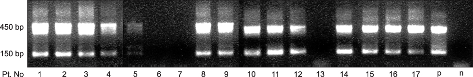
Fig. 1 Electrophoresis of the 1st PCR products (450 bp) and the nested PCR products (150 bp) (p = positive control, n = negative control).

Fig. 2 DNA chip scanning of patients No. 2, 5, 8.
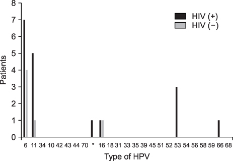
Fig. 3 HPV type distribution in anal condyloma according to the HIV status (* = other type).

Fig. 4 Multiplicity of the HPV infection according to the HIV status.
Cited by 1 articles
-
Immunohistochemistry and Polymerase Chain Reaction for Detection Human Papilloma Virus in Warts: A Comparative Study
Hong Sun Lee, Ji Hyun Lee, Ji Yoon Choo, Hee Jin Byun, Jin Hyun Jun, Jun Young Lee
Ann Dermatol. 2016;28(4):479-485. doi: 10.5021/ad.2016.28.4.479.
Reference
-
1. Brown DR, Schroeder JM, Bryan JT, Stoler MH, Fife KH. Detection of multiple human papillomavirus types in Condylomata acuminata lesions from otherwise healthy and immunosuppressed patients. J Clin Microbiol. 1999. 37:3316–3322.2. Brown DR, Bryan JT, Cramer H, Fife KH. Analysis of human papillomavirus types in exophytic condylomata acuminata by hybrid capture and Southern blot techniques. J Clin Microbiol. 1993. 31:2667–2673.3. Gissmann L, Wolnik L, Ikenberg H, Koldovsky U, Schnurch HG, zur Hausen H. Human papillomavirus types 6 and 11 DNA sequences in genital and laryngeal papillomas and in some cervical cancers. Proc Natl Acad Sci U S A. 1983. 80:560–563.4. Kim TJ, Joo JH, Kim HR, Kim DY. Human papillomavirus infection in anal carcinoma, anal condylomata and rectalcarcinoma. J Korean Soc Coloproctol. 1997. 13:7–14.5. Critchlow CW, Hawes SE, Kuypers JM, Goldbaum GM, Holmes KK, Surawicz CM, et al. Effect of HIV infection on the natural history of anal human papillomavirus infection. AIDS. 1998. 12:1177–1184.6. Anderson CA, Boller AM, Richardson CJ, Balcos EG, Zera RT. Anal condyloma: a comparison between HIV positive and negative patients. Am Surg. 2004. 70:1014–1018.7. Palefsky JM, Holly EA, Ralston ML, Jay N, Berry JM, Darragh TM. High incidence of anal high-grade squamous intra-epithelial lesions among HIV-positive and HIV-negative homosexual and bisexual men. AIDS. 1998. 12:495–503.8. Palefsky JM, Holly EA, Hogeboom CJ, Ralston ML, DaCosta MM, Botts R, et al. Virologic, immunologic, and clinical parameters in the incidence and progression of anal squamous intraepithelial lesions in HIV-positive and HIV-negative homosexual men. J Acquir Immune Defic Syndr Hum Retrovirol. 1998. 17:314–319.9. Munoz N, Bosch FX, de Sanjose S, Herrero R, Castellsague X, Shah KV, et al. Epidemiologic classification of human papillomavirus types associated with cervical cancer. N Engl J Med. 2003. 348:518–527.10. Jung WW, Chun T, Sul D, Hwang KW, Kang HS, Lee DJ, et al. Strategies against human papillomavirus infection and cervical cancer. J Microbiol. 2004. 42:255–266.11. Choi YD, Jung WW, Nam JH, Choi HS, Park CS. Detection of HPV genotypes in cervical lesions by the HPV DNA Chip and sequencing. Gynecol Oncol. 2005. 98:369–375.
- Full Text Links
-
- Actions
-
Cited
- CITED
-
- Close
- Share
-
- Similar articles
-
- Detection of Human Papillomavirus DNA in Genital and Laryngeal Papilloma Using the Polymerase Chain Reaction
- Association of Immune Status with Recurrent Anal Condylomata in Human Immunodeficiency Virus-Positive Patients
- A Case of unusual condyloma Acuminatum in an Immunosuppressed Patient
- Nd:YAG Laser Therapy for Intraurethral Condyloma Acuminatum in Men
- Two cases of human immunodeficiency virus infection associated with condyloma acuminatum
