Ascorbic acid insufficiency induces the severe defect on bone formation via the down-regulation of osteocalcin production
- Affiliations
-
- 1Labratory of Vitamin C and Immunology, Department of Anatomy, Seoul National University College of Medicine, Seoul, Korea. kinglee@snu.ac.kr genius29@snu.ac.kr
- 2Department of Rehabilitation Medicine, Seoul National University College of Medicine, Seoul, Korea.
- 3Department of Psychology, Boston College, Chestnut Hill, MA, USA.
- 4Division of Hematology-Oncology, Department of Internal Medicine, Ewha Womans University School of Medicine, Seoul, Korea.
- KMID: 2046766
- DOI: http://doi.org/10.5115/acb.2013.46.4.254
Abstract
- The L-gulono-gamma-lactone oxidase gene (Gulo) encodes an essential enzyme in the synthesis of ascorbic acid from glucose. On the basis of previous findings of bone abnormalities in Gulo-/- mice under conditions of ascorbic acid insufficiency, we investigated the effect of ascorbic acid insufficiency on factors related to bone metabolism in Gulo-/- mice. Four groups of mice were raised for 4 weeks under differing conditions of ascorbic acid insufficiency, namely, wild type; ascorbic acid-sufficient Gulo-/- mice, 3-week ascorbic acid-insufficient Gulo-/- mice, and 4-week ascorbic acid-insufficient Gulo-/- mice. Four weeks of ascorbic acid insufficiency resulted in significant weight loss in Gulo-/- mice. Interestingly, average plasma osteocalcin levels were significantly decreased in Gulo-/- mice after 3 weeks of ascorbic acid insufficiency. In addition, the tibia weight in ascorbic acid-sufficient Gulo-/- mice was significantly higher than that in the other three groups. Moreover, significant decreases in trabecular bone volume near to the growth plate, as well as in trabecular bone attachment to the growth plate, were evident in 3- or 4-week ascorbic acid-insufficient Gulo-/-. In summary, ascorbic acid insufficiency in Gulo-/- mice results in severe defects in normal bone formation, which are closely related to a decrease in plasma osteocalcin levels.
MeSH Terms
Figure
-
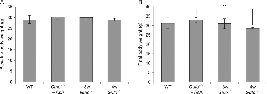
Fig. 1 Mice weight at baseline and at the conclusion of the experiment. The weight of the wild-type (WT), ascorbic acid-sufficient Gulo-/- mice (Gulo-/-+AsA), and ascorbic acid-insufficient Gulo-/- mice (Gulo-/-) was measured at baseline and after 3 or 4 weeks of ascorbic acid withdrawal. (A) No significant differences in weight were observed among the groups at baseline. (B) Weight was significantly decreased in Gulo-/- mice after 4 weeks of ascorbic acid insufficiency compared with ascorbic acid-sufficient (-/-) mice. Data are presented as the means±SD, and each group included 5 animals. **P<0.01.
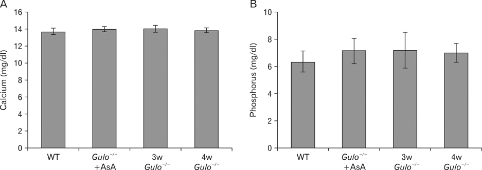
Fig. 2 Plasma levels of calcium and phosphorus after ascorbic acid withdrawal. Plasma levels of calcium (A) and phosphorus (B) were measured in 3-week (3w Gulo-/-) and 4-week (4w Gulo-/-) ascorbic acid-insufficient Gulo-/- mice. No significant differences were observed among the groups. The data are presented as the means±SD, and each group included 5 animals. WT, wild-type.
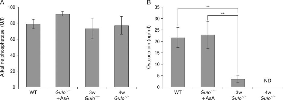
Fig. 3 The plasma levels of alkaline phosphatase and osteocalcin after ascorbic acid withdrawal. Plasma levels of alkaline phosphatase (A) and osteocalcin (B) were measured in 3-week (3w Gulo-/-) and 4-week (4w Gulo-/-) ascorbic acid-insufficient Gulo-/- mice. (A) The plasma level of alkaline phosphatase was not significantly different among the groups. (B) The plasma level of osteocalcin was significantly lower in 3w Gulo-/- mice than in wild-type (WT) and ascorbic acid-sufficient Gulo-/- mice (Gulo-/-+AsA). Osteocalcin was undetectable in the plasma of 4w Gulo-/- mice. Data are presented as the means±SD, ND, not detected, each group included 5 animals. **P<0.01.
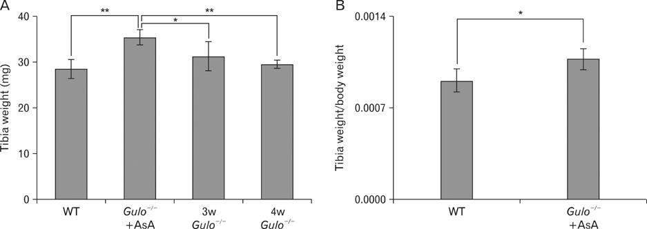
Fig. 4 Tibia dry weight and the ratio of the tibia weight to the final body weight at the end of the experiment. Tibia dry weight was measured in 3-week (3w Gulo-/-) and 4-week (4w Gulo-/-) ascorbic acid-insufficient Gulo-/- mice. Tibia dry weight (A) and ratio of the tibia weight to the final body weight (B) was significantly higher in the ascorbic acid-sufficient Gulo-/- mice (Gulo-/-+AsA) than in the other three groups. Data are shown as the means±SD, each group included 5 animals. *P<0.05, **P<0.01.
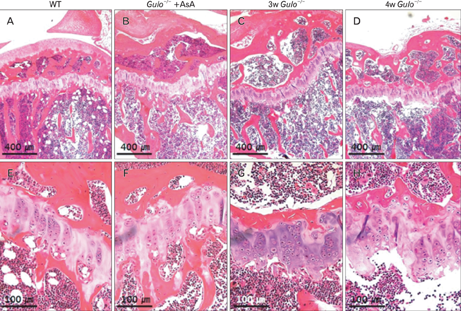
Fig. 5 The proximal metaphysis and growth plate of the proximal tibia after ascorbic acid withdrawal. Mouse tibias from 3-week (3w Gulo-/-) and 4-week (4w Gulo-/-) ascorbic acid-insufficient Gulo-/- mice were harvested, fixed, decalcified, sectioned at 4 µm and stained with H&E. Compared with the wild-type (WT) (A, E) and ascorbic acid-sufficient Gulo-/- mice (Gulo-/-+AsA) (B, F), 3w Gulo-/- (C, G), 4w Gulo-/- (D, H) mice had decreased trabecular bone volume near the growth plate, and decreased trabecular bone attachment to the growth plate. Scale bars=400 µm (A-D), 100 µm (E-H).
Cited by 1 articles
-
Nutrition and growth: assessing the impact of regional nutritional intake on childhood development and metacarpal parameters
Christian Moro, Jessica Covino
Anat Cell Biol. 2018;51(1):31-40. doi: 10.5115/acb.2018.51.1.31.
Reference
-
1. Peterkofsky B. Ascorbate requirement for hydroxylation and secretion of procollagen: relationship to inhibition of collagen synthesis in scurvy. Am J Clin Nutr. 1991; 54:6 Suppl. 1135S–1140S.2. Fain O. Musculoskeletal manifestations of scurvy. Joint Bone Spine. 2005; 72:124–128.3. Niyibizi C, Eyre DR. Structural characteristics of cross-linking sites in type V collagen of bone: chain specificities and heterotypic links to type I collagen. Eur J Biochem. 1994; 224:943–950.4. Cummine J, Armstrong L, Nade S. Osteogenesis after bone and bone marrow transplantation: studies of cellular behaviour using combined myelo-osseous grafts in the subscorbutic guinea pig. Acta Orthop Scand. 1983; 54:235–241.5. Bourne G. The effect of graded doses of vitamin C upon the regeneration of bone in guinea-pigs on a scorbutic diet. J Physiol. 1942; 101:327–336.6. Léone J, Delhinger V, Maes D, Scheer C, Pennaforte JL, Eschard JP, Etienne JC. Rheumatic manifestations of scurvy: a report of two cases. Rev Rhum Engl Ed. 1997; 64:428–431.7. Shetty AK, Buckingham RB, Killian PJ, Girdany D, Meyerowitz R. Hemarthrosis and femoral head destruction in an adult diet faddist with scurvy. J Rheumatol. 1988; 15:1878–1880.8. Wapnick AA, Lynch SR, Seftel HC, Charlton RW, Bothwell TH, Jowsey J. The effect of siderosis and ascorbic acid depletion on bone metabolism, with special reference to osteoporosis in the Bantu. Br J Nutr. 1971; 25:367–376.9. Hall SL, Greendale GA. The relation of dietary vitamin C intake to bone mineral density: results from the PEPI study. Calcif Tissue Int. 1998; 63:183–189.10. Sahni S, Hannan MT, Gagnon D, Blumberg J, Cupples LA, Kiel DP, Tucker KL. High vitamin C intake is associated with lower 4-year bone loss in elderly men. J Nutr. 2008; 138:1931–1938.11. Ohta Y, Nishikimi M. Random nucleotide substitutions in primate nonfunctional gene for L-gulono-gamma-lactone oxidase, the missing enzyme in L-ascorbic acid biosynthesis. Biochim Biophys Acta. 1999; 1472:408–411.12. Nishikimi M, Fukuyama R, Minoshima S, Shimizu N, Yagi K. Cloning and chromosomal mapping of the human nonfunctional gene for L-gulono-gamma-lactone oxidase, the enzyme for L-ascorbic acid biosynthesis missing in man. J Biol Chem. 1994; 269:13685–13688.13. Nishikimi M, Kawai T, Yagi K. Guinea pigs possess a highly mutated gene for L-gulono-gamma-lactone oxidase, the key enzyme for L-ascorbic acid biosynthesis missing in this species. J Biol Chem. 1992; 267:21967–21972.14. Maeda N, Hagihara H, Nakata Y, Hiller S, Wilder J, Reddick R. Aortic wall damage in mice unable to synthesize ascorbic acid. Proc Natl Acad Sci U S A. 2000; 97:841–846.15. Bae S, Cho CH, Kim H, Kim Y, Kim HR, Hwang YI, Yoon JH, Kang JS, Lee WJ. In vivo consequence of vitamin C insufficiency in liver injury: vitamin C ameliorates T-cell-mediated acute liver injury in Gulo(-/-) mice. Antioxid Redox Signal. 2013; 19:2040–2053.16. Kim H, Bae S, Yu Y, Kim Y, Kim HR, Hwang YI, Kang JS, Lee WJ. The analysis of vitamin C concentration in organs of gulo(-/-) mice upon vitamin C withdrawal. Immune Netw. 2012; 12:18–26.17. Alers JC, Krijtenburg PJ, Vissers KJ, van Dekken H. Effect of bone decalcification procedures on DNA in situ hybridization and comparative genomic hybridization: EDTA is highly preferable to a routinely used acid decalcifier. J Histochem Cytochem. 1999; 47:703–710.18. Sakamoto Y, Takano Y. Morphological influence of ascorbic acid deficiency on endochondral ossification in osteogenic disorder Shionogi rat. Anat Rec. 2002; 268:93–104.19. Hasegawa T, Li M, Hara K, Sasaki M, Tabata C, de Freitas PH, Hongo H, Suzuki R, Kobayashi M, Inoue K, Yamamoto T, Oohata N, Oda K, Akiyama Y, Amizuka N. Morphological assessment of bone mineralization in tibial metaphyses of ascorbic acid-deficient ODS rats. Biomed Res. 2011; 32:259–269.20. Tsuchiya H, Bates CJ. Ascorbic acid deficiency in guinea pigs: contrasting effects of tissue ascorbic acid depletion and of associated inanition on status indices related to collagen and vitamin D. Br J Nutr. 1994; 72:745–752.21. Mahmoodian F, Gosiewska A, Peterkofsky B. Regulation and properties of bone alkaline phosphatase during vitamin C deficiency in guinea pigs. Arch Biochem Biophys. 1996; 336:86–96.22. Mohan S, Kapoor A, Singgih A, Zhang Z, Taylor T, Yu H, Chadwick RB, Chung YS, Donahue LR, Rosen C, Crawford GC, Wergedal J, Baylink DJ. Spontaneous fractures in the mouse mutant sfx are caused by deletion of the gulonolactone oxidase gene, causing vitamin C deficiency. J Bone Miner Res. 2005; 20:1597–1610.23. Kipp DE, McElvain M, Kimmel DB, Akhter MP, Robinson RG, Lukert BP. Scurvy results in decreased collagen synthesis and bone density in the guinea pig animal model. Bone. 1996; 18:281–288.24. Civitelli R, Armamento-Villareal R, Napoli N. Bone turnover markers: understanding their value in clinical trials and clinical practice. Osteoporos Int. 2009; 20:843–851.25. Kim DY. Biochemical markers of bone turnover. Korean J Nucl Med. 1999; 33:341–351.26. Green S, Anstiss CL, Fishman WH. Automated differential isoenzyme analysis. II. The fractionation of serum alkaline phosphatases into "liver", "intestinal" and "other" components. Enzymologia. 1971; 41:9–26.27. Wolf RL, Cauley JA, Pettinger M, Jackson R, Lacroix A, Leboff MS, Lewis CE, Nevitt MC, Simon JA, Stone KL, Wactawski-Wende J. Lack of a relation between vitamin and mineral antioxidants and bone mineral density: results from the Women's Health Initiative. Am J Clin Nutr. 2005; 82:581–588.28. Leveille SG, LaCroix AZ, Koepsell TD, Beresford SA, Van Belle G, Buchner DM. Dietary vitamin C and bone mineral density in postmenopausal women in Washington State, USA. J Epidemiol Community Health. 1997; 51:479–485.29. Tsunenari T, Fukase M, Fujita T. Bone histomorphometric analysis for the cause of osteopenia in vitamin C-deficient rat (ODS rat). Calcif Tissue Int. 1991; 48:18–27.
- Full Text Links
-
- Actions
-
Cited
- CITED
-
- Close
- Share
-
- Similar articles
-
- Effects of Ascorbic Acid on Keratinocyte and Epidermalization of Skin
- An Implant for Bone Formation using Autologous Mesenchymal Stem Cells
- The effect of preosteoblast graft on osseous regeneration of the cranial bone defect in mouse
- The Effect of Ascorbic acid on Endotoxin-induced Fibrosis
- An overview of the endocrine functions of osteocalcin
