Significance of Expression of Cell Cycle Related Proteins and Apoptosis Related Proteins in Follicular Adenoma and Follicular Carcinoma of Thyroid
- Affiliations
-
- 1Department of Surgery, Chung-Ang University School of Medicine, Seoul, Korea. ushinchoi@hotmail.com
- KMID: 2040598
- DOI: http://doi.org/10.4174/jkss.2009.76.1.7
Abstract
- PURPOSE
To explore the role of cell cycle regulators and apoptosis regulators in carcinogenesis of thyroid, the expression of cell cycle related proteins (cyclin D1, Ki-67) and apoptosis related proteins (survivin, caspase 3, bcl-2, p53) were investigated in follicular adenoma and follicular carcinoma of thyroid.
METHODS
The following formalin-fixed paraffin embedded surgical specimens were immunohistochemically stained by avidin-biotin complex method for cyclin D1, Ki-67, survivin, caspase 3, bcl-2, p53; 15 cases of follicular adenoma (FA), 31 cases of minimally invasive follicular carcinoma (MIFC) and 12 cases of widely invasive follicular carcinoma (WIFC).
RESULTS
The overexpression of six gene products in follicular neoplasms of thyroid was noted in varying frequency. Among them, increased Ki-67, caspase 3 index and overexpression of bcl-2 were noted in statistically significant, widely invasive follicular carcinoma than that of follicular adenoma and minimally invasive follicular carcinoma.
CONCLUSION
These results suggest that the overexpression of Ki-67, caspase 3, bcl-2 appear to play an important role during follicular carcinogenesis of thyroid. In addition, the overexpression of these proteins is related to the differentiation of MIFC and WIFC. However, further molecular genetic studies are required to determine the interrelationships between the expression of cell cycle related proteins and apoptosis related proteins.
Keyword
MeSH Terms
Figure
-
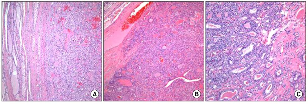
Fig. 1 Hematoxylin and eosin staining. (A) Thyroid follicular adenoma shows microfollicle, prominent nuclei, intact capsule (H&E stain, ×100). (B) Thyroid minimally invasive follicular carcinoma shows mushroom pattern capsular invasion (H&E stain, ×100). (C) Thyroid widely invasive follicular carcinoma shows destructed capsule, aberrant cellular invasion (H&E stain, ×200).
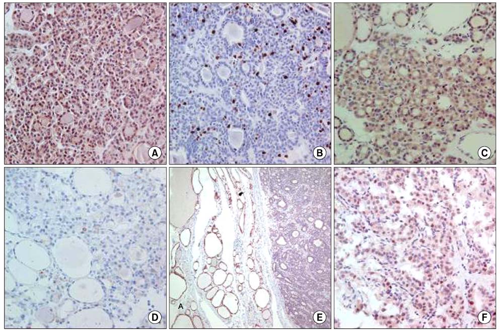
Fig. 2 Immunohistochemical staining. (A) Cyclin D1 in WIFC (IHC stain, ×200), (B) Ki-67 in WIFC (IHC stain, ×400), (C) Survivin in WIFC (IHC stain, ×200), (D) Caspase 3 in WIFC (IHC stain, ×400), (E) Bcl-2 in normal thyroid (A) and MIFC (B) (IHC stain, ×200), (F) p53 in WIFC (IHC stain, ×200).
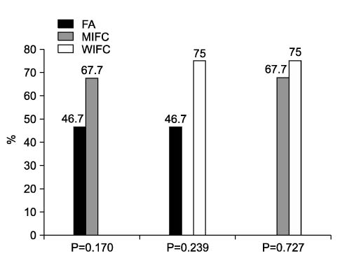
Fig. 3 Proportion of tumors expressing cyclin D1 according to histologic type.
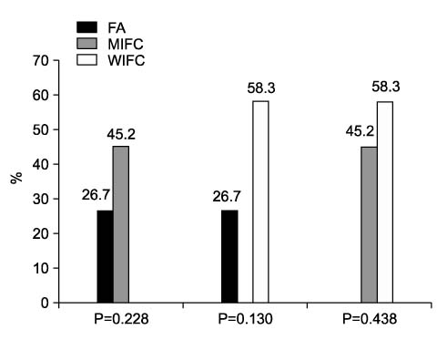
Fig. 4 Proportion of tumors expressing survivin according to histologic type.
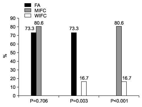
Fig. 5 Proportion of tumors expressing bcl-2 according to histologic type.
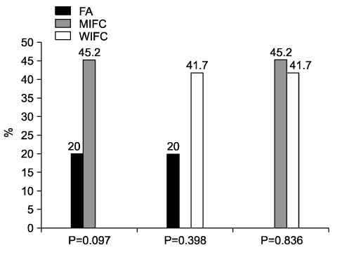
Fig. 6 Proportion of tumors expressing p53 according to histologic type.
Reference
-
1. Rojeski MT, Gharib H. Nodular thyroid disease. Evaluation and management. N Engl J Med. 1985. 313:428–436.2. Siironen P, Nordling S, Louhimo J, Haapiainen R, Haglund C. Immunohistochemical expression of Bcl-2, Ki-67, and p21 in patients with papillary thyroid cancer. Tumour Biol. 2005. 26:50–56.3. Hoshi T, Sasano H, Kato K, Yabuki N, Ohara S, Konno R, et al. Immunohistochemistry of Caspase3/CPP32 in human stomach and its correlation with cell proliferation and apoptosis. Anticancer Res. 1998. 18:4347–4353.4. Basolo F, Pollina L, Fontanini G, Fiore L, Pacini F, Baldanzi A. Apoptosis and proliferation in thyroid carcinoma: correlation with bcl-2 and p53 protein expression. Br J Cancer. 1997. 75:537–541.5. Lu X, Toki T, Konishi I, Nikaido T, Fujii S. Expression of p21WAF1/CIP1 in adenocarcinoma of the uterine cervix: a possible immunohistochemical marker of a favorable prognosis. Cancer. 1998. 82:2409–2417.6. Grossman D, McNiff JM, Li F, Altieri DC. Expression and targeting of the apoptosis inhibitor, survivin, in human melanoma. J Invest Dermatol. 1999. 113:1076–1081.7. Kang YK, Kim WH, Jang JJ. Expression of G1-S modulators (p53, p16, p27, cyclin D1, Rb) and Smad4/Dpc4 in intrahepatic cholangiocarcinoma. Hum Pathol. 2002. 33:877–883.8. Wang S, Lloyd RV, Hutzler MJ, Safran MS, Patwardhan NA, Khan A. The role of cell cycle regulatory protein, cyclin D1, in the progression of thyroid cancer. Mod Pathol. 2000. 13:882–887.9. Goto A, Sakamoto A, Machinami R. An immunohistochemical analysis of cyclin D1, p53, and p21waf1/cip1 proteins in tumors originating from the follicular epithelium of the thyroid gland. Pathol Res Pract. 2001. 197:217–222.10. Gerdes J, Lemke H, Baisch H, Wacker HH, Schwab U, Stein H. Cell cycle analysis of a cell proliferation-associated human nuclear antigen defined by the monoclonal antibody Ki-67. J Immunol. 1984. 133:1710–1715.11. Müller-Höcker J. Immunoreactivity of p53, Ki-67, and Bcl-2 in oncocytic adenomas and carcinomas of the thyroid gland. Hum Pathol. 1999. 30:926–933.12. Augustynowicz A, Dziecioł J, Barwijuk-Machała M, Dadan J, Puchalski Z, Sulkowski S. Assessment of proliferative activity of thyroid Hürthle cell tumors using PCNA, Ki-67 and AgNOR methods. Folia Histochem Cytobiol. 2004. 42:165–168.13. Altieri DC, Marchisio PC. Survivin apoptosis: an interloper between cell death and cell proliferation in cancer. Lab Invest. 1999. 79:1327–1333.14. Ambrosini G, Adida C, Altieri DC. A novel anti-apoptosis gene, survivin, expressed in cancer and lymphoma. Nat Med. 1997. 3:917–921.15. Jiang X, Wilford C, Duensing S, Munger K, Jones G, Jones D. Participation of Survivin in mitotic and apoptotic activities of normal and tumor-derived cells. J Cell Biochem. 2001. 83:342–354.16. Kim TH, Lee TJ, Kim BG, Cha SJ, Park SJ, Lim HM, et al. Expression of survivin and HSP90 in colorectal cancer and its relationship with clinicopathologic factors. J Korean Sur Soc. 2006. 70:113–119.17. Haghpanah V, Shooshtarizadeh P, Heshmat R, Larijani B, Tavangar SM. Immunohistochemical analysis of survivin expression in thyroid follicular adenoma and carcinoma. Appl Immunohistochem Mol Morphol. 2006. 14:422–425.18. Ito Y, Yoshida H, Uruno T, Nakano K, Miya A, Kobayashi K, et al. Survivin expression is significantly linked to the dedifferentiation of thyroid carcinoma. Oncol Rep. 2003. 10:1337–1340.19. Alnemri ES, Livingston DJ, Nicholson DW, Salvesen G, Thornberry NA, Wong WW, et al. Human ICE/CED-3 protease nomenclature. Cell. 1996. 87:171.20. Li YH, Wang C, Meng K, Chen LB, Zhou XJ. Influence of survivin and caspase-3 on cell apoptosis and prognosis in gastric carcinoma. World J Gastroenterol. 2004. 10:1984–1988.21. Sohn JH, Chae SW, Choi KC, Shin HS. Caspase 3 and ki-67 immunoreactivity of apoptosis in gastric adenomas and carcinomas. Korean J Pathol. 2001. 35:286–290.22. Oltvai ZN, Milliman CL, Korsmeyer SJ. Bcl-2 heterodimerizes in vivo with a conserved homolog, Bax, that accelerates programmed cell death. Cell. 1993. 74:609–619.23. Gaffney EF, O'Neil AJ, Staunton MJ. Bcl-2 and prognosis in non-small-cell lung carcinoma. N Engl J Med. 1994. 330:1757–1758.24. Haynik DM, Prayson RA. Immunohistochemical expression of Bcl-2, Bcl-x, and Bax in follicular carcinomas of the thyroid. Appl Immunohistochem Mol Morphol. 2006. 14:417–421.25. Saltman B, Singh B, Hedvat CV, Wreesmann VB, Ghossein R. Patterns of expression of cell cycle/apoptosis genes along the spectrum of thyroid carcinoma progression. Surgery. 2006. 140:899–905.26. Wiseman SM, Griffith OL, Deen S, Rajput A, Masoudi H, Gilks B, et al. Identification of molecular markers altered during transformation of differentiated into anaplastic thyroid carcinoma. Arch Surg. 2007. 142:717–727.27. Fields S, Jang SK. Presence of a potent transcription activating sequence in the p53 protein. Science. 1990. 249:1046–1049.28. Lane DP. Cancer. p53, guardian of the genome. Nature. 1992. 358:15–16.29. Vogelstein B, Kinzler KW. p53 function and dysfunction. Cell. 1992. 70:523–526.30. Li JH, Jeon HJ, Lee MJ. Significance of p53, cyclin D1 and c-myc expressions in thyroid tumors. Korean J Pathol. 2004. 38:29–34.
- Full Text Links
-
- Actions
-
Cited
- CITED
-
- Close
- Share
-
- Similar articles
-
- SPARC Expression in Thyroid Follicular Adenomas and Carcinomas
- A Case of Follicular Adenoma Occurring in Congenital Goiter due to Dyshormogensis
- Demonstration of TCM-9 Monoclonal Antibody in Follicular Neoplasm of Thyroid
- Expression of p53 and MDM2 Proteins in Thyroid Carcinomas
- Ki-67 Labelling Index and Bax Expression According to the Capsular Invasion in the Follicular Neoplasms of the Thyroid
